


ABSTRACT
Objective
Huntington's disease (HD) speech/language disorders have typically been attributed to motor and executive impairment due to striatal dysfunction. In‐depth study of linguistic skills and the role of extrastriatal structures in HD is scarce. This study aimed to explore the profile of language compromise in HD and identify the structural neuroimaging correlates.
Methods
Language and structural correlates were assessed using the Mini Linguistic State Examination (MLSE) in 81 participants (20 HD‐ISS 0‐1, 40 HD‐ISS 2‐3 and 21 controls). Clinical and global cognition measures were also obtained. Imaging data included computed gray matter volume (GMV) and cortical thickness (CTh) values extracted from a general linear model with the MLSE. Correlation analyses were performed with the language components of the MLSE. Multivariate regression analyses were used to explore the predictive ability of the language components on GMV and CTh loss.
Results
HD individuals showed impaired MLSE performance (84.5 ± 12.8), particularly in syntax, motor speech, and to a lesser extent, semantics and phonology. Significant associations were found between linguistic performance and the structural integrity of nodes within the temporo‐parietal, fronto‐parietal, and fronto‐striatal lexical‐semantic and syntactic networks. Correlation analyses linked motor speech and syntax with predominantly left fronto‐striatal GMV and CTh clusters, while semantics had a bilateral fronto‐parietal topography. Multivariate regression analyses showed language domains as independent contributing factors of GMV and CTh loss in classical language‐related regions.
Interpretation
Language impairment is an integral part of the HD cognitive phenotype, with severity associated with structural disintegration in extensive cortico‐subcortical territories involved in language production and processing.
Keywords: brain structure, cognitive phenotype, Huntington's disease, language, neuroimaging
1. Introduction
Huntington's disease (HD) is a monogenic, autosomal dominant neurodegenerative disease caused by a mutation in the HTT gene [1]. While motor abnormalities are the leading symptoms for the clinical diagnosis of HD, it is well known that cognitive and neuropsychiatric abnormalities can appear up to 15 years before the diagnosis [2].
The predominant cognitive disturbances in HD are associated with striatal atrophy, which progressively disrupts the frontal–striatal networks. Accordingly, the progressive decline in attention, processing speed, and executive functions characterizes the cognitive profile of the majority of HD patients [3]. Although atrophy and dysfunction of the basal ganglia play an essential role in the cognitive phenotype of HD, both neuroimaging studies and comprehensive neuropsychological assessments of affected individuals show that neurodegeneration impacts the whole brain. As a result, the neurocognitive profile associates several deficits extending beyond those typically expected as a consequence of basal ganglia dysfunction [4, 5, 6, 7]. Moreover, longitudinal neuropsychological studies have also highlighted the significant heterogeneity observed in HD, with variability in both the rate of cognitive decline and the specific affected domains. Patients who developed a more aggressive pattern of cognitive decline and neurodegeneration were associated with a significantly worse performance in visuoperceptual, linguistic, and memory processes [5, 7, 8]. This highlights the need to emphasize the assessment of processes not restricted to the basal ganglia and the frontal–subcortical circuitry. In this regard, while numerous studies have explored visuoperceptual, spatial, and memory processes in HD, current understanding of the precise neurolinguistic profile of HD, its heterogeneity, neural correlates, and prognostic implications remains limited [9, 10, 11].
Although motor‐related speech symptoms, such as dysarthria, are well documented in nearly all HD patients at some point during the disease [12, 13], less attention has been given to the profile of language impairment and its subcomponents [14]. Early investigations in HD concluded that language impairment could be detected in dementia stages, but probably secondary to the underlying executive dysfunction [15, 16]. Later, thanks to the use of more sophisticated linguistic analysis tools, it was found that linguistic disturbances, including sentence production and comprehension, semantic processing, lexical retrieval, and the application of morphosyntactical rules, were impaired even in the early stages of the disease [5, 17, 18, 19, 20, 21, 22, 23, 24, 25]. The few studies that have addressed the association between language disturbances in HD and brain structure and function restricted their findings to the striatum, showing no evidence of cortical or non‐striatal involvement in the occurrence of these symptoms [17, 19, 22, 25]. In this sense, there is compelling evidence on the key role of the striatum and other subcortical structures on language processes such as manipulation of linguistic rules and units through verbal components of executive function, working memory, and attention, and on how these processes are disrupted in HD [26, 27]. However, we hypothesize that, similarly to what is found with other neurocognitive processes and in alignment with the fact that HD is likely associated with distinct cognitive phenotypes, HD may associate different profiles of language impairment that may in turn relate to additional cortical degeneration and a more severe cognitive profile [5, 28, 29].
In accordance with this idea and in alignment with our previous works on cognitive phenotypes, the present work aims to deepen the profiles of language impairment that HD may associate and to investigate the neural basis of these profiles. By doing so, we aim to not only enhance the clinical understanding of HD but also to explore the role of language assessment as a potentially prognostic tool.
2. Methods
2.1. Participants
We included 60 huntingtin gene expansion carriers (CAG > 39) and 21 healthy controls (HC) from the HD‐Clinic of the Movement Disorders Unit at Hospital de la Santa Creu i Sant Pau. Inclusion criteria were: being right‐handed, having no history of neurological diseases other than HD, and having no history of traumatic brain injury, brain surgery, epilepsy, drug abuse, or uncompensated systemic disease.
Informed consent was obtained from all participants. All procedures were conducted in accordance with the 1964 Helsinki Declaration and its later amendments and approved by the ethics committee from the Hospital de la Santa Creu i Sant Pau.
2.2. Assessments
Clinical and sociodemographic data regarding age, sex, and education were recorded. The severity of motor symptoms was assessed with the Unified Huntington's Disease Rating Scale—Total Motor Score (UHDRS‐TMS) [30]. Total Functional Capacity (TFC) [31] was also recorded. All patients were assigned to Stages 0–3 according to the biological classification of the Huntington's Disease Integrated Staging System [32] (HD‐ISS calculator can be found at https://enroll‐hd.org/calc/html_basic.htm). The CAG age product (CAP score) was calculated based on age and CAG repeat length with the following formula: age x (CAG‐33.66) [33]. The Symbol Digit Modalities Test (SDMT) and Stroop Word Reading Test were administered as measures of processing speed and executive functions [34], as well as to calculate the composite UHDRS score (cUHDRS) [35]. Global cognition was assessed using the Parkinson's Disease—Cognitive Rating Scale (PD‐CRS) [36] an instrument with excellent psychometric properties for HD [37]. Language was assessed using the Spanish version of the Mini Linguistic State Examination (MLSE) [38, 39], a brief screening instrument designed to detect and discriminate types of language impairment in the context of neurodegenerative disorders, especially in primary progressive aphasias (PPA). In the absence of a specific instrument to assess language in HD, we considered the MLSE an excellent option due to its comprehensive assessment of several linguistic processes and its usefulness in other movement disorders [40]. To ensure consistency and reliability, a neuropsychologist with expertise in language disorders (A.P.‐D.) administered the MLSE to all participants.
The test is comprised of 11 subtests, each targeting different aspects of language competence affected by primary progressive aphasias (PPA). The MLSE assesses the nature of language impairment by analyzing the type and frequency of errors made during responses. The test has a maximum overall score of 100, reflecting the severity of linguistic impairment and a profile score representing performance in five domains of linguistic competence: motor‐speech (0–30), phonology (0–30), semantics (0–20), syntax (0–10) and auditory‐verbal working memory (0–10).
The 11 subtests include: (1) picture naming, (2) syllable and multisyllable repetition, (3) word repetition combined with single‐word comprehension, (4) non‐word repetition, (5) non‐verbal semantic association, (6) verbal sentence comprehension, (7) pictorial sentence comprehension, (8) word and non‐word reading, (9) sentence repetition, (10) writing, and (11) picture description. Multiple errors can occur in a single response; for instance, in the naming task, if a participant makes a semantic substitution that also contains a phonological error, both a semantic and a phonological error would be recorded. For more information on tasks and scoring, refer to the original publication [39].
In previous investigations in the context of PPA [38, 39], the average [median (IQR)] MLSE total scores were: semantic variant = 79 (76–82), logopenic variant = 78 (71–84) and non‐fluent variant = 67 (55–76). The optimal cut‐off to differentiate between HC and PPA was 95 points.
2.3. Neuroimaging Acquisition and Preprocessing
T1‐weighted MRI images were acquired on a 3T Philips Achieva using an MPRAGE sequence (TR/TE = 12.65/7.08 milliseconds, flip‐angle = 8°, field of view = 23 cm, matrix = 256 × 256 and slice thickness = 1 mm). We applied standard voxel‐based morphometry (VBM) procedures using the statistical parametric mapping software package (SPM12, http://www.fil.ion.ucl.ac.uk/spm). Tissue probability maps of gray matter volume (GMV) were generated using T1‐weighted MRI scans. These maps were subsequently normalized into the Montreal Neurological Institute (MNI) space using the DARTEL algorithm. To minimize inter‐individual differences, the normalized GMV maps were smoothed using an isotropic spatial filter with a full width at half maximum (FWHM) of 8 × 8 × 8 mm. Surface‐based CTh analysis was performed using the FreeSurfer 6.0 software package (https://surfer.nmr.mgh.harvard.edu). The specific methods used for cortical reconstruction of T1‐weighted MRI brain images have been fully described elsewhere [41]. In short, optimized surface deformation models following intensity gradients accurately identify white matter and gray matter boundaries in the cerebral cortex, from which cortical thickness is computed at each vertex of the resulting surfaces. The resulting vertex‐wise CTh data were normalized to freesurfer's average space and smoothed using a Gaussian kernel of 15 mm FWHM.
2.4. Statistical Analysis
Clinical and demographic data were subjected to independent two‐tailed t‐tests and ANOVA with HSD Tukey's post hoc test for continuous variables with normal distribution. Kruskal–Wallis rank sum test with Conover–Iman post hoc test was used for non‐normal distribution variables. χ 2 was used for categorical variables.
Data are expressed as mean (standard deviation, SD) or median (interquartile range, IQR). Statistics are expressed as t‐stats for t‐student analysis, F for ANOVA, and H for Kruskal–Wallis.
Voxel‐wise and vertex‐wise measures derived from VBM and CTh analyses were introduced into a general linear model (GLM) to explore the structural brain correlates of the MLSE total score. These GLMs included age, CAG, years of education, and sex as nuisance covariates. Only clusters surviving p < 0.05 and family‐wise error (FWE) correction for multiple comparisons were considered significant. FWE correction was performed using cluster‐level RFT for the VBM analyses as implemented in SPM12, and a Monte Carlo simulation with 10,000 repeats for the CTh analysis as implemented in FreeSurfer. To investigate the potential associations between the MLSE total score‐related imaging measures and the performance in the specific linguistic domains, partial correlations using the same covariates and adding the UHDRS‐TMS were performed with the computed average GMV and CTh values at the identified clusters where we observed significant effects. Subsequently, multivariate linear regression models were used to explore the predictive value of linguistic impairment over atrophy in key cortical and subcortical linguistic structures [26, 42]. Assumptions of linearity between predictors and the outcome, as well as normality and homoscedasticity of residuals, were checked and met. Covariates showed no multicollinearity. Model selection was refined using the Akaike Information Criterion (AIC) to compare different models, and the model with the lowest AIC value was chosen. p‐values were adjusted using the Benjamini–Hochberg (BH) method to control the false discovery rate. The significance level was set at α = 0.05.
Analysis was performed in IBM‐SPSS software (version 24; SPSS. Inc. Armonk, NY) and R version 4.2.2 (R Project for Statistical Computing).
3. Results
3.1. Sample Characteristics
Clinical and sociodemographic characteristics of the different groups are presented in Table 1. Presymptomatic patients (HD‐ISS Stages 0–1) had a mean age of 38 (9.08) years, with a female representation of 70%. HD patients (HD‐ISS Stages 2–3) had a mean age of 49.35 (7.96) years, with a female representation of 45%. Healthy Controls (HC) had a mean age of 48.24 (12.43) years, with a female representation of 47.62%. All the participants were bilinguals (Spanish–Catalan) with a proficiency level for Spanish. There was a consistent Spanish‐dominant profile across all groups, with an average of 75.06%, χ 2(4,81) = 1.27, p = 0.866.
TABLE 1.
Demographic and clinical characteristics.
| Healthy controls, n = 21 | HD‐ISS stage 0, n = 11 | HD‐ISS stage 1, n = 9 | HD‐ISS stage 2, n = 13 | HD‐ISS stage 3, n = 27 | Statistic (p‐value) | |
|---|---|---|---|---|---|---|
| Age | 48.24 (12.43) a | 31.73 (4.22) b – d | 45.67 (7.31) | 47.77 (7.33) | 50.11 (8.28) | 8.81 (< 0.001) |
| Education | 15 (11–18) a | 18 (17–20) b | 17 (14–18) e , f | 13 (10.50–17) | 13 (8–16) | 16.71 (0.002) |
| Sex (male/female) | 10/11 | 2/9 | 4/5 | 8/5 | 14/13 | 5.05 (0.282) |
| CAG | — | 42.64 (2.16) | 42.33 (2.06) | 43 (2.35) | 44.33 (2.72) | 2.33 (0.085) |
| CAP score | — | 281.51 (58.95) c , d | 387.42 (62.42) e , f | 438.16 (95.77) | 519.63 (96.84) | 21 (< 0.001) |
| UHDRS‐TMS | 0 (0–0) g – i | 0 (0–0) b – d | 2 (0–4.50) e , f | 9 (4–16.50) j | 40 (30–56) | 71.92 (< 0.001) |
| TFC | 13 (13–13) i | 13 (13–13) d | 13 (13–13) f | 13 (13–13) j | 11 (10–11) | 76.30 (< 0.001) |
| PD‐CRS | 106.62 (13.29) h , i | 112.73 (9.71) c , d | 113.44 (10.84) e , f | 88.54 (18.39) j | 72.26 (18.34) | 24.78 (< 0.001) |
| SDMT | 53.29 (14.97) h , i | 57.73 (8) c , d | 55.89 (8.89) f | 40 (11) j | 23.41 (11.97) | 28.71 (< 0.001) |
| SWRT | 110 (100–126) i | 114 (105–120) b – d | 113 (103–119) e , f | 92 (73.50–102) j | 53 (44–75) | 49.59 (< 0.001) |
| cUHDRS | 18.16 (16.07–19.84) i | 18.50 (17.30–18.95) b – d | 18.10 (17.40–18.47) e , f | 14.42 (13.22–16.72) j | 8.32 (5.74–10.16) | 58.80 (< 0.001) |
| MLSE | 99 (97.50–100) h , i | 100 (98–100) b – d | 98 (95.50–98.50) e , f | 93 (88.5–97) j | 85 (72–92) | 50.9 (< 0.001) |
| Motor speech | 30 (29–30) i | 30 (29–30) b , d | 30 (28.50–30) e , f | 29 (26–29.50) j | 23 (16–28) | 37.26 (< 0.001) |
| Phonology | 30 (29–30) i | 30 (30–30) b – d | 30 (28.50–30) e , f | 29 (27.50–30) | 28 (24–29) | 32.42 (< 0.001) |
| Semantics | 20 (20–20) i | 20 (20–20) d | 20 (19–20) e | 20 (18–20) j | 19 (16–20) | 19.10 (< 0.001) |
| Syntax | 10 (9.50–10) h , i | 10 (10–10) b – d | 10 (9–10) e , f | 8 (8–9) j | 6 (3–8) | 51.63 (< 0.001) |
| Working memory | 10 (9.50–10) | 10 (10–10) d | 10 (6.50–10) e | 9 (8.50–10) | 9 (7–10) | 13.25 (0.010) |
Note: Data are expressed as mean (standard deviation, SD) or median (interquartile range, IQR). ANOVA Post hoc analysis conducted with the Tukey test. Kruskal–Wallis post hoc analysis conducted with the Conover–Iman test. The Benjamini–Hochberg method was applied for multiple comparison correction, yielding adjusted p‐values < 0.05.
Abbreviations: cUHDRS, Composite UHDRS; MLSE, Mini Linguistic State Examination; PD‐CRS, Parkinson's Disease Cognitive Rating Scale; SDMT, Symbol Digit Modalities Test; SWRT, Stroop Word Reading Test; TFC, Total Functional Capacity; UHDRS‐TMS, Unified Huntington's Disease Rating Scale—Total Motor Score.
Healthy Controls vs. HD‐ISS 0; p < 0.05.
HD‐ISS 0 vs. HD‐ISS 1; p < 0.05.
HD‐ISS 0 vs. HD‐ISS 2; p < 0.05.
HD‐ISS 0 vs. HD‐ISS 3; p < 0.05.
HD‐ISS 1 vs. HD‐ISS 2; p < 0.05.
HD‐ISS 1 vs. HD‐ISS 3; p < 0.05.
Healthy Controls vs. HD‐ISS 1; p < 0.05.
Healthy Controls vs. HD‐ISS 2; p < 0.05.
Healthy Controls vs. HD‐ISS 3; p < 0.05.
HD‐ISS 2 vs. HD‐ISS 3; p < 0.05.
Differences among groups were identified in all the studied clinical variables. In general, HD‐ISS 2–3 showed significantly worse scores in cognitive and motor variables than HD‐ISS 0–1 groups and HC. Focusing on linguistic scores, the HD‐ISS 0–1 groups showed no significant differences in linguistic errors compared to HC. HD‐ISS 2–3 patients scored significantly worse than HC in all the domains studied except working memory. However, the FDR‐corrected Connover–Iman post hoc test only revealed differences with the HD‐ISS 3 group, and in MLSE total score and Syntax with the HD‐ISS 2 group. Post hoc analysis also revealed significant differences in all linguistic domains between HD‐ISS Stages 0–1 and HD‐ISS 2–3. Finally, with respect to the HD‐ISS 2 group, the HD‐ISS 3 group performed significantly worse in Motor speech, Semantics, and Syntax, whereas no differences were found in Phonology and Working memory (Figure 1).
FIGURE 1.
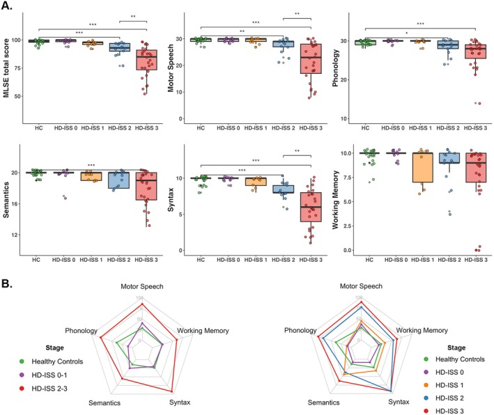
(A) Boxplots of MLSE score domains. The figure shows the differences (p < 0.05) between the different groups. HC, Healthy controls. * = p < 0.05; ** = p < 0.01; *** = p < 0.001. (B) Group radar charts with the percentage of participants with > 1 error in each linguistic domain.
The language profile in symptomatic stages showed compromised syntax, motor speech, and, to a lesser extent, semantics and phonology. Motor‐speech errors were mainly distortions and repetitions. Phonological errors were primarily due to phonological paraphasias and phoneme substitutions in pseudoword reading and repetition tasks. Syntactic processing errors were observed in verbal and pictorial sentence comprehension and syntactic production in writing. Finally, semantic errors displayed as regularization errors in word reading, anomia in picture description, and less frequently in semantic association tasks (for detailed results on the type of errors, refer to Figure S1 and Material S1).
3.2. Neuroimaging Analysis
Given that only the HD‐ISS 2–3 groups presented an impaired MLSE performance compared to the HC group, the neuroimaging GLM analyses were only applied to this combined group to explore the structural brain correlates of the MLSE total score.
Voxel‐wise VBM analysis revealed significant associations between lower MLSE total score and reduced GMV in the bilateral striatum, left insula, left inferior frontal gyrus, left superior and inferior temporal gyrus, left supplementary motor area, and right precentral and middle temporal gyrus (Figure 2). Errors in the different language domains were associated with distinct topographical patterns (Table S1): Motor speech errors were related to lower GMV in the left striatum, left premotor cortex, and left insula. Semantic errors were related to lower GMV in bilateral temporal clusters in regions like the left superior temporal gyrus or the right middle temporal gyrus. Syntactic errors were related to lower GMV in the bilateral striatum and left fronto‐temporal clusters such as the insula or the superior temporal gyrus.
FIGURE 2.

The upper figure shows the GMV correlates of MLSE total score (FWE corrected; p < 0.05) and the association between MLSE domains and GMV. The bottom figure shows the CTh correlates of MLSE total score (FWE corrected; p < 0.05) and the association between MLSE domains and CTh.
The vertexwise CTh analysis revealed a pattern of predominantly left‐lateralized cortical thinning in several fronto‐temporo‐parietal and occipital clusters, including bilateral pars opercularis, left transverse temporal gyrus, right paracentral lobule, right supramarginal gyrus, and bilateral superior and inferior parietal lobules (Figure 2).
Linguistic domains were also associated with distinct topographical patterns in the CTh analysis (Table S2): Motor speech errors were related to thinning in the left hemisphere transverse temporal and postcentral gyrus, right hemisphere supramarginal gyrus, and right rostral middle frontal gyrus. Phonological errors were associated with thinning in the left postcentral gyrus, right inferior parietal, and right precentral gyrus. Semantic errors were related to thinning in the left caudal middle frontal, left rostral middle frontal gyrus, left precuneus, left inferior parietal lobule, and right superior parietal and inferior parietal lobules. Syntactic errors were associated with cortical thinning in the left transverse temporal gyrus, left precentral gyrus, left superior frontal gyrus, left pars opercularis, right banks of the superior temporal sulcus, right superior parietal, right pars opercularis, and right paracentral lobule.
Multivariate regression models, adjusting for UHDRS‐TMS, sex, CAG, and age, identified linguistic components as independent predictors of brain atrophy and loss of GMV in different structures (Tables 2 and 3). Motor speech errors were predictors of GMV loss in the left striatum, left insula, and bilateral supplementary motor area. Syntax errors were predictors of GMV loss in the bilateral striatum, left inferior frontal gyrus, and left posterior superior temporal gyrus. Semantic errors predicted GMV loss in the left posterior superior temporal gyrus and right middle temporal gyrus.
TABLE 2.
Multivariate linear regression models for computed GMV clusters.
| Regression coefficient (95% confidence interval) | Standard error | t‐value | FDR corrected p‐value | |
|---|---|---|---|---|
| L Caudate putamen | ||||
| Motor speech | 0.013 (0.001 to 0.025) | 0.006 | 2.238 | 0.073 |
| UHDRS‐TMS | −0.006 (−0.010 to −0.002) | 0.002 | −2.774 | 0.028 |
| Sex | −0.022 (−0.158 to 0.113) | 0.067 | −0.335 | 0.881 |
| CAG | −0.058 (−0.092 to −0.024) | 0.017 | −3.428 | 0.017 |
| Age | −0.010 (−0.021 to 0.001) | 0.005 | −1.930 | 0.124 |
| L Caudate putamen | ||||
| Syntax | 0.049 (0.018 to 0.079) | 0.015 | 3.222 | 0.021 |
| UHDRS‐TMS | −0.005 (−0.009 to −0.001) | 0.002 | −2.650 | 0.032 |
| Sex | −0.027 (−0.153 to 0.098) | 0.062 | −0.446 | 0.844 |
| CAG | −0.055 (−0.088 to −0.023) | 0.016 | −3.483 | 0.013 |
| Age | −0.011 (−0.022 to −0.001) | 0.005 | −2.329 | 0.062 |
| R Caudate putamen | ||||
| Syntax | 0.029 (0.011 to 0.047) | 0.009 | 3.339 | 0.017 |
| UHDRS‐TMS | −0.003 (−0.006 to −0.001) | 0.001 | −2.850 | 0.025 |
| Sex | −0.031 (−0.104 to 0.042) | 0.036 | −0.871 | 0.557 |
| CAG | −0.036 (−0.055 to −0.017) | 0.009 | −3.912 | 0.003 |
| Age | −0.006 (−0.012 to 0.000) | 0.003 | −2.051 | 0.104 |
| L Frontal inferior | ||||
| Syntax | 0.023 (0.007 to 0.038) | 0.008 | 2.978 | 0.025 |
| UHDRS‐TMS | −0.002 (−0.002 to 0.002) | 0.001 | −0.009 | 0.974 |
| Sex | 0.057 (−0.006 to 0.121) | 0.031 | 1.835 | 0.139 |
| CAG | 0.000 (−0.016 to 0.017) | 0.008 | 0.027 | 0.979 |
| Age | −0.007 (−0.012 to −0.002) | 0.002 | −2.946 | 0.025 |
| L Insula | ||||
| Motor speech | 0.002 (0.000 to 0.003) | 0.001 | 2.673 | 0.031 |
| UHDRS‐TMS | 0.000 (−0.001 to 0.000) | 0.000 | −0.170 | 0.903 |
| Sex | 0.013 (−0.001 to 0.026) | 0.007 | 1.839 | 0.139 |
| CAG | −0.003 (−0.006 to 0.001) | 0.002 | −1.634 | 0.181 |
| Age | −0.001 (−0.002 to 0.000) | 0.001 | −0.386 | 0.863 |
| L Brodmann area 6 | ||||
| Motor speech | 0.004 (0.001 to 0.008) | 0.002 | 2.589 | 0.035 |
| UHDRS‐TMS | −0.001 (−0.002 to 0.001) | 0.001 | −0.184 | 0.903 |
| Sex | −0.033 (−0.071 to 0.005) | 0.019 | −1.740 | 0.163 |
| CAG | 0.001 (−0.009 to 0.010) | 0.005 | 0.032 | 0.903 |
| Age | −0.002 (−0.005 to 0.001) | 0.001 | −0.260 | 0.903 |
| L Superior temporal gyrus | ||||
| Semantics | 0.016 (0.004 to 0.029) | 0.006 | 2.704 | 0.031 |
| UHDRS‐TMS | −0.001 (−0.002 to 0.000) | 0.001 | −0.193 | 0.903 |
| Sex | 0.056 (0.017 to 0.095) | 0.019 | 2.909 | 0.025 |
| CAG | −0.006 (−0.015 to 0.004) | 0.005 | −1.184 | 0.371 |
| Age | −0.004 (−0.007 to −0.001) | 0.001 | −0.376 | 0.863 |
| L Superior temporal gyrus | ||||
| Syntax | 0.018 (0.010 to 0.026) | 0.004 | 4.591 | 0.003 |
| UHDRS‐TMS | 0.000 (−0.001 to 0.001) | 0.000 | −0.067 | 0.785 |
| Sex | 0.058 (0.026 to 0.090) | 0.016 | 3.691 | 0.013 |
| CAG | −0.007 (−0.015 to 0.001) | 0.004 | −1.676 | 0.172 |
| Age | −0.004 (−0.006 to −0.001) | 0.001 | −0.348 | 0.027 |
| R Middle temporal gyrus | ||||
| Semantics | 0.009 (0.003 to 0.016) | 0.003 | 2.868 | 0.025 |
| UHDRS‐TMS | 0.000 (−0.001 to 0.000) | 0.000 | −0.230 | 0.903 |
| Sex | 0.012 (−0.009 to 0.033) | 0.010 | 1.137 | 0.387 |
| CAG | −0.001 (−0.007 to 0.004) | 0.003 | −0.087 | 0.804 |
| Age | −0.002 (−0.004 to −0.001) | 0.001 | −0.448 | 0.025 |
| R Brodmann area 22 | ||||
| Motor speech | 0.085 (0.029 to 0.141) | 0.028 | 3.062 | 0.025 |
| UHDRS‐TMS | −0.013 (−0.033 to 0.007) | 0.010 | −1.323 | 0.305 |
| Sex | 0.626 (−0.014 to 1.267) | 0.315 | 1.987 | 0.115 |
| CAG | −0.055 (−0.218 to 0.107) | 0.080 | −0.693 | 0.685 |
| Age | −0.042 (−0.092 to 0.009) | 0.025 | −1.673 | 0.172 |
Note: The Benjamini–Hochberg method was applied for multiple comparison correction, yielding adjusted p‐values < 0.05.
Abbreviations: L, left; R, right.
TABLE 3.
Multivariate linear regression models for cortical clusters and subcortical volumes.
| Regression coefficient (95% confidence interval) | Standard error | t‐value | FDR corrected p‐value | |
|---|---|---|---|---|
| Putamen volume | ||||
| Motor speech | 0.057 (0.016 to 0.098) | 0.020 | 2.820 | 0.045 |
| UHDRS‐TMS | −0.019 (−0.034 to −0.004) | 0.007 | −2.603 | 0.047 |
| Sex | −0.910 (−1.379 to −0.441) | 0.231 | −3.946 | 0.002 |
| CAG | −0.052 (−0.171 to −0.067) | 0.059 | −0.889 | 0.502 |
| Age | −0.004 (−0.041 to −0.033) | 0.018 | −0.230 | 0.889 |
| Caudate volume | ||||
| Motor speech | 0.050 (0.009 to 0.090) | 0.020 | 2.510 | 0.047 |
| UHDRS‐TMS | −0.020 (−0.034 to −0.005) | 0.007 | −2.773 | 0.045 |
| Sex | −0.896 (−1.353 to −0.440) | 0.225 | −3.988 | 0.002 |
| CAG | −0.087 (−0.203 to 0.029) | 0.057 | −1.532 | 0.216 |
| Age | −0.001 (−0.037 to 0.036) | 0.018 | −0.032 | 0.975 |
| L Transverse temporal gyrus | ||||
| Motor speech | 0.015 (0.007 to 0.024) | 0.004 | 3.628 | 0.011 |
| UHDRS‐TMS | −0.002 (−0.005 to 0.001) | 0.001 | −1.259 | 0.305 |
| Sex | −0.125 (−0.222 to −0.029) | 0.048 | −2.639 | 0.045 |
| CAG | −0.015 (−0.040 to 0.009) | 0.012 | −1.258 | 0.305 |
| Age | −0.009 (−0.016 to −0.001) | 0.004 | −2.312 | 0.063 |
| L Caudal middle frontal gyrus | ||||
| Syntax | 0.026 (0.005 to 0.048) | 0.011 | 2.462 | 0.047 |
| UHDRS‐TMS | −0.002 (−0.005 to 0.001) | 0.001 | −1.539 | 0.216 |
| Sex | −0.070 (−0.159 to 0.018) | 0.044 | −1.609 | 0.202 |
| CAG | −0.004 (−0.027 to 0.019) | 0.011 | −0.344 | 0.845 |
| Age | −0.010 (−0.017 to −0.003) | 0.003 | −2.929 | 0.038 |
| L Rostral middle frontal gyrus | ||||
| Semantics | 0.030 (0.001 to 0.060) | 0.014 | 2.107 | 0.090 |
| UHDRS‐TMS | −0.003 (−0.005 to 0.000) | 0.001 | −2.088 | 0.090 |
| Sex | −0.101 (−0.194 to −0.008) | 0.046 | −2.215 | 0.076 |
| CAG | 0.002 (−0.021 to 0.026) | 0.011 | 0.216 | 0.889 |
| Age | −0.003 (−0.010 to 0.004) | 0.003 | −0.138 | 0.562 |
| L Inferior parietal lobule | ||||
| Semantics | 0.044 (0.008 to 0.081) | 0.018 | 2.453 | 0.047 |
| UHDRS‐TMS | −0.003 (−0.006 to 0.000) | 0.002 | −1.876 | 0.129 |
| Sex | −0.144 (−0.259 to −0.029) | 0.057 | −2.538 | 0.047 |
| CAG | 0.004 (−0.025 to 0.032) | 0.014 | 0.255 | 0.889 |
| Age | −0.003 (−0.012 to 0.005) | 0.004 | −0.804 | 0.549 |
| L Pars opercularis | ||||
| Syntax | 0.032 (0.007 to 0.057) | 0.012 | 2.638 | 0.045 |
| UHDRS‐TMS | −0.002 (−0.005 to 0.001) | 0.002 | −0.205 | 0.321 |
| Sex | −0.074 (−0.176 to 0.028) | 0.050 | −1.478 | 0.221 |
| CAG | −0.006 (−0.032 to 0.021) | 0.013 | −0.080 | 0.795 |
| Age | −0.007 (−0.015 to 0.001) | 0.004 | −0.316 | 0.140 |
| R Inferior parietal lobule | ||||
| Semantics | 0.041 (0.007 to 0.074) | 0.016 | 2.488 | 0.047 |
| UHDRS‐TMS | −0.002 (−0.005 to 0.001) | 0.001 | −0.227 | 0.221 |
| Sex | −0.173 (−0.278 to −0.068) | 0.052 | −3.353 | 0.018 |
| CAG | −0.002 (−0.028 to 0.025) | 0.013 | −0.021 | 0.926 |
| Age | −0.010 (−0.018 to −0.002) | 0.004 | −0.435 | 0.045 |
| R Paracentral lobule | ||||
| Syntax | 0.048 (0.022 to 0.075) | 0.013 | 3.698 | 0.011 |
| UHDRS‐TMS | −0.001 (−0.004 to 0.002) | 0.002 | −0.096 | 0.679 |
| Sex | −0.168 (−0.276 to −0.060) | 0.053 | −3.152 | 0.022 |
| CAG | 0.026 (−0.002 to 0.054) | 0.014 | 0.342 | 0.127 |
| Age | 0.001 (−0.008 to 0.009) | 0.004 | 0.031 | 0.891 |
Note: The Benjamini–Hochberg method was applied for multiple comparison correction, yielding adjusted p‐values < 0.05.
Abbreviations: L, left; R, right.
Regarding CTh, motor speech errors were predictors of atrophy in the bilateral striatum and the left transverse temporal gyrus. Syntactic errors predicted atrophy in the left caudal middle frontal gyrus, the left pars opercularis, and the right paracentral lobule. Semantic errors predicted CTh loss in the left rostral middle frontal gyrus and the left inferior parietal lobule. Sex also appeared to be a significant predictor of atrophy in most linear regression models.
4. Discussion
In the present study, we investigated the profile of language impairment in HD alongside its structural brain correlates. The main findings were that patients in HD‐ISS Stages 2 and 3 exhibited significant language impairments, mainly involving syntactic processing and production, together with motor speech, and, to a lesser extent, semantic and phonological errors. These alterations were associated with a loss of gray matter volume and cortical thickness in cortico‐subcortical regions predominantly lateralized to the left hemisphere, aligning with classic language areas [42]. In contrast, HD‐ISS Stages 0 and 1 did not demonstrate language impairments on the MLSE compared to HC. It is important to note that the clinical expression of language impairment in neurodegenerative diseases results from structural and/or functional disruption of the nodes that compose the language networks [43]. Although several structural and functional changes have been described in presymptomatic gene‐mutation carriers [4, 6, 44], these alterations typically show limited association with cognitive performance on standardized measures. Therefore, it cannot be expected to find overt language alterations in this population. Addressing potential language dysfunction in presymptomatic HD probably requires more complex methods of assessment of subtle linguistic compromise. Focusing on functional brain imaging and neurophysiological data could help explore the possible brain mechanisms underlying these subtle linguistic deficits.
From a neurolinguistic perspective, HD patients showed linguistic errors in syntactic processing from HD‐ISS Stage 2, with an increased number and variety of errors, including motor speech, semantic, and phonological errors, at HD‐ISS Stage 3. Syntactic and motor speech errors were clearly more prominent than semantic and phonological errors. At a descriptive level, this linguistic profile suggests certain similarities to the patterns observed in the non‐fluent forms of primary progressive aphasias (nfPPA), though with much milder severity (median MLSE score nfPPA = 67, median MLSE score HD‐ISS 3 = 85). While the imaging correlates of language impairment in our sample also showed some overlap with the typical findings of atrophy and hypometabolism reported in nfPPA [45, 46], we also found involvement of posterior left temporo‐parietal regions, often associated with phonological loop disruptions and lexical retrieval deficits through the involvement of the superior longitudinal fasciculus, anomalies more characteristic of the logopenic variant APP [45]. This observation raises the possibility of the coexistence of subtle difficulties related to lexical access and working memory, more attributable to posterior temporo‐parietal anomalies [45, 46, 47]. Beyond attempting to draw parallels between clearly distinct pathological entities, this comparative description in relation to previous evidence may be useful for understanding the cognitive mechanisms underlying the type of linguistic errors in the context of its structural topography.
The type of errors made by HD patients throughout the tasks were characterized by prominent deficits in syntactic processing in verbal and pictorial sentence comprehension and syntactic production in writing, together with motor‐speech distortions and repetitions, as reported in previous HD studies [12, 20, 25]. The topography of this type of errors had been traditionally circumscribed to the striatum [19, 22, 25] for its role in the manipulation of linguistic rules through the verbal components of executive functions, working memory, and attention [27]. However, the presence of other types of errors, such as those in the semantic domain, with problems of regularization during reading and semantic association, had not been previously reported [14]. Given the type of cognitive processes dependent on the integrity of the semantic network, the presence of this type of errors suggests a fronto‐temporo‐parietal topography that may extend beyond typical language‐related cortical regions [43, 48]. Future research should explore the role of structural and functional connectivity of these complex language networks. In parallel, the presence of phonological paraphasias and paralexias in the repetition and reading of pseudowords suggests the involvement of a network that includes regions responsible for phonological processing and decoding, along with regions responsible for planning of speech [47, 49].
Overall, language components do not act in isolation, making it challenging to determine whether the observed errors were strictly linguistic or influenced by other cognitive processes or motor‐speech disturbances. Differentiating between phonological and motor‐speech errors can be challenging in a population with a predominance of dysarthria, as both errors can often co‐occur [50]. Moreover, the origin of phonological errors may involve overlapping linguistic and working memory mechanisms [24, 51]. For example, phonological errors identified in tasks like pseudoword repetition could arise from deficits in working memory, such as impairments in the phonological loop, rather than purely from disruptions in the retrieval of phonological representations or phonological processing. Given the limitations of a brief screening test in distinguishing between these features, we ensured to strictly adhere to the scoring guidelines and only score a phonological error when it was evident (e.g., substitutions, repetitions or omissions) and still conformed to the phonotactic rules of the language.
Similarly, syntactic errors may not exclusively reflect deficits in syntactic processing but could also be influenced by impairments in working memory or executive dysfunction. Although research has shown that syntactic processing errors persist even when working memory demands are controlled [20], the tools used in this study do not allow us to definitively isolate syntactic deficits from these broader cognitive influences.
What seems clear is that, given the type of errors identified and the cognitive processes involved, it is plausible to consider that language problems in HD transcend striatal involvement. Future research should study the prognostic role of language‐dependent cognitive processes in longitudinal cognitive decline, with a focus on disentangling the contributions of linguistic, cognitive, and motor speech components to better understand the complexity of language impairments in this population.
From a structural neuroimaging perspective, we showed that language impairment in HD was associated with a left‐hemisphere‐lateralized topography that included clusters of GMV or CTh loss in frontal regions (e.g., pars opercularis, middle frontal gyrus), temporal regions (e.g., temporal transverse gyrus, bilateral middle temporal gyrus), bilateral inferior parietal lobule clusters, and left striatum, most of which were central nodes of the main language networks [27, 42, 43, 47, 48]. Previous neuroimaging and language studies in HD have implicated the striatum in syntactic processing and grammatical rule application [22, 25]. In a study of sentence grammaticality judgments using fMRI in healthy individuals, an anterior–posterior gradient of cognitive control was found in the dorsomedial striatum, with greater cognitive load leading to greater recruitment of anterior regions, which were functionally connected to frontal regions [52]. In our study, the role of the striatum was central to processes related to syntax and motor speech. Indeed, these processes, together with UHDRS‐TMS and CAG, were independent predictors of striatal volume loss. But beyond striatal volume loss and its disruption of corticostriatal circuitry [26], we identified a number of neuroanatomical correlates related to different language processes not previously described in HD. Motor speech errors were independent predictors of GMV loss and cortical atrophy in left frontal‐subcortical motor regions, syntactic errors predicted structural compromise of left fronto‐temporal and bilateral striatal topography, and semantic errors predicted bilateral medial and superior temporal compromise. On the other hand, sex also appeared to be an important predictor of atrophy in some models of our sample. Thus, further investigations should take into account the possible role of sex in the expression of the linguistic impairment profile and the degree of atrophy.
From a clinical perspective, our study also suggests the usefulness of the MLSE for the assessment of language in HD, given the brain correlates of each language domain with some specific brain regions.
As a strength of the study, we explored language impairment in a representative sample of HD unambiguously classified using the HD‐ISS classification. Moreover, we obtained structural MRI data from all participants and compared the performance of all groups against HC using a validated language measure to detect aphasic‐like errors. To our knowledge, this represents the largest study to date with available MRI data.
The present study also has some limitations. Having only structural measures limits the possible analyses and interpretations. Incorporating functional MRI and DTI measures would have provided additional insights into the progressive disruption of language network nodes across HD‐ISS stages. Diffusion‐based metrics could have further elucidated the structural connectivity alterations within language networks. Moreover, a longitudinal analysis of a bigger cohort may have contributed to deepening the possible heterogeneity in terms of severity of these symptoms in HD patients and on the prognostic implications of language disturbances in the prediction of different rates of cognitive or clinical progression. Finally, obtaining biological biomarkers would have strengthened the hypothesis of the role that language may play in association with a more aggressive cognitive phenotype [7].
In conclusion, HD patients exhibit evident linguistic errors with a predominance of syntax and motor‐speech disturbances, but also some degree of semantic and phonological compromise. The topography of these impairments was associated with damage in several structures related to language networks, mostly in the left hemisphere. Overall, this study highlights the need to incorporate language assessment as part of the comprehensive neuropsychological evaluation of individuals with HD, as a tool to deepen understanding of the underlying dysfunctional mechanisms with possible prognostic implications in the expression of the cognitive phenotype.
Author Contributions
A.P.‐D., J.K., and S.M.‐H. contributed to conception and design of the study. A.P.‐D., C.F.‐M., I.R.‐B., F.S., J.P.‐P., G.O.‐S., L.P.‐C., A.H.‐B., I.A.‐B., and S.M.‐H. contributed to data acquisition. A.P.‐D., I.R.‐B., F.S., and S.M.‐H. contributed to the design, execution and review of the statistical analysis. A.P.‐D., I.R.‐B., and S.M.‐H. wrote the first draft. A.P.‐D., C.F.‐M., I.R.‐B., F.S., J.P.‐P., J.A.M.‐G., F.C., G.O.‐S., L.P.‐C., A.H.‐B., I.A.‐B., J.P., J.K., and S.M.‐H. contributed to the revision of the final version of the manuscript.
Conflicts of Interest
The authors declare no conflicts of interest.
Supporting information
Data S1.
Acknowledgments
This study was funded by Fondo de Investigaciones Sanitarias from the Instituto de Salud Carlos III (ISCIII) and Fondo Europeo de Desarrollo Regional grant #PI21/01758. A.P.‐D. is the recipient of a predoctoral grant from the Government of Andorra (ATC027‐AND/2021), I.R.‐B. is the recipient of a Rio Hortega contract from ISCIII (CM22/00072), and I.A.‐B. is the recipient of a Juan Rodés contract from ISCIII (JR22/00059). None of the funders played any role at any stage of the study. We thank patients and caregivers for their effort and commitment to the study.
Funding: This study was funded by Fondo de Investigaciones Sanitarias from the Instituto de Salud Carlos III (ISCIII) and Fondo Europeo de Desarrollo Regional grant PI21/01758. A.P.‐D. is the recipient of a predoctoral contract from the Government of Andorra (ATC027‐AND/2021), I.R.‐B. is the recipient of a Rio Hortega contract from ISCIII (CM22/00072), and I.A.‐B. is the recipient of a Juan Rodés contract from ISCIII (JR22/00059).
Funding Statement
This work was funded by Instituto de Salud Carlos III grant PI21/01758.
Contributor Information
Jaime Kulisevsky, Email: jaime.kulisevsky@uab.cat.
Saul Martinez‐Horta, Email: smartinezho@santpau.cat.
Data Availability Statement
The data sets generated or analyzed during the study are available from the corresponding authors upon reasonable request.
References
- 1. Ross C. A. and Tabrizi S. J., “Huntington's Disease: From Molecular Pathogenesis to Clinical Treatment,” Lancet Neurology 10, no. 1 (2011): 83–98. [DOI] [PubMed] [Google Scholar]
- 2. Paulsen J. S., Miller A. C., Hayes T., and Shaw E., “Cognitive and Behavioral Changes in Huntington Disease Before Diagnosis,” Handbook of Clinical Neurology 144 (2017): 69–91. [DOI] [PubMed] [Google Scholar]
- 3. Baake V., Reijntjes R. H. A. M., Dumas E. M., et al., “Cognitive Decline in Huntington's Disease Expansion Gene Carriers,” Cortex; A Journal Devoted to the Study of the Nervous System and Behavior 95 (2017): 51–62. [DOI] [PubMed] [Google Scholar]
- 4. Tabrizi S. J., Scahill R. I., Owen G., et al., “Predictors of Phenotypic Progression and Disease Onset in Premanifest and Early‐Stage Huntington's Disease in the TRACK‐HD Study: Analysis of 36‐Month Observational Data,” Lancet Neurology 12, no. 7 (2013): 637–649, 10.1016/S1474-4422(13)70088-7. [DOI] [PubMed] [Google Scholar]
- 5. Martinez‐Horta S., Sampedro F., Horta‐Barba A., et al., “Structural Brain Correlates of Dementia in Huntington's Disease,” NeuroImage: Clinical 28 (2020): 102415. [DOI] [PMC free article] [PubMed] [Google Scholar]
- 6. Wijeratne P. A., Garbarino S., Gregory S., et al., “Revealing the Timeline of Structural MRI Changes in Premanifest to Manifest Huntington Disease,” Neurology Genetics 7, no. 5 (2021): e617. [DOI] [PMC free article] [PubMed] [Google Scholar]
- 7. Martinez‐Horta S., Perez‐Perez J., Perez‐Gonzalez R., et al., “Cognitive Phenotype and Neurodegeneration Associated With Tau in Huntington's Disease,” Annals of Clinical Translational Neurology 11, no. 5 (2024): 1160–1171. [DOI] [PMC free article] [PubMed] [Google Scholar]
- 8. Martinez‐Horta S., Perez‐Perez J., Oltra‐Cucarella J., et al., “Divergent Cognitive Trajectories in Early Stage Huntington's Disease: A 3‐Year Longitudinal Study,” European Journal of Neurology 30, no. 7 (2023): 1871–1879. [DOI] [PubMed] [Google Scholar]
- 9. Coppen E. M., Jacobs M., van der Zwaan K. F., et al., “Visual Object Perception in Premanifest and Early Manifest Huntington's Disease,” Archives of Clinical Neuropsychology 34, no. 8 (2019): 1320–1328. [DOI] [PMC free article] [PubMed] [Google Scholar]
- 10. Labuschagne I., Cassidy A. M., Scahill R. I., et al., “Visuospatial Processing Deficits Linked to Posterior Brain Regions in Premanifest and Early Stage Huntington's Disease,” Journal of the International Neuropsychological Society 22, no. 6 (2016): 595–608. [DOI] [PubMed] [Google Scholar]
- 11. Harris K. L., Armstrong M., Swain R., et al., “Huntington's Disease Patients Display Progressive Deficits in Hippocampal‐Dependent Cognition During a Task of Spatial Memory,” Cortex; A Journal Devoted to the Study of the Nervous System and Behavior 119 (2019): 417–427. [DOI] [PubMed] [Google Scholar]
- 12. Diehl S. K., Mefferd A. S., Lin Y.‐C., et al., “Motor Speech Patterns in Huntington Disease,” Neurology 93, no. 22 (2019): e2042–e2052, 10.1212/WNL.0000000000008541. [DOI] [PMC free article] [PubMed] [Google Scholar]
- 13. Kouba T., Frank W., Tykalova T., et al., “Speech Biomarkers in Huntington's Disease: A Cross‐Sectional Study in Pre‐Symptomatic, Prodromal and Early Manifest Stages,” European Journal of Neurology 30, no. 5 (2023): 1262–1271. [DOI] [PubMed] [Google Scholar]
- 14. Gagnon M., Barrette J., and Macoir J., “Language Disorders in Huntington Disease: A Systematic Literature Review,” Cognitive and Behavioral Neurology: Official Journal of the Society for Behavioral and Cognitive 31, no. 4 (2018): 179–192. [DOI] [PubMed] [Google Scholar]
- 15. Podoll K., Caspary P., Lange H. W., and Noth J., “Language Functions in Huntington's Disease,” Brain: A Journal of Neurology 111, no. Pt 6 (1988): 1475–1503. [DOI] [PubMed] [Google Scholar]
- 16. Wallesch C. W. and Fehrenbach R. A., “On the Neurolinguistic Nature of Language Abnormalities in Huntington's Disease,” Journal of Neurology, Neurosurgery, and Psychiatry 51, no. 3 (1988): 367–373. [DOI] [PMC free article] [PubMed] [Google Scholar]
- 17. De Diego‐Balaguer R., Couette M., Dolbeau G., et al., “Striatal Degeneration Impairs Language Learning: Evidence From Huntington's Disease,” Brain: A Journal of Neurology 131, no. Pt 11 (2008): 2870–2881. [DOI] [PMC free article] [PubMed] [Google Scholar]
- 18. Giavazzi M., Daland R., Palminteri S., et al., “The Role of the Striatum in Linguistic Selection: Evidence From Huntington's Disease and Computational Modeling,” Cortex; A Journal Devoted to the Study of the Nervous System and Behavior 109 (2018): 189–204. [DOI] [PubMed] [Google Scholar]
- 19. Hinzen W., Rosselló J., Morey C., et al., “A Systematic Linguistic Profile of Spontaneous Narrative Speech in Pre‐Symptomatic and Early Stage Huntington's Disease,” Cortex; A Journal Devoted to the Study of the Nervous System and Behavior 100 (2018): 71–83. [DOI] [PMC free article] [PubMed] [Google Scholar]
- 20. Sambin S., Teichmann M., de Diego Balaguer R., et al., “The Role of the Striatum in Sentence Processing: Disentangling Syntax From Working Memory in Huntington's Disease,” Neuropsychologia 50, no. 11 (2012): 2625–2635. [DOI] [PubMed] [Google Scholar]
- 21. Teichmann M., Dupoux E., Cesaro P., and Bachoud‐Lévi A.‐C., “The Role of the Striatum in Sentence Processing: Evidence From a Priming Study in Early Stages of Huntington's Disease,” Neuropsychologia 46, no. 1 (2008): 174–185. [DOI] [PubMed] [Google Scholar]
- 22. Teichmann M., Dupoux E., Kouider S., et al., “The Role of the Striatum in Rule Application: The Model of Huntington's Disease at Early Stage,” Brain: A Journal of Neurology 128, no. Pt 5 (2005): 1155–1167. [DOI] [PubMed] [Google Scholar]
- 23. Teichmann M., Dupoux E., Kouider S., and Bachoud‐Lévi A.‐C., “The Role of the Striatum in Processing Language Rules: Evidence From Word Perception in Huntington's Disease,” Journal of Cognitive Neuroscience 18, no. 9 (2006): 1555–1569. [DOI] [PubMed] [Google Scholar]
- 24. Teichmann M., Darcy I., Bachoud‐Lévi A.‐C., and Dupoux E., “The Role of the Striatum in Phonological Processing: Evidence From Early Stages of Huntington's Disease,” Cortex; A Journal Devoted to the Study of the Nervous System and Behavior 45, no. 7 (2009): 839–849. [DOI] [PubMed] [Google Scholar]
- 25. Teichmann M., Gaura V., Démonet J.‐F., et al., “Language Processing Within the Striatum: Evidence From a PET Correlation Study in Huntington's Disease,” Brain: A Journal of Neurology 131, no. Pt 4 (2008): 1046–1056. [DOI] [PubMed] [Google Scholar]
- 26. Copland D. A., Brownsett S., Iyer K., and Angwin A. J., “Corticostriatal Regulation of Language Functions,” Neuropsychology Review 31, no. 3 (2021): 472–494. [DOI] [PubMed] [Google Scholar]
- 27. Jacquemot C. and Bachoud‐Lévi A.‐C., “Striatum and Language Processing: Where Do We Stand?,” Cognition 213 (2021): 104785. [DOI] [PubMed] [Google Scholar]
- 28. Camerino I., Ferreira J., Vonk J. M., et al., “Systematic Review and Meta‐Analyses of Word Production Abilities in Dysfunction of the Basal Ganglia: Stroke, Small Vessel Disease, Parkinson's Disease, and Huntington's Disease,” Neuropsychology Review 34, no. 1 (2024): 1–26. [DOI] [PubMed] [Google Scholar]
- 29. Clarke A. J., Manser D., Fleischer R., Fulham M., and Ahmed R. M., “Pearls & oy‐Sters: Huntington Disease Presenting as Primary Progressive Aphasia: A Case of Semantics,” Neurology 101, no. 9 (2023): 414–417. [DOI] [PMC free article] [PubMed] [Google Scholar]
- 30. Huntington Study Group , “Unified Huntington's Disease Rating Scale: Reliability and Consistency. Huntington Study Group,” Movement Disorders, Official Journal of the Movement Disorder Society 11, no. 2 (1996): 136–142. [DOI] [PubMed] [Google Scholar]
- 31. Shoulson I. and Fahn S., “Huntington Disease: Clinical Care and Evaluation,” Neurology 29, no. 1 (1979): 1–3. [DOI] [PubMed] [Google Scholar]
- 32. Tabrizi S. J., Schobel S., Gantman E. C., et al., “A Biological Classification of Huntington's Disease: The Integrated Staging System,” Lancet Neurology 21, no. 7 (2022): 632–644, 10.1016/S1474-4422(22)00120-X. [DOI] [PubMed] [Google Scholar]
- 33. Zhang Y., Long J. D., Mills J. A., et al., “Indexing Disease Progression at Study Entry With Individuals At‐Risk for Huntington Disease,” American Journal of Medical Genetics, Part B: Neuropsychiatric Genetics, Official Publication of the International Society of Psychiatric Genetics 156B, no. 7 (2011): 751–763. [DOI] [PMC free article] [PubMed] [Google Scholar]
- 34. Landwehrmeyer G. B., Fitzer‐Attas C. J., Giuliano J. D., et al., “Data Analytics From Enroll‐HD, a Global Clinical Research Platform for Huntington's Disease,” Movement Disorders Clinical Practice 4, no. 2 (2017): 212–224. [DOI] [PMC free article] [PubMed] [Google Scholar]
- 35. Schobel S. A., Palermo G., Auinger P., et al., “Motor, Cognitive, and Functional Declines Contribute to a Single Progressive Factor in Early HD,” Neurology 89, no. 24 (2017): 2495–2502. [DOI] [PMC free article] [PubMed] [Google Scholar]
- 36. Pagonabarraga J., Kulisevsky J., Llebaria G., et al., “Parkinson's Disease‐Cognitive Rating Scale: A New Cognitive Scale Specific for Parkinson's Disease,” Movement Disorders, Official Journal of the Movement Disorder Society 23, no. 7 (2008): 998–1005. [DOI] [PubMed] [Google Scholar]
- 37. Martinez‐Horta S., Horta‐Barba A., Perez‐Perez J., et al., “Utility of the Parkinson's Disease‐Cognitive Rating Scale for the Screening of Global Cognitive Status in Huntington's Disease,” Journal of Neurology 267, no. 5 (2020): 1527–1535. [DOI] [PubMed] [Google Scholar]
- 38. Matias‐Guiu J. A., Pytel V., Hernández‐Lorenzo L., et al., “Spanish Version of the Mini‐Linguistic State Examination for the Diagnosis of Primary Progressive Aphasia,” Journal of Alzheimer's Disease 83, no. 2 (2021): 771–778. [DOI] [PubMed] [Google Scholar]
- 39. Patel N., Peterson K. A., Ingram R. U., et al., “A ‘Mini Linguistic State Examination’ to Classify Primary Progressive Aphasia,” Brain Communications 4, no. 2 (2021): fcab299. [DOI] [PMC free article] [PubMed] [Google Scholar]
- 40. Peterson K. A., Jones P. S., Patel N., et al., “Language Disorder in Progressive Supranuclear Palsy and Corticobasal Syndrome: Neural Correlates and Detection by the MLSE Screening Tool,” Frontiers in Aging Neuroscience 13 (2021): 675739. [DOI] [PMC free article] [PubMed] [Google Scholar]
- 41. Puig‐Davi A., Martinez‐Horta S., Sampedro F., et al., “Cognitive and Affective Empathy in Huntington's Disease,” Journal of Huntington's Disease 10, no. 3 (2021): 323–334. [DOI] [PubMed] [Google Scholar]
- 42. Lipkin B., Tuckute G., Affourtit J., et al., “Probabilistic Atlas for the Language Network Based on Precision fMRI Data From >800 Individuals,” Scientific Data 9, no. 1 (2022): 529. [DOI] [PMC free article] [PubMed] [Google Scholar]
- 43. Fedorenko E., Ivanova A. A., and Regev T. I., “The Language Network as a Natural Kind Within the Broader Landscape of the Human Brain,” Nature Reviews. Neuroscience 25, no. 5 (2024): 289–312. [DOI] [PMC free article] [PubMed] [Google Scholar]
- 44. Estevez‐Fraga C., Scahill R. I., Durr A., et al., “Composite UHDRS Correlates With Progression of Imaging Biomarkers in Huntington's Disease,” Movement Disorders, Official Journal of the Movement Disorder Society 36, no. 5 (2021): 1259–1264. [DOI] [PubMed] [Google Scholar]
- 45. Routier A., Habert M.‐O., Bertrand A., et al., “Structural, Microstructural, and Metabolic Alterations in Primary Progressive Aphasia Variants,” Frontiers in Neurology 9 (2018): 766. [DOI] [PMC free article] [PubMed] [Google Scholar]
- 46. Mesulam M. M., Coventry C. A., Bigio E. H., et al., “Neuropathological Fingerprints of Survival, Atrophy and Language in Primary Progressive Aphasia,” Brain: A Journal of Neurology 145, no. 6 (2022): 2133–2148. [DOI] [PMC free article] [PubMed] [Google Scholar]
- 47. Hickok G., “The Dual Stream Model of Speech and Language Processing,” Handbook of Clinical Neurology 185 (2022): 57–69. [DOI] [PubMed] [Google Scholar]
- 48. Lau E. F., Phillips C., and Poeppel D., “A Cortical Network for Semantics: (de)constructing the N400,” Nature Reviews. Neuroscience 9, no. 12 (2008): 920–933. [DOI] [PubMed] [Google Scholar]
- 49. Wilson S. M., Henry M. L., Besbris M., et al., “Connected Speech Production in Three Variants of Primary Progressive Aphasia,” Brain: A Journal of Neurology 133, no. Pt 7 (2010): 2069–2088. [DOI] [PMC free article] [PubMed] [Google Scholar]
- 50. Galluzzi C., Bureca I., Guariglia C., and Romani C., “Phonological Simplifications, Apraxia of Speech and the Interaction Between Phonological and Phonetic Processing,” Neuropsychologia 71 (2015): 64–83. [DOI] [PubMed] [Google Scholar]
- 51. Perrachione T. K., Ghosh S. S., Ostrovskaya I., et al., “Phonological Working Memory for Words and Nonwords in Cerebral Cortex,” Journal of Speech, Language, and Hearing Research 60, no. 7 (2017): 1959–1979. [DOI] [PMC free article] [PubMed] [Google Scholar]
- 52. Mestres‐Missé A., Turner R., and Friederici A. D., “An Anterior‐Posterior Gradient of Cognitive Control Within the Dorsomedial Striatum,” NeuroImage 62, no. 1 (2012): 41–47. [DOI] [PubMed] [Google Scholar]
Associated Data
This section collects any data citations, data availability statements, or supplementary materials included in this article.
Supplementary Materials
Data S1.
Data Availability Statement
The data sets generated or analyzed during the study are available from the corresponding authors upon reasonable request.