

Abstract
Introduction and importance
While MoM tribology gained popularity in the late 20th and early 21st centuries mainly due to the low wear rates and excellent durability, in the following years, it has been the cause of many implant failures due to adverse reactions to metal debris (ARMD) of different entities.
Case presentation
We presented the case of a 61 years old female with failure of a metal-on-metal (MoM) THA, due to an improper positioning of the cup. The excessive verticality of the component generated the failure.
Clinical discussion
Since acetabular cup orientation is one of the most critical factors influencing the performance and longevity of MoM total hip prostheses, this study highlights how incorrect cup orientation can cause a negative and sometimes catastrophic outcome, necessitating revision.
Conclusion
The precise acetabular cup positioning in MoM hip prosthesis geometry is essential in preventing functional overloading, release of metal ions and adverse tissue reactions. When evaluating patients with metal-on-metal hip prostheses, it is necessary to consider the orientation of the cup as a predictive factor for failure and wear of the components.
Keywords: Acetabular orientation, Metal-On-Metal (MoM), Edge loading, Metallosis, Pseudotumor, Revision total hip arthroplasty, Case report
Highlights
-
•
An improper inclination of the acetabular cup can cause a variety of complications.
-
•
We present the case of a MoM THA with a vertical tilt of the acetabular cup, who come to our attention due to hip dislocation.
-
•
A bulky pseudotumor was discovered during surgery, infiltrating soft tissues.
-
•
Cup orientation is essential in preventing functional overloading, excessive release of metal ions and adverse tissue reactions.
1. Introduction
Total hip arthroplasty (THA) is one of the most performed orthopedic surgeries [1]. Through the years, this procedure has changed from a technical point of view, along with the instruments and components employed [2,3]. Among various bearing surface options, metal-on-metal (MoM) hip prostheses have gained popularity in the late 20th and early 21st centuries, mainly thanks to the low wear rates and more excellent durability than traditional couplings. In the following years, MoM THAs have caused high rates of implant failures due to adverse reactions to metal debris (ARMD) ranging from local tissue asymptomatic lesions to serious complications such as osteolysis, solid or cystic pseudotumor and necrosis, with high blood levels of chrome (Cr) and cobalt (Co), with a consequent risk of systemic manifestations [[4], [5], [6], [7]]. All these concerns appeared, leading to a decline in MoM implant usage and increased scrutiny of implant design, surgical technique, and patient selection [8].
Previous studies highlight that acetabular cup orientation is one of the most critical factors influencing the performance and longevity of MoM hip prostheses [9]. Implant failure, increased wear, enhanced metal ion release, and edge loading may all arise from improper positioning of the cup component. To improve patient outcomes and ensure the long-term success of MoM hip replacements, fully understanding and regulating acetabular cup inclination and anteversion angles is essential.
This study highlights how incorrect cup orientation can cause a negative and sometimes catastrophic outcome.
2. Case presentation
The case presented is a 61-year-old female patient with a BMI of 40.2 (103 kg of body weight), with an active lifestyle. Silent anamnesis for significant issues, except for hypertension in treatment with Ramipril (30 mg/die); two previous surgeries for a bilateral hip arthroplasty (left MoM THA in 2010, right metal-on-polyethylene in 2020). She arrived at the Emergency Department in March 2022, complaining of acute left hip pain associated with giving way of his left leg and complete functional impairment. The limb appeared swollen, shortened and externally rotated. Neurologic and vascular exams were regular. X-rays showed a dislocation of the left THA, and the leading cause was attributed to the vertical orientation of the acetabular cup (68.9°), which did not guarantee adequate coverage to the prosthetic head; apparently, the components did not appear to be loosened. At first, closed reduction techniques were attempted but unsuccessful, so the THA was closely reduced in the operating room under anesthesia (Fig. 1).
Fig. 1.

X-ray check after reduction of the dislocation. Note the eccentricity of the femoral head and the vertical orientation of the acetabular cup.
Metallosis among the soft tissues was suspected, considering the coupling and the components' suboptimal orientation.
Consequently, the patient was admitted to the Orthopedic and Traumatology Department for a planned THA revision.
Before the surgery, a blood sample was obtained to evaluate the concentrations of metal ions, which detected a 4.1 μg/L concentration of chromium and a 12.6 μg/L concentration of cobalt.
Two days later, under general anesthesia and after administering antibiotic prophylaxis (2 g Cefazolin), the surgery was performed with the patient in right lateral decubitus and through a standard posterolateral left hip approach.
After the superficial tissues, a bulky pseudotumor at the level of the subtrochanteric bursa was found (Fig. 2). It appeared bluish-grayish colored, roundish, and infiltrating the gluteus medius and the quadriceps femoris. A histological examination of the removed tissue was performed.
Fig. 2.
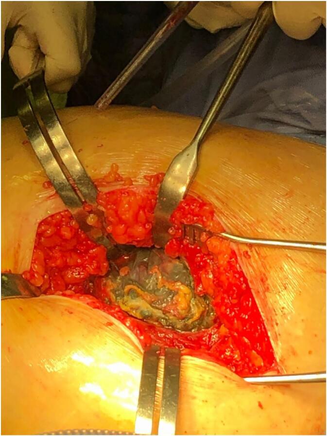
Bulky pseudotumor at the level of the subtrochanteric bursa.
A solid osteointegration of the stem and the cup complicated the removal of both components. No osteolysis was detected. After the components removal and an adequate debridement of pathological tissues which also involved the upper part of the greater trochanter (Fig. 3), an anterior column fracture of the acetabulum was highlighted, along with a bone defect in the acetabular fundus.
Fig. 3.

Part of the pseudotumor removed.
Bone substitutes were used to fill the gaps: MgHA/collagen-based scaffolds (RegenOss®, Finceramica, Faenza, Italy) and fully resorbable MgHA nanocrystals synthetic grafts (SINTlife®, Finceramica, Faenza, Italy). For the stem revision, a monoblock revision femoral stem 240 mm in length with high offset (REDAPT revision femoral stem®, Smith and Nephew, London, UK) was employed. A 52 mm fully porous shell fixed with four screws (70 mm - 40 mm - 20 mm - 15 mm in length x 6.5 mm in diameter), a 20° overhang polyethylene liner, and a 36 mm + 0 Oxinium head were utilized (Fig. 4). Finally, we inserted the remaining part of the gluteus medius on the tensor fasciae latae.
Fig. 4.

Postoperative check. Monoblock revision femoral stem 240 mm with high offset, 52 mm fully porous shell with four 6.5 mm screws, 20° overhang polyethylene liner, and 36 mm + 0 Oxinium head.
Hispopathological analysis showed dense perivascular lymphocytic infiltration and intracellular metallic debris in macrophages. These are characteristics of a chronic immune-mediated inflammatory reaction, which match a pattern of aseptic lymphocyte-dominated vasculitis-associated lesion (ALVAL). Microbiological examination showed sterile cultures of synovial fluid and periprosthetic tissue, with no growth of aerobic or anaerobic bacteria, mycobacteria, or fungi. Systemic inflammatory markers, including ESR and CRP, were slightly elevated. Synovial fluid analysis showed an inflammatory cell count not inkeeping with bacterial infection. Overall, the clinical, histological, and microbiological findings are in keeping with a diagnosis of ARMD in the absence of active infection.
In the immediate postoperative period specific exercises for restoration of the ROM and muscular strengthening without weight bearing where prescribed from the first postoperative day. The sitting position was permitted after the first week. Toe-touch weight bearing was prescribed up to the first x-rays 30 days after the surgery; then, the patient was allowed partial weight bearing. Full weight bearing was achieved in a week.
Stitches were removed at approximately three weeks, and antithrombotic prophylaxis was administered for the first six weeks after the surgery.
Follow-up radiographs at 3–6-12 months after surgery, together with clinical evaluation.
No complications were reported throughout the period, and the patient returned to daily activities.
The patient had recovered a good level of muscle strength and was able to walk without aids at the 6-month clinical check-up. Due to the gluteus medius weakness, a slight limp persisted. The patient was able to stay sitting for extended periods of time and walk eight blocks pain-free. The range of motion was somewhat limited in internal rotation but acceptable in flexion-extension, external rotation and abduction.
This case report was held in line with the SCARE criteria [24].
3. Discussion
The acetabular cups should be set at specific inclination and anteversion angles to ensure optimal system functionality [10]. Proper cup positioning aids in load distribution, while suboptimal angles can cause increased implant work and lead to wear and elevated ions levels, along with early failure due to dislocation, impingement, pelvic osteolysis, and acetabular migration [11]. In case of significant vertical inclination of the cup, the so-called edge loading can be generated, with the consequent increase in the release of cobalt‑chromium ions. For this reason, an appropriate preoperative measure of cup orientation is crucial. Indeed, the most accurate method to verify it is to perform a CT scan, but standard anteroposterior pelvis X-rays can also provide an inclination and version angle with sufficient accuracy. Nonetheless, new technologies have been introduced to increase the accuracy of hip replacement procedures, such as robot-assisted total hip arthroplasty [12].
Lewinnek et al. determined that 45° ± 10° inclination and 15 ± 10 anteversion were the safe range for cup inclination and version for minimizing the dislocation rate, respectively [13]. Most surgeons plan their THA procedures on these angle ranges [14]. On the other hand, significant differences in acetabular cup alignment between standing and sitting positions were demonstrated by spinal-pelvic motion analysis, specifically in patients with pelvic hypermobility [15]. The fact that Lewinnek's safe zone ignored joint motion dynamics and individual anatomical differences was questioned, and a new “safe zone” consisting of combined anteversion was favored [16].
An extremely vertical acetabular cup tilt can cause substantial local wear, as demonstrated by various studies [17]. When the acetabular and femoral components' contact zone extends over the edge of the cup, edge loading can occur. This causes higher contact pressures, which in turn produces increased edge wear [18]. Furthermore, latest studies reveal that vertical orientation coupled with an incorrect reconstruction of the center of rotation produces the worst outcomes [19].
Cup positioning becomes even more critical when addressing THA revisions [20]. Even though there are several contributing factors to postoperative dislocation, Sharma et al. revealed that using computer navigation significantly lowers the rate of component malalignment and, thus, the rate of dislocation after THA revision [20].
De Haan et al. investigated the inclination angle of the acetabular component in 214 MoM resurfacing hip replacements, as well as the serum concentrations of cobalt and chromium ions [21]. They found that patients with prominent sloped components had much higher levels of metal ions, and this tends to occur when the component has an abduction angle greater than 55°.
The case presented confirms this phenomenon and demonstrates that increased metal ions can also be accompanied by the formation of pseudotumors around the implant. Moreover, this study corroborates that blood metal ions (particularly cobalt and chromium) could effectively distinguish well-functioning from failed implants [22].
According to Lavigne et al., metal ion level ranges widely in THA with large femoral heads, with the most significant values observed in young male patients probably due to stressing activities on the joint [23].
The failure of a MoM THA due to edge loading is a multifactorial event in which obesity and physical activity play a significant role. While malpositioning of the acetabular component, especially the excessive inclination or anteversion, is usually the primary cause, patient-related factors can greatly exacerbate the problem. Obesity increases the mechanical load on the prosthetic joint, contributing to micromotion and instability that promote misalignment and peripheral wear. This accelerates the release of metal ions and raises the risk of adverse local tissue reactions, such as pseudotumors or soft tissue necrosis. High levels of physical activity, particularly in sports or physically demanding work, increase the frequency of joint cycles and the likelihood of off-axis movements, further intensifying the effects of edge loading. The combination of a poorly positioned implant, obesity, and high physical activity constitutes a major risk factor for early prosthetic failure, as occurred in the case described.
One of the limitations of this case is the absence of a recent preoperative CT scan, which could have provided more detailed insight into the bony morphology and potential contributing factors to edge loading. However, the patient had undergone a CT several years prior, which had already revealed abnormalities involving both the acetabulum and the greater trochanter. These findings were consistent with the intraoperative observations. Due to concerns related to the COVID-19 pandemic, the patient had declined any form of surgical or diagnostic intervention at that time, delaying definitive treatment. Although this limits the documentation, the case was still evaluated using thorough radiographic analysis and clinical examination, which allowed for an accurate understanding of the underlying pathology. Future cases would benefit from updated cross-sectional imaging whenever feasible.
4. Conclusion
The precise acetabular cup positioning in MoM hip prosthesis geometry is essential in preventing functional overloading, excessive release of metal ions and adverse tissue reactions. When evaluating patients with metal-on-metal hip prostheses, it is necessary to consider the orientation of the cup as a predictive factor for failure and wear of the components.
Consent
Written informed consent was obtained from the patient for publication of this case report and accompanying images.
Ethical approval
The study is exempt from ethnical approval since it is retrospective case report, whose data is totally anonymized.
Guarantor
Corrado Ciatti.
Funding
The authors declare that they did not receive any funding for this study.
CRediT authorship contribution statement
Corrado Ciatti: Conceptualization, Methodology, Validation, Investigation, Writing – original draft, Writing – review & editing, Visualization. Luca Andriollo: Conceptualization, Methodology, Writing – original draft, Writing – review & editing. Francesco Pisanu: Validation, Supervision. Virginia Masoni: Conceptualization, Methodology, Writing – original draft, Writing – review & editing. Edoardo Bori: Conceptualization, Methodology, Writing – original draft, Writing – review & editing. Fabrizio Quattrini: Investigation, Visualization, Supervision.
Declaration of competing interest
All other authors declare that they have no existing conflicts of interest.
References
- 1.Descamps J., Teissier V., Graff W., Mouton A., Bouché P.A., Marmor S. Managing early complications in total hip arthroplasty: the safety of immediate revision. J. Orthop. Traumatol. Jul 31 2023;24(1):38. doi: 10.1186/s10195-023-00719-1. [DOI] [PMC free article] [PubMed] [Google Scholar]
- 2.Ciatti C., Moschella M., Bori E., Doria C., Caggiari G., Innocenti B., et al. Gross taper failure and fracture of the true neck in total hip arthroplasty: retrieval scanning electron microscope analysis. Medicina. Mar 9 2024;60(3):458. doi: 10.3390/medicina60030458. [DOI] [PMC free article] [PubMed] [Google Scholar]
- 3.Khalifa A.A., Bakr H.M. Updates in biomaterials of bearing surfaces in total hip arthroplasty. Arthroplasty. 2021;3(1):32. doi: 10.1186/s42836-021-00092-6. (35236490) [DOI] [PMC free article] [PubMed] [Google Scholar]
- 4.Latteier M.J., Berend K.R., Lombardi A.V., Ajluni A.F., Seng B.E., Adams J.B. Gender is a significant factor for failure of metal-on-metal total hip arthroplasty. J. Arthroplast. 2011;26(6):19–23. doi: 10.1016/j.arth.2011.04.012. [DOI] [PubMed] [Google Scholar]
- 5.Sangaletti R., Spreafico A., Barbieri F., Ferrari R., Castelli C.C. Metal ion trend in patients with metal-on-metal total HIP arthroplasty: a 10-year prospective study. HIP Int. Nov 2018;28(2_suppl):43–47. doi: 10.1177/1120700018812991. [DOI] [PubMed] [Google Scholar]
- 6.Corten K., MacDonald S.J. Hip resurfacing data from national joint registries: what do they tell us? What do they not tell us? Clin. Orthop. Relat. Res. Feb 2010;468(2):351–357. doi: 10.1007/s11999-009-1157-3. [DOI] [PMC free article] [PubMed] [Google Scholar]
- 7.Ciatti C., Maniscalco P., Bosio S., Pagliarello C.P., Bianchi G., Quattrini F. Pseudotumor from ceramic-on-ceramic total hip arthroplasty. Int. J. Surg. Case Rep. Feb 13 2024 doi: 10.1016/j.ijscr.2024.109374. [DOI] [PMC free article] [PubMed] [Google Scholar]
- 8.New Zealand Orthopaedic Association; 2022 AOANJRR annual report and supplementary reports. https://www.nzoa.org.nz/2022-aoanjrr-annual-report-and-supplementary-reports Available from: (Internet, cited 2024 Feb 28)
- 9.Ciatti C., Andriollo L., Asti C., Morsia D., Quattrini F., Cosentino M., et al. The role of femoral head size in metal-on-metal hip arthroplasty: analysis of a cohort of 3813 patients with long term follow-up. Arch. Orthop. Trauma Surg. Nov 5 2024;144(11):4809–4818. doi: 10.1007/s00402-024-05567-0. [DOI] [PMC free article] [PubMed] [Google Scholar]
- 10.Perticarini L., Andriollo L., Rossi S.M.P., Sangaletti R., Benazzo F. Severe acetabular bone loss management: is there still a role for titanium cages and cemented cups? Hip Int. Feb 2 2025 doi: 10.1177/11207000251315837. (Epub ahead of print. PMID: 39894957) [DOI] [PubMed] [Google Scholar]
- 11.Harrison C.L., Thomson A.I., Cutts S., Rowe P.J., Riches P.E. Research synthesis of recommended acetabular cup orientations for total hip arthroplasty. J. Arthroplast. Feb 2014;29(2):377–382. doi: 10.1016/j.arth.2013.06.026. [DOI] [PubMed] [Google Scholar]
- 12.Zhang X., Shen X., Zhang R., Chen M., Ma R., Zhang Z., et al. Radiographic evaluation of robot-assisted versus manual total hip arthroplasty: a multicenter randomized controlled trial. J. Orthop. Traumatol. Jun 26 2024;25(1):33. doi: 10.1186/s10195-024-00773-3. [DOI] [PMC free article] [PubMed] [Google Scholar]
- 13.Lewinnek G.E., Lewis J.L., Tarr R., Compere C.L., Zimmerman J.R. Dislocations after total hip-replacement arthroplasties. J. Bone Joint Surg. Am. Mar 1978;60(2):217–220. [PubMed] [Google Scholar]
- 14.Meermans G., Fawley D., Zagra L., Ten Broeke R.H.M., Johnson K., Bernard T., et al. Accuracy of cup placement compared with preoperative surgeon targets in primary total hip arthroplasty using standard instrumentation and techniques: a global, multicenter study. J. Orthop. Traumatol. May 10 2024;25(1):25. doi: 10.1186/s10195-024-00766-2. [DOI] [PMC free article] [PubMed] [Google Scholar]
- 15.Pierrepont J., Hawdon G., Miles B.P., Connor B.O., Baré J., Walter L.R., et al. Variation in functional pelvic tilt in patients undergoing total hip arthroplasty. Bone Joint J. 2017;99-B(2):184–191. doi: 10.1302/0301-620X.99B2.BJJ-2016-0098.R1. [DOI] [PubMed] [Google Scholar]
- 16.O’Connor P.B., Thompson M.T., Esposito C.I., Poli N., McGree J., Donnelly T., et al. The impact of functional combined anteversion on hip range of motion: a new optimal zone to reduce risk of impingement in total hip arthroplasty. Bone Jt Open. Oct 2021;2(10):834–841. doi: 10.1302/2633-1462.210.BJO-2021-0117.R1. [DOI] [PMC free article] [PubMed] [Google Scholar]
- 17.Langton D.J., Joyce T.J., Jameson S.S., Lord J., Van Orsouw M., Holland J.P., et al. Adverse reaction to metal debris following hip resurfacing: the influence of component type, orientation and volumetric wear. J. Bone Joint Surg. (Br.) Feb 2011;93-B(2):164–171. doi: 10.1302/0301-620X.93B2.25099. [DOI] [PubMed] [Google Scholar]
- 18.Underwood R.J., Zografos A., Sayles R.S., Hart A., Cann P. Edge loading in metal-on-metal hips: low clearance is a new risk factor. Proc. Inst. Mech. Eng. H. Mar 2012;226(3):217–226. doi: 10.1177/0954411911431397. [DOI] [PMC free article] [PubMed] [Google Scholar]
- 19.Nieschk C., Abelmann-Brockmann J., Lisitano L., Fenwick A., Röttinger H., Ecker M., et al. Clinical effects of different center of rotation reconstructions in total hip arthroplasty after femoral neck fractures: a cohort study including a follow-up analysis on patient’s mobility and daily living ability. J. Orthop. Traumatol. Nov 9 2023;24(1):58. doi: 10.1186/s10195-023-00738-y. [DOI] [PMC free article] [PubMed] [Google Scholar]
- 20.Sharma A.K., Cizmic Z., Carroll K.M., Jerabek S.A., Paprosky W.G., Sculco P.K., et al. Computer navigation for revision total hip arthroplasty reduces dislocation rates. JOIO. Jun 2022;56(6):1061–1065. doi: 10.1007/s43465-022-00606-7. [DOI] [PMC free article] [PubMed] [Google Scholar]
- 21.De Haan R., Pattyn C., Gill H.S., Murray D.W., Campbell P.A., De Smet K. Correlation between inclination of the acetabular component and metal ion levels in metal-on-metal hip resurfacing replacement. J. Bone Joint Surg. (Br.) Oct 2008;90-B(10):1291–1297. doi: 10.1302/0301-620X.90B10.20533. [DOI] [PubMed] [Google Scholar]
- 22.Hart A.J., Sabah S.A., Bandi A.S., Maggiore P., Tarassoli P., Sampson B., et al. Sensitivity and specificity of blood cobalt and chromium metal ions for predicting failure of metal-on-metal hip replacement. J. Bone Joint Surg. (Br.) Oct 2011;93-B(10):1308–1313. doi: 10.1302/0301-620X.93B10.26249. [DOI] [PubMed] [Google Scholar]
- 23.Lavigne M., Belzile E.L., Roy A., Morin F., Amzica T., Vendittoli P.A. Comparison of whole-blood metal ion levels in four types of metal-on-metal large-diameter femoral head total hip arthroplasty: the potential influence of the adapter sleeve. J. Bone Joint Surg. May 4 2011;93(Supplement_2):128–136. doi: 10.2106/JBJS.J.01885. [DOI] [PubMed] [Google Scholar]
- 24.Kerwan A., Al-Jabir A., Mathew G., Sohrabi C., Rashid R., Franchi T., Nicola M., Agha M., Agha R.A. Revised surgical CAse REport (SCARE) guideline: an update for the age of artificial intelligence. Premier Journal of Science. 2025;10(100079) [Google Scholar]