



Abstract
Background
Quantitative flow ratio (QFR) is a non-invasive angiographic tool that provides functional assessment of coronary stenosis without the need for pressure wires or hyperemia. This prospective study aimed to evaluate the procedural and inpatient treatment outcomes of QFR-guided percutaneous coronary intervention (PCI) compared with that of angiography-guided PCI in patients with ST-elevation myocardial infarction (STEMI) undergoing staged revascularization of non-culprit lesions.
Material/Methods
This randomized prospective single-center study was conducted at the Hospital of the Lithuanian University of Health Sciences Kaunas Clinics (July 2020–June 2021). After successful culprit-lesion PCI for STEMI, 124 participants with residual angiographically significant non-culprit stenosis (50–75%) were randomized to QFR-guided (n=62) or angiography-guided PCI (n=62). Procedural characteristics, fluoroscopy time, contrast usage, stent number/length, and inpatient treatment outcomes were compared between groups using SPSS 28.0 software.
Results
Compared with PCI guided by visual estimation alone, the QFR-guided PCI group showed significant reductions in fluoroscopy time (median 6.2 vs 8.0 min, P=0.009), contrast volume (median 100 vs 120 mL, P=0.038), number of stents implanted (median 1.5 vs 2.0, P=0.002), and stent length (median 28 vs 45 mm, P<0.001). No significant differences were found between the groups in terms of periprocedural complications or length of inpatient stay.
Conclusions
QFR-guided PCI of the non-culprit lesion resulted in shorter fluoroscopy time, lower contrast volume, and a smaller number and average length of implanted stents. These findings highlight the potential of QFR to enhance procedural efficiency and reduce unnecessary stenting in clinical practice without compromising patient outcomes.
Keywords: Cardiovascular Diseases, Myocardial Infarction, Coronary Disease, Coronary Artery Disease, Physiology
Introduction
Quantitative Flow Ratio (QFR) is a diagnostic tool used in the field of interventional cardiology to assess the severity of coronary artery stenosis. It is a noninvasive method that uses angiographic images to calculate the pressure drop across a coronary artery lesion and estimate the fractional flow reserve, without the need for a pressure wire or hyperemic agent [1].
QFR can potentially shorten stenting procedures, because interventional cardiologists can plan the intervention more effectively, knowing which lesions are physiologically significant and require treatment. The FAVOR III Europe trial suggested that the current QFR method can offer diagnostic clinical value that might be positioned between angiography and fractional flow reserve [2].
Traditional assessment methods, like fractional flow reserve, require the insertion of a pressure wire and administration of hyperemic agents, which extends fluoroscopic time [3]. QFR can provide similar diagnostic information without these steps [4]. With QFR providing detailed physiological data upfront, interventional cardiologists can plan stent placement more accurately and avoid unnecessary prolonged fluoroscopic imaging due to indecision or multiple adjustments during the procedure [5]. QFR has the potential to reduce the use of radiographic contrast agents during percutaneous coronary intervention (PCI) procedures [3]. The use of QFR can potentially lead to fewer stents being placed during PCI. In some cases, angiographic imaging alone may suggest that a lesion is severe and needs stenting, while in reality, it may not significantly impair blood flow [8]. QFR provides additional functional information that can prevent over-treatment by showing that some lesions do not require stenting [9]. Avoiding unnecessary stents can reduce the risk of stent-related complications, such as restenosis and stent thrombosis [10]. This can lead to better long-term outcomes for patients.
QFR enables precise localization of functionally significant lesions, allowing interventional cardiologists to identify the exact artery segments needing stenting [1,9]. This avoids unnecessary extension of the stent beyond the diseased area. By providing an accurate assessment of the functional significance and exact length of coronary lesions, QFR can lead to the use of shorter stents during PCI [11]. This precision helps prevent unnecessary stenting, reduces the risk of complications, and enhances both the cost-effectiveness and overall outcomes of the procedure [10].
We conducted a prospective experimental study to evaluate the diagnostic efficacy and limitations of QFR during primary and staged angiographies in patients who had already undergone successful culprit artery PCI for STEMI, but who still had angiographically significant stenoses in non-infarct-related arteries. We hope that the obtained results will encourage more frequent use of the QFR method in daily clinical practice, to reduce the number of unnecessary PCIs and associated complications. Therefore, this study aimed to evaluate outcomes from QFR-guided complete revascularization and angiography-guided PCI in 124 patients with STEMI.
Material and Methods
Ethical Statement
Authorization for the study was obtained from the Regional Biomedical Research Ethics Committee, under permission No. BE-2–14. Our study included only those patients who provided informed consent for participation in a biomedical study. During the study, the selected patients were examined and treated according to the research and treatment algorithms applied by the Hospital of Lithuanian University of Health Sciences Kaunas Clinics, recommended by international organizations.
Patient Selection
This was a randomized prospective experimental single center study, conducted in the Hospital of the Lithuanian University of Health Sciences Kaunas Clinics (July 2020 to June 2021). All patients admitted to our PCI center within 120 min after onset of symptoms who were diagnosed with STEMI according to the criteria outlined in the European Society of Cardiology guidelines for management of patients presenting with ST-segment elevation, which was released in 2017 [12], underwent index culprit lesion PCI to restore blood flow in the occluded artery, and presented with one or more residual non-culprit stenosis (50–75%) were enrolled into this study (Figure 1). Patients were excluded from the study if mechanical reopening and stenting of the infarct-related artery were unsuccessful; it was decided by the Heart Team that coronary artery bypass grafting would be the most appropriate treatment strategy. In addition, patients with serious comorbidities with unfavorable prognosis that could influence survival within the 1 year were excluded. Finally, patients with circulatory instability, cardiogenic shock, pulmonary edema, and other serious conditions at the time of inclusion in the study were not enrolled in the study. The total of 124 patients met all inclusion and exclusion criteria.
Figure 1.

Patient selection into this study with a case example. Figure represents a case from our study of a 72-year-old man who was admitted to our center with acute chest pain. Electrocardiography confirmed an inferior ST-elevation myocardial infarction (A). Coronary angiography revealed a total occlusion of the right coronary artery (B), and the patient underwent an index culprit lesion percutaneous coronary intervention (PCI) to restore blood flow in the occluded artery (C). Coronary angiography also identified an approximately 75% stenosis in the left anterior descending artery (LAD) (white arrowhead) (D). A quantitative flow ratio of the LAD was performed, yielding a result of 0.74, indicating that the stenosis was hemodynamically significant (E). The LAD was treated during a staged procedure 3 months after the index PCI (F).
Randomization
After the enrollment to the study, 124 patients were randomized into 1 of 2 groups: (1) QFR-guided PCI (n=62; QFR group) and (2) visual estimation-only guided PCI (n=62; angiography group). Data on patient demographic information, medical history, procedure duration, the amount of contrast used, the number and length of stents, fluoroscopy time were collected from PCI procedure protocols and medical records.
Angiography-Quided PCI
In the visual estimation-only guided PCI, the coronary arteries were assessed by the interventional cardiologist performing the index procedure, and the angiographies were additionally discussed in interventional cardiologists’ meetings. Angiography-guided non-culprit coronary lesions PCI were performed as a staged procedure at least 3 months after the index PCI.
QFR-Guided Complete Revascularization
In the QFR-guided PCI group, QFR analyses were performed using the certified software QAngio-XA 3D, version 2.0 (Medis Medical Imaging Systems, Leiden, The Netherlands) from coronary artery angiograms. In this study, stenoses were deemed hemodynamically significant when the QFR was less than 0.8. All QFR analyses were performed offline twice by an experienced and internationally certified QFR observer, with the results averaged. If the 2 measurements did not match (difference >0.02), a third measurement was conducted, and the 3 results were averaged. All patients were suitable for QFR analyses; hence, none were excluded from this group. Residual hemodynamically significant stenoses, according to QFR, were treated during a staged procedure at least 3 months after the index PCI.
Statistical Analysis
Statistical analysis was conducted using SPSS 28.0 software. Descriptive statistics were used to summarize the data, with the Kolmogorov-Smirnov test assessing normality. Continuous variables, including the duration of the stenting procedure, fluoroscopy time, contrast volume, number and length of stents implanted, and inpatient treatment time, were analyzed using means, standard deviations, and medians. To compare these variables between the 2 groups, the Mann-Whitney U test (a non-parametric test) was applied. Receiver operating characteristic (ROC) curve analysis was used to determine thresholds for fluoroscopy time and contrast usage, along with calculating odds ratios (OR) and 95% confidence intervals (CI) to assess the likelihood of exceeding these thresholds between groups. The chi-square test was used to compare categorical variables, such as the number of stents implanted and periprocedural complication rates. A significance level of P<0.05 was considered statistically significant.
Results
In assessing the characteristics of coronary artery stenting procedures among 124 stented patients, divided into angiography (n=62) and QFR (n=62) method groups, the evaluation included the duration of the stenting procedure and fluoroscopy, average amount of contrast used, number and total length of implanted stents, periprocedural complications rate, and the duration of hospitalization.
Evaluation of the Duration of the Stenting Procedure
When comparing the duration of the stenting procedure between the angiography and QFR method groups, no significant differences were found (U=1857.0, P=0.75). The average duration of the stenting procedure in the angiography group was 40.42±3.76 min (median 32.50 min, interquartile range (IQR) 20–46.25 min), while in the QFR method group, the average duration was 33.63±1.64 min (median 30.0 min, IQR 25–40 min).
Evaluation of the Duration of Fluoroscopy
When comparing the duration of fluoroscopy during the stenting procedure between the angiography and QFR method groups, statistically significant differences were found (U=1399.5, P=0.009). The average duration of fluoroscopy during the stenting procedure in the angiography group was 10.88±1.24 min (median 8.0 min, IQR 5.2–13.5 min), whereas in the QFR method group, the average duration was 6.59±0.42 min (median 6.2 min, IQR 4.1–8.4 min) (Figure 2). Based on the ROC test, we determined a fluoroscopy time threshold of 10.4 min for the second stage procedure, considering the groups. The likelihood of the fluoroscopy procedure duration exceeding 10.4 min in the angiography group was, on average, 6.3 times higher than in the QFR method group (OR 6.31, 95% CI 2.36–16.85, P=0.001).
Figure 2.
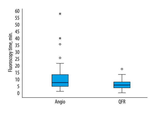
Fluoroscopy time (min) in quantitative flow ratio (QFR)-guided and angiography-guided percutaneous coronary intervention (PCI) groups during second-stage stenting procedure. This box plot illustrates the significantly shorter fluoroscopy time in the QFR-guided group than in the angiography group during the second-stage stenting procedure (6.59±0.42 min vs 10.88±1.24 min). P=0.009, based on the nonparametric Mann-Whitney test.
Evaluation of the Amount of Contrast Used
Statistically significant differences were found in the amount of contrast used during the stenting procedure between the angiography and QFR groups (U=1524.0, P=0.038). The average amount of contrast used in the angiography group was 148.23±9.19 mL (median 120.0 mL, IQR 100–200 mL), while in the QFR method group, the amount of contrast used was 116.45±4.38 mL (median 100 mL, IQR 100–122.5 mL; Figure 3). Based on the ROC test, we determined a threshold for the amount of contrast used during the second stage of the stenting procedure, considering the groups (160 mL). The likelihood of using more than 160 mL of contrast in the angiography group was, on average, 10.8 times higher than in the QFR method group (OR 10.82, 95% CI 3.03–38.57, P=0.001).
Figure 3.
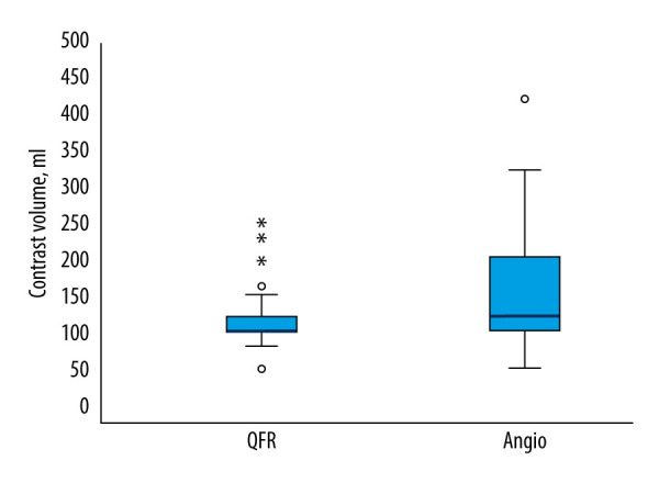
Contrast volume (mL) in quantitative flow ratio (QFR)-guided and angiography-guided percutaneous coronary intervention (PCI) groups during second-stage stenting procedure. This box plot illustrates the significantly lower contrast volume used in the QFR-guided group than in the angiography group during the second-stage stenting procedure (116.45±4.38 mL vs 148.23±9.19 mL). P=0.038, based on the nonparametric Mann-Whitney test.
Evaluation of the Number of Stents Implanted
Statistically significant differences were identified in the number of stents implanted during the stenting procedure between the angiography and QFR groups (U=1343.5, P=0.002). In the angiography group, an average of 2.26±0.14 stents were implanted (median 2.0 stents, IQR 1–3 stents). In the QFR method group, an average of 1.69±1.0 stents were implanted during the procedure (median 1.5 stents, IQR 1–2 stents; Figure 4). In the QFR group, 50.0% of patients had only 1 stent used during non-culprit lesion PCI, compared with 25.8% of patients in the angiography-guided PCI group (P<0.05). Five stents were used significantly more often in the angiography group than in the QFR group, at 6.5% and 0%, respectively (P<0.05).
Figure 4.
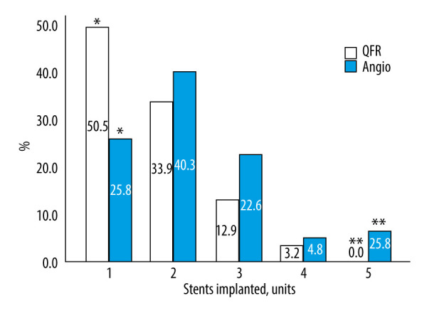
Stents implanted (units) in quantitative flow ratio (QFR)-guided and angiography-guided percutaneous coronary intervention (PCI) groups during second-stage stenting procedure. This bar chart illustrates the statistically significant difference in the number of stents implanted between the QFR-guided and angiography-guided PCI groups during the second-stage stenting procedure, showing that the QFR method was associated with fewer stents per procedure (1.69±1.0 vs 2.26±0.14). χ2=10.971; lls=4; P=0.027; *, ** P<0.05, based on the χ2 independence test.
Evaluation of the Length of Implanted Stents
Statistically significant differences were found in the length of implanted stents during the stenting procedure between the angiography and QFR groups (U=1203.0, P<0.001). In the angiography group, the average stent length was 47.69±3.11 mm (median 45 mm, IQR 28–60.25 mm; Figure 5). In the QFR method group, the average stent length was 33.18±2.26 mm (median 28 mm, IQR 21.25–43.0 mm).
Figure 5.
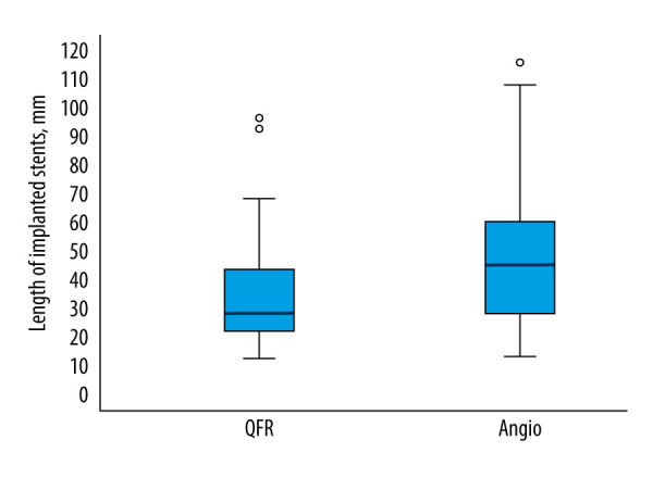
Length of implanted stents (mm) in quantitative flow ratio (QFR)-guided and angiography-guided percutaneous coronary intervention (PCI) groups during second-stage stenting procedure. This box plot illustrates the significantly shorter implanted stent length in the QFR-guided group than in the angiography group during the second-stage stenting procedure (33.18±2.26 mm vs 47.69±3.11 mm). P<0.001, based on the nonparametric Mann-Whitney test.
Evaluation of Procedural and Inpatient Treatment Outcomes
In our study, we evaluated periprocedural complications, such as coronary artery dissection, perforation, no-reflow phenomenon, stent thrombosis (acute or subacute), and unexpected hemodynamic collapse due to various etiological causes. In assessing the frequency of periprocedural complications during PCI between the angiography and QFR method groups, no significant differences were found, with rates of 3.2% and 1.6%, respectively (P>0.05). No significant differences were found in inpatient treatment time between the angiography and QFR method groups (U=1690.0, P=0.24). In the angiography group, the average inpatient treatment time was 7.47±0.52 days (median – 6.5 days, IQR 5.75–9.0 days). In the QFR method group, the average inpatient treatment time was 7.37±0.70 days (median 6 days, IQR 5–8 days).
Discussion
QFR is a diagnostic method used in the field of interventional cardiology to estimate angiographically derived fractional flow reserve in real time by analyzing the ratio of coronary pressure distal to a stenosis and aortic pressure under conditions of maximum myocardial hyperemia. In this randomized, prospective, single-center experimental study, QFR-guided PCI improved the effectiveness of revascularization by reducing fluoroscopy time, lowering contrast volume, and decreasing both the number and average length of implanted stents in patients with STEMI and extensive coronary artery atherosclerosis who had already undergone successful recanalization and stenting of the culprit vessel.
Historically, revascularization based on visual assessment often overestimated the severity of stenosis, leading to overtreatment and suboptimal long-term outcomes [13]. However, avoiding overtreatment remains a relevant issue, due to associated costs, potential complications, and the emphasis on minimally invasive approaches. To address this, functional evaluation of intermediate stenoses using pressure measurements during invasive coronary angiography has been recommended in revascularization guidelines since 2005 [14]. Despite robust data, the use of pressure measurements in routine clinical practice remains limited, with adoption estimated at only 10% of cases [15]. The reasons include inconsistent reimbursement, the need for intracoronary instrumentation with harder-to-manipulate wires and associated risks of complications, and, in the case of pressure wire–derived fractional flow reserve, the requirement to induce hyperemia, which adds patient discomfort, time, and costs. QFR is an excellent technology that allows the evaluation of the hemodynamic significance of coronary artery stenosis while eliminating the inconveniences associated with pressure wires and pharmacological agents. It also holds a class 1B recommendation for the assessment of intermediate stenosis in the 2024 European Society of Cardiology guidelines [16]. However, since the majority of previous studies demonstrating the benefits of QFR focused on patients with chronic coronary artery disease, our aim was to evaluate the capabilities and limitations of QFR in patients with STEMI.
In our study population, almost two-thirds underwent stenting of non-infarct-related coronary arteries in a second-stage procedure. Among these patients, more than two-thirds had a single coronary artery stented, while the remaining had 2 coronary arteries stented. In the remaining third of patients, non-infarct-related coronary artery stenoses were assessed as hemodynamically insignificant and were therefore not treated with stenting.
When evaluating the specifics of revascularization, we found that the average duration of the stenting procedure was slightly longer in the angiography group; however, the difference was not significant. In our study, the average fluoroscopy time was 8.7 min, and in the QFR-guided PCI group, this duration was on average 4 min shorter than in the angiography group. The likelihood of exceeding 10.4 min of fluoroscopy was 6.3 times higher in the angiography group than in the QFR group. It is known that interventional cardiologists are exposed to the highest cumulative radiation among health professionals. Due to the effects of cumulative radiation, cataracts, thyroid cancer, and a disproportionate incidence of left-sided brain tumors have been reported in interventional cardiologists [17–19]. Clinical studies are most often focused on patient outcomes. However, given the occupational hazards faced by interventional cardiologists, including prolonged radiation exposure, the benefits of QFR application are undeniable. In addition, on average, 132 mL of contrast was used during stenting procedures, with significantly more (32 mL on average) used in the angiography group than in the QFR method group. The aim to reduce the amount of contrast during interventional procedures is very important, knowing that approximately 7% of patients undergoing PCI experience contrast-induced nephropathy [20]. These findings of less radiation exposure and less contrast used in the QFR-guided PCI approach align with similar results observed in the Angiographic Quantitative Flow Ratio-Guided Coronary Intervention (FAVOR III China) trial [21].
Studies have demonstrated that complete revascularization, guided by angiography or physiological assessment, outperforms the culprit-only approach in younger, low-risk STEMI patients [22,23]. This also applies to older patients, as shown by the FIRE trial, in which physiology-guided complete revascularization reduced the composite risk of death, myocardial infarction, stroke, or repeat ischemia-driven procedures by 27%, compared with a culprit-only strategy [24]. Zhang et al, in a randomized trial, demonstrated the benefits of QFR-guided complete revascularization of the non-infarct-related artery in patients with STEMI, compared with no further invasive treatment, by reducing the incidence of major adverse cardiovascular events and unstable angina [25]. Mentioned studies prove that evaluating non-culprit lesions in STEMI patients with extensive coronary artery atherosclerosis physiologically is crucial to avoid excessive stenting, thus reducing higher risks of restenosis, stent thrombosis, and major adverse cardiovascular events. In our study, the average number of stents implanted was close to 2, but the number was significantly higher in the angiography group than in the QFR group. In the QFR method group, a single stent was implanted in twice as many patients as in the angiography group. The likelihood of using more than 1 stent was 2.9 times higher in the angiography group, and the likelihood of using stents longer than 44 mm was 4.3 times higher in the angiography group than in the QFR method group. Other studies also indicate fewer stents were used in the QFR group, but the stents were slightly shorter, averaging 30 to 35 mm [26,27]. This can be related to the number of affected coronary arteries, anatomical characteristics, stenosis severity, QFR index, and other factors.
When evaluating the frequency of periprocedural complications during PCI, no significant differences were found between the angiography and QFR method groups. The average hospital stay for all stented patients with a diagnosis of STEMI was 7.4 days, with no significant differences observed between the patient groups.
Limitations
While our study offers valuable insights, certain limitations should be acknowledged. First, this was a single-center study, which can limit generalizability. Second, the study’s relatively small sample size can reduce the statistical power. Third, the precision and consistency of QFR measurements rely on the technique and quality of angiographic imaging, which currently requires 2 projections for each vessel. Fourth, procedural bias may have been introduced, as PCI operators were aware of the group assignments. Fifth, the angiography-guided PCI group relied on angiography guidance for PCI lesion assessment, which, despite current guideline recommendations, remains the prevailing standard of care in most catheterization laboratories.
Conclusions
In this randomized, prospective, experimental, single-center study, patients with STEMI who had undergone successful recanalization and stenting of the culprit lesion using QFR-guided PCI of the non-culprit lesion resulted in shorter fluoroscopy time, lower contrast volume, and a smaller number and average length of implanted stents. These findings highlight the potential of QFR to enhance procedural efficiency and reduce unnecessary stenting in clinical practice without compromising patient outcomes.
Footnotes
Conflict of interest: None declared
Publisher’s note: All claims expressed in this article are solely those of the authors and do not necessarily represent those of their affiliated organizations, or those of the publisher, the editors and the reviewers. Any product that may be evaluated in this article, or claim that may be made by its manufacturer, is not guaranteed or endorsed by the publisher
Patient Permission/Consent Declarations: Informed consent to participate in the study was obtained for all human study participants.
Declaration of Figures’ Authenticity: All figures submitted have been created by the authors, who confirm that the images are original with no duplication and have not been previously published in whole or in part.
Financial support: None declared
References
- 1.Barauskas M, Žiubrytė G, Barauskienė G, et al. Systemic analysis of diagnostic performance and agreement between fractional flow reserve and quantitative flow ratio. Cor Vasa. 2021;63(6):683–87. [Google Scholar]
- 2.Andersen BK, Sejr-Hansen M, Maillard L, et al. Quantitative flow ratio versus fractional flow reserve for coronary revascularisation guidance (FAVOR III Europe): A multicentre, randomised, non-inferiority trial. Lancet. 2024;404(10465):1835–46. doi: 10.1016/S0140-6736(24)02175-5. [published correction appears in Lancet. 2025;404(10471):2542] [DOI] [PubMed] [Google Scholar]
- 3.Antoniadis M, Blum M, Ussat M, et al. Standardized angiographic projections allow evaluation of coronary artery side branches with quantitative flow ratio (QFR) Int J Cardiol Heart Vasc. 2024;50:101349. doi: 10.1016/j.ijcha.2024.101349. [DOI] [PMC free article] [PubMed] [Google Scholar]
- 4.Ziubryte G, Jarusevicius G. Fractional flow reserve, quantitative flow ratio, and instantaneous wave-free ratio: A comparison of the procedure-related dose of ionising radiation. Postepy Kardiol Interwencyjnej. 2021;17(1):33–38. doi: 10.5114/aic.2021.104765. [DOI] [PMC free article] [PubMed] [Google Scholar]
- 5.Barauskas M, Žiubrytė G, Jodka N, Unikas R. Quantitative flow ratio for assessment of non-culprit coronary artery lesions during percutaneous coronary intervention (PCI) in 79 patients diagnosed with ST-elevation myocardial infarction (STEMI): A study from a single center in Lithuania. Med Sci Monit. 2023;29:e939360. doi: 10.12659/MSM.939360. [DOI] [PMC free article] [PubMed] [Google Scholar]
- 6.Xu B, Tu S, Qiao S, et al. Diagnostic accuracy of angiography-based quantitative flow ratio measurements for online assessment of coronary stenosis. J Am Coll Cardiol. 2017;70(25):3077–87. doi: 10.1016/j.jacc.2017.10.035. [DOI] [PubMed] [Google Scholar]
- 7.Liu ZY, Yin ZH, Liang CY, et al. Zero contrast optical coherence tomography-guided percutaneous coronary intervention in patients with non-ST segment elevation myocardial infarction and chronic kidney disease. Catheter Cardiovasc Interv. 2021;97(Suppl 2):1072–79. doi: 10.1002/ccd.29655. [DOI] [PubMed] [Google Scholar]
- 8.Erriquez A, Campo G, Guiducci V, et al. QFR for the revascularization of nonculprit vessels in MI patients: Insights from the FIRE trial. JACC Cardiovasc Interv. 2024;17(12):1425–36. doi: 10.1016/j.jcin.2024.04.022. [DOI] [PubMed] [Google Scholar]
- 9.Barauskas M, Žiubrytė G, Jodka N, Unikas R. Quantitative flow ratio vs. angiography-only guided PCI in STEMI patients: One-year cardiovascular outcomes. BMC Cardiovasc Disord. 2023;23(1):136. doi: 10.1186/s12872-023-03153-7. [published correction appears in BMC Cardiovasc Disord. 2023;23(1):174] [DOI] [PMC free article] [PubMed] [Google Scholar]
- 10.Pan C, Han Y, Lu J. Structural design of vascular stents: A review. Micromachines (Basel) 2021;12(7):770. doi: 10.3390/mi12070770. [DOI] [PMC free article] [PubMed] [Google Scholar]
- 11.Strauss BH, Tanguay JF, Picard F, et al. Coronary stenting: Reflections on a 35-year journey. Can J Cardiol. 2022;38(10 Suppl 1):S17–S29. doi: 10.1016/j.cjca.2021.07.224. [DOI] [PubMed] [Google Scholar]
- 12.Ibanez B, James S, Agewall S, et al. 2017 ESC Guidelines for the management of acute myocardial infarction in patients presenting with ST-segment elevation: The Task Force for the management of acute myocardial infarction in patients presenting with ST-segment elevation of the European Society of Cardiology (ESC) Eur Heart J. 2018;39(2):119–77. doi: 10.1093/eurheartj/ehx393. [DOI] [PubMed] [Google Scholar]
- 13.Zimmermann FM, Ferrara A, Johnson NP, et al. Deferral vs. performance of percutaneous coronary intervention of functionally non-significant coronary stenosis: 15-year follow-up of the DEFER trial. Eur Heart J. 2015;36(45):3182–88. doi: 10.1093/eurheartj/ehv452. [DOI] [PubMed] [Google Scholar]
- 14.Silber S, Albertsson P, Avilés FF, et al. Guidelines for percutaneous coronary interventions. The Task Force for Percutaneous Coronary Interventions of the European Society of Cardiology. Eur Heart J. 2005;26(8):804–47. doi: 10.1093/eurheartj/ehi138. [DOI] [PubMed] [Google Scholar]
- 15.Peper J, van Hamersvelt RW, Rensing BJWM, et al. Diagnostic performance and clinical implications for enhancing a hybrid quantitative flow ratio-FFR revascularization decision-making strategy. Sci Rep. 2021;11(1):6425. doi: 10.1038/s41598-021-85933-9. [DOI] [PMC free article] [PubMed] [Google Scholar]
- 16.Vrints C, Andreotti F, Koskinas KC, et al. 2024 ESC Guidelines for the management of chronic coronary syndromes. Eur Heart J. 2024;45(36):3415–37. doi: 10.1093/eurheartj/ehae177. [published correction appears in Eur Heart J. 2025:ehaf079] [DOI] [PubMed] [Google Scholar]
- 17.Elmaraezy A, Ebraheem Morra M, Tarek Mohammed A, et al. Risk of cataract among interventional cardiologists and catheterization lab staff: A systematic review and meta-analysis. Catheter Cardiovasc Interv. 2017;90(1):1–9. doi: 10.1002/ccd.27114. [DOI] [PubMed] [Google Scholar]
- 18.Roguin A, Goldstein J, Bar O, Goldstein JA. Brain and neck tumors among physicians performing interventional procedures. Am J Cardiol. 2013;111(9):1368–72. doi: 10.1016/j.amjcard.2012.12.060. [DOI] [PubMed] [Google Scholar]
- 19.Nikiforov YE. Is ionizing radiation responsible for the increasing incidence of thyroid cancer? Cancer. 2010;116(7):1626–28. doi: 10.1002/cncr.24889. [DOI] [PMC free article] [PubMed] [Google Scholar]
- 20.Tsai TT, Patel UD, Chang TI, et al. Contemporary incidence, predictors, and outcomes of acute kidney injury in patients undergoing percutaneous coronary interventions: Insights from the NCDR Cath-PCI registry. JACC Cardiovasc Interv. 2014;7(1):1–9. doi: 10.1016/j.jcin.2013.06.016. [DOI] [PMC free article] [PubMed] [Google Scholar]
- 21.Xu B, Tu S, Song L, et al. Angiographic quantitative flow ratio-guided coronary intervention (FAVOR III China): A multicentre, randomised, sham-controlled trial. Lancet. 2021;398(10317):2149–59. doi: 10.1016/S0140-6736(21)02248-0. [DOI] [PubMed] [Google Scholar]
- 22.Pavasini R, Biscaglia S, Barbato E, et al. Complete revascularization reduces cardiovascular death in patients with ST-segment elevation myocardial infarction and multivessel disease: Systematic review and meta-analysis of randomized clinical trials. Eur Heart J. 2020;41(42):4103–10. doi: 10.1093/eurheartj/ehz896. [DOI] [PubMed] [Google Scholar]
- 23.Joshi FR, Lønborg J, Sadjadieh G, et al. The benefit of complete revascularization after primary PCI for STEMI is attenuated by increasing age: Results from the DANAMI-3-PRIMULTI randomized study. Catheter Cardiovasc Interv. 2021;97(4):E467–E74. doi: 10.1002/ccd.29131. [DOI] [PubMed] [Google Scholar]
- 24.Biscaglia S, Guiducci V, Escaned J, et al. Complete or culprit-only PCI in older patients with myocardial infarction. N Engl J Med. 2023;389(10):889–98. doi: 10.1056/NEJMoa2300468. [DOI] [PubMed] [Google Scholar]
- 25.Zhang J, Yao M, Jia X, et al. The efficacy and safety of quantitative flow ratio-guided complete revascularization in patients with ST-segment elevation myocardial infarction and multivessel disease: A pilot randomized controlled trial. Cardiol J. 2023;30(2):178–87. doi: 10.5603/CJ.a2021.0111. [DOI] [PMC free article] [PubMed] [Google Scholar]
- 26.Tang J, Chu J, Hou H, et al. Clinical implication of QFR in patients with ST-segment elevation myocardial infarction after drug-eluting stent implantation. Int J Cardiovasc Imaging. 2021;37(3):755–66. doi: 10.1007/s10554-020-02068-0. [DOI] [PubMed] [Google Scholar]
- 27.van Diemen PA, de Winter RW, Schumacher SP, et al. Residual quantitative flow ratio to estimate post-percutaneous coronary intervention fractional flow reserve. J Interv Cardiol. 2021;2021:4339451. doi: 10.1155/2021/4339451. [DOI] [PMC free article] [PubMed] [Google Scholar]