Abstract
Background
The cyclin-dependent kinase inhibitor p27 is a putative tumor suppressor that is downregulated in the majority of human prostate cancers. The mechanism of p27 down-regulation in prostate cancers in unknown, but presumably involves increased proteolysis mediated by the SCFSKP2 ubiquitin ligase complex. Here we used the human prostate cancer cell line LNCaP, which undergoes G1 cell cycle arrest in response to androgen, to examine the role of the SKP2 F-box protein in p27 regulation in prostate cancer.
Results
We show that androgen-induced G1 cell cycle arrest of LNCaP cells coincides with inhibition of cyclin-dependent kinase 2 activity and p27 accumulation caused by reduced p27 ubiquitylation activity. At the same time, androgen decreased expression of SKP2, but did not affect other components of SCFSKP2. Adenovirus-mediated overexpression of SKP2 led to ectopic down-regulation of p27 in asynchronous cells. Furthermore, SKP2 overexpression was sufficient to overcome p27 accumulation in androgen arrested cells by stimulating cellular p27 ubiquitylation activity. This resulted in transient activation of CDK2 activity, but was insufficient to override the androgen-induced G1 block.
Conclusions
Our studies suggest that SKP2 is a major determinant of p27 levels in human prostate cancer cells. Based on our in vitro studies, we suggest that overexpression of SKP2 may be one of the mechanisms that allow prostate cancer cells to escape growth control mediated by p27. Consequently, the SKP2 pathway may be a suitable target for novel prostate cancer therapies.
Background
A plethora of circumstantial evidence implicates downregulation of the cyclin-dependent kinase (CDK) inhibitor p27 in prostate cancer. While greater than 85% of terminally differentiated secretory cells in normal human prostate display strong nuclear staining for p27, all cases of high-grade prostatic intraepithelial neoplasia, invasive carcinoma, and pelvic lymph node metastases studied by DeMarzo et al. showed down-regulation of p27 [1]. In addition, low p27 expression correlates with higher mean Gleason scores, a number of prognostic morphological features, and decreased survival [2-4]. Thus, p27 may be a prostate tumor suppressor.
In support of this notion, the p27 protein has been identified as a target of viral oncoproteins [5,6]. However, unlike traditional tumor suppressors, the p27 gene rarely shows homozygous inactivation in cancer cells [7-9], a finding that points towards alternative mechanisms of p27 inactivation.
p27 specifically inhibits CDKs, which mediate entry into S phase [10,11]. The level of p27 is higher in quiescent than in proliferating cells, and this increase in p27 abundance is required for an effective cell cycle exit [12]. The cell cycle-dependent variations in p27 levels are not reflected by similar changes in p27 mRNA [13]. Many aggressive prostate cancers display decreased p27 protein levels in the presence of high p27 mRNA [14], suggesting that p27 depletion may result from ectopic proteolysis. In fact, p27 depletion in several cancers was shown to result from increased proteolysis via the ubiquitin/proteasome system [15-18].
This system employs a cascade of enzymatic reactions that covalently attach a ubiquitin chain to substrate proteins, thereby targeting them to the proteasome [19]. The ubiquitin transfer reaction involves three enzymes: E1, which mediates the ATP-dependent activation of ubiquitin, and E2, or ubiquitin conjugating enzyme (UBC), which, together with an E3 ubiquitin ligase, transfers ubiquitin to the target protein.
Biochemical studies identified SCFSKP2, an E3 that mediates p27 ubiquitylation in vitro[20,21]. This complex consists of at least four proteins: SKP1, CUL1, HRT1 (=RBX1/ROC1), and SKP2. SKP2 contains a so-called F-box, which mediates binding to SKP1, and C-terminal leucine-rich repeats that recognize p27. CUL1, in turn binds to SKP1, and together with HRT1, mediates the interaction with the ubiquitin-conjugating enzyme CDC34/UBC3. CKS1, a small protein that associates with CDKs and greatly stimulates p27 ubiquitylation, was recently identified as a forth SCFSKP2 component [22,23].
Two rate-limiting steps for p27 ubiquitylation were defined: (1.) phosphorylation of p27 by CDK2 at threonine 187 [24-26], and (2.) binding of phosphorylated p27 to SKP2 [20,21]. SKP2 is down-regulated in resting cells with stable p27, but strongly up-regulated in cells, which progress into S phase [27,28]. In some tissue culture cells, overexpression of SKP2 is sufficient to induce p27 degradation and S phase entry [29-32], and can cooperate with ras in transformation [33,34]. Significantly, overexpression of SKP2 has been observed in many cancer cell lines [28,35] as well as in primary cancer specimens and many of these tumors also display down-regulation of p27 [33,36-38].
Here, we used the androgen-sensitive human prostate cancer cell line LNCaP [39] as a model system to address the role of SKP2 in androgen-mediated cell cycle control. This cell line undergoes reversible G1 arrest in response to androgens [40-44]. The G1 arrest is accompanied by p27 upregulation [42,43,45], however the pathway leading to p27 upregulation is unknown. We show that androgen-induced p27 upregulation is paralleled by p27 stabilization and SKP2 downregulation. SKP2 overexpression is sufficient to overcome androgen-mediated p27 accumulation, indicating that it is a major mediator of androgen-mediated cell cycle control.
Results
Androgen-induced G1 arrest correlates with inhibition of cyclin E kinase activity
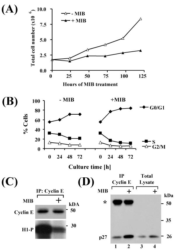
Consistent with our previous studies, the synthetic androgen 7α-17α-dimethyl-19-nortestosterone (mibolerone, MIB) inhibits the proliferation and suppresses the transformed phenotype of LNCaP cells ([46] and Fig. 1A). No cytotoxic or apoptosis-inducing activity was associated with this growth inhibition ([46], and data not shown). To determine whether MIB caused arrest in a particular phase of the cell cycle, MIB treated cells were examined by flow cytometry. MIB induced a time-dependent accumulation of cells in G1 at the expense of both S and G2/M phases (Fig. 1B). Consistent with G1 arrest, MIB induced inhibition of cyclin E-associated H1 kinase activity (Fig. 1C). This inhibition correlated with increased recovery of the CDK2 inhibitor p27 in cyclin E immunocomplexes isolated from MIB-treated LNCaP cells (Fig. 1D). Similar finding were previously obtained with the synthetic androgen R1881 and the natural androgen dihydrotestosterone (DHT) [42,43]. The latter study also showed that p27 is quantitatively bound to cyclin E complexes in DHT-treated cells, indicating that p27 upregulation was sufficient to saturate and inhibit cyclinE/CDK2 complexes [43].
Figure 1.
MIB induces G1 cell cycle arrest and inhibition of CDK2 activity (A) LNCaP cells were grown in the absence or presence of 10 ng/ml MIB and harvested after the times indicated. Cell numbers were determined by counting in a hemacytometer. (B) LNCaP cells were maintained in the absence or presence of 10 ng/ml MIB for the indicated times, and cells were harvested for flow cytometry. (C) Cyclin E immunoprecipitates were retrieved from LNCaP cells treated with 10 ng/ml MIB for 72 h and examined for associated H1 kinase activity in vitro. The precipitated amount of cyclin E (top) and the associated kinase activity (H1-P) are shown (bottom). (D) Cell lysate was prepared from LNCaP cells maintained in the absence or presence of 10 ng/ml MIB for 72 h. Lysates were precipitated with cyclin E antibodies and immunoprecipitates were examined for co-precipitation of p27 by immunoblotting (lanes 1 and 2). The asterisk denotes the immunoglobulin heavy chains. Total cell lysates are shown in lanes 3 and 4.
MIB-induced cell cycle arrest and CDK2 inhibition coincide with upregulation of p27 and downregulation of SKP2
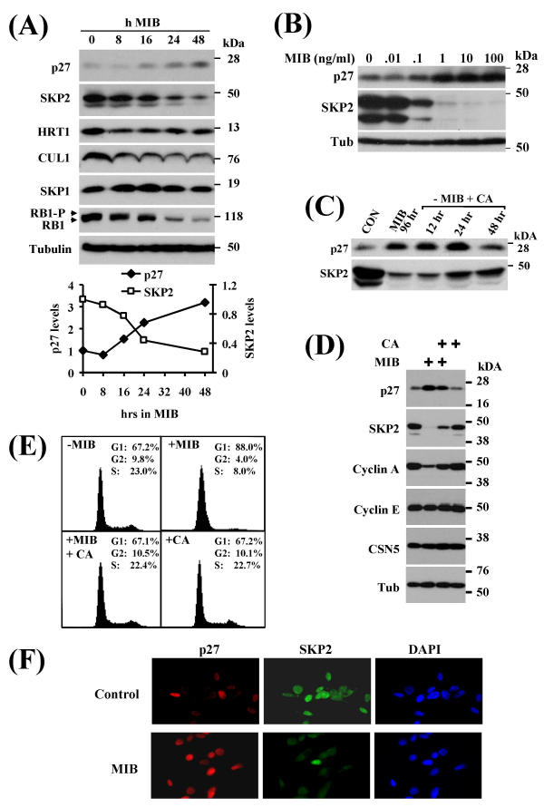
Consistent with increased recovery of p27 in cyclin E complexes, MIB caused a concentration and time-dependent increase in steady-state p27 protein levels (Fig. 2A,2B). This increase was reversible, as p27 levels were gradually restored to near control levels after removal of MIB and addition of a 500-fold molar excess of the antiandrogen cyproterone acetate (CA) (Fig. 2C and 2D). Simultaneous administration of MIB and CA partially prevented p27 accumulation, indicating that it was mediated by the androgen receptor (AR) (Fig. 2D).
Figure 2.
Effect of MIB on p27, SKP2, and other cell cycle regulators (A) LNCaP cells were incubated with 10 ng/ml MIB for the indicated periods. Cell lysate was harvested and expression of the indicated proteins was assessed by immunoblotting with respective antibodies. Tubulin is shown as a loading control. SKP2 and p27 protein levels were quantitated using a demonstration copy of the Totallab software from Nonlinear Dynamics. (B) LNCaP cells were incubated with the indicated concentrations of MIB for 72 h, followed by preparation of total cell lysate and immunoblotting with antibodies against p27, SKP2, and tubulin. The effects of androgen on p27 and SKP2 were maximal at 1 ng/ml MIB. (C) LNCaP cells were maintained in the presence of 1 ng/ml MIB for 96 h after which the medium was replaced and 750 ng/ml of the antiandrogen cyproterone acetate (CA) was added. Cell lysates were prepared after the indicated times, and p27 and SKP2 levels were assessed by immunoblotting. (D) LNCaP cells were incubated with 1 ng/ml MIB and/or 750 ng/ml CA for 72 h and cell lysates were prepared. Expression of the indicated proteins was assessed by immunoblotting. Tubulin levels are shown as loading controls. (E) LNCaP cells were incubated with 1 ng/ml MIB and/or 750 ng/ml CA for 72 h and cells were fixed for flow cytometry. Cell cycle profiles and the fraction of cells in each cell cycle phase are shown. (F) LNCaP cells were maintained in the absence or presence of 10 ng/ml MIB for 72 h. Cells were fixed and double-stained with antibodies against p27 (red) and SKP2 (green). Cell nuclei were counterstained with DAPI.
Since the level of SKP2 is a rate-limiting determinant of p27 levels [20,21,29], we examined the effect of MIB on SKP2 protein levels. SKP2 levels were down-regulated nearly four-fold by MIB at concentrations and with kinetics that closely paralleled p27 accumulation (Fig. 2A,2B). In addition, downregulation of RB1 and accumulation in the underphosphorylated form paralleled SKP2 down-regulation (Fig. 2A). In contrast, only minor changes were detected in the expression of the SKP2-associated SCFSKP2 subunits HRT1, CUL1, and SKP1 (Fig. 2A). MIB-dependent SKP2 down-regulation was efficiently counteracted by CA (Fig. 2C and 2D), again indicating an involvement of the AR. Cyclin A, but not cyclin E levels were also suppressed by MIB in an AR-dependent manner, while the levels of the COP9/signalosome subunit CSN5/JAB1 and tubulin remained constant (Fig. 2D). Finally, consistent with SKP2 and p27 being integral components of androgen-mediated growth control of LNCaP cells, CA also efficiently reversed the MIB-induced G1 cell cycle arrest (Fig. 2E).
To confirm the effect of MIB on p27 and SKP2 at the level of individual cells, we performed immunofluorescence staining. In untreated controls, most cells displayed strong staining for SKP2 with few cells positive for p27 (Fig. 2F). The nuclear staining patterns of SKP2 and p27 appeared mutually exclusive. (Fig. 2F). In contrast, the majority of MIB-treated cells showed strong p27 expression, while only few cells were positive for SKP2.
MIB-induced p27 upregulation correlates with increased p27 protein stability and decreased ubiquitylation
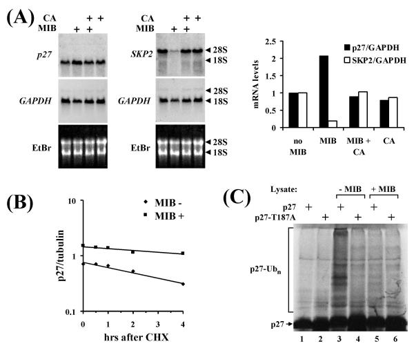
Northern blot analysis revealed that MIB-induced SKP2 downregulation at the protein level is reflected by quantitatively similar changes in steady state RNA levels (Fig 3A). In contrast, p27 mRNA levels were upregulated by MIB only two-fold (Fig. 3A). This suggested that the effect of MIB on p27 RNA levels can not fully account for the accumulation of p27 protein (compare Figs. 2B and 3A)
Figure 3.
Effect of MIB on p27 protein stability and ubiquitylation (A) LNCaP cells were maintained in the presence of 1 ng/ml MIB and/or 750 ng/ml CA for 72 h. Total cellular RNA was isolated and p27 and SKP2 RNA levels were determined by Northern blotting as described in Materials and methods. Glycerol aldehyde phosphate dehydrogenase (GAPDH) RNA levels are shown as loading controls. Ribosomal RNA is indicated as size markers. The ethidium bromide (EtBr) stained gel before transfer is show to demonstrate the integrity of the RNA. Blots were quantitated using a phosphoimager and RNA levels normalized to the GAPDH reference are shown in a block diagram (right). (B) LNCaP cells maintained in the absence or presence of 10 ng/ml MIB for 72 h were treated with cycloheximide (CHX, 100 ug/ml) to inhibit protein synthesis. Samples were taken after the indicated times, and p27 abundance was determined by immunoblotting. Blots were scanned and data normalized to the signal of tubulin were blotted in the diagram. (C) In vitro ubiquitylation of p27. Wildtype p27 or a point mutant in which threonine 187 was replaced by alanine (p27-T187A) was radiolabeled with 35S-methionine by coupled in vitro transcription/translation. The labeled substrate was incubated with total protein lysate prepared from LNCaP cells maintained in the absence or presence of 10 ng/ml MIB for 72 hours. The reaction also contained ATP and ubiquitin as described in Materials and methods. Polyubiquitylated p27 species generated in the reaction are indicated (p27-Ubn).
We therefore determined the effect of MIB on p27 half-life. Protein synthesis was inhibited by cycloheximide in control cells and in cells pretreated with MIB for 72 h. Cells were harvested after various times and the effect on p27 protein levels was assessed by immunoblotting. Using this assay, we determined a p27 half-life of 4 h in control cells, which was increased to more than 6 h in MIB treated cells (Fig. 3B). This experiment likely overestimates p27 half-life in untreated cells, as SKP2 was itself downregulated by CHX (data not shown). Nevertheless, the data suggest that p27 upregulation in response to MIB is partially mediated by changes in protein stability.
To confirm this conjecture, we used an in vitro assay to determine p27 ubiquitylation activity in LNCaP cell lysate. Similar to previously described protocols [21,22,30], cell lysates were prepared by hypotonic lysis of untreated LNCaP cells or cells treated with MIB for 96 h. Ubiquitylation substrate was prepared by in vitro transcription and translation of p27 in the presence of 35S-methionine. A point mutant of p27 (p27T187A), in which the critical CDK2 phosphorylation site in position 187 was replaced by alanine [24] was also prepared. Both substrates were incubated with recombinant cyclin E/CDK2 complexes in the presence of ATP, ubiquitin, and LNCaP cell lysate. Conversion of p27 into polyubiquitylated high molecular weight species was monitored by autoradiography of 35S-labeled p27 following gel electrophoresis. Polyubiquitylation of wildtype, but not mutant p27 was observed in untreated LNCaP cell lysate (Fig. 3C). This activity was completely abolished in MIB-treated cells (Fig. 3C), suggesting that p27 accumulation results from reduced p27 ubiquitylation.
Overexpression of SKP2 induces F-box-dependent p27 ubiquitylation
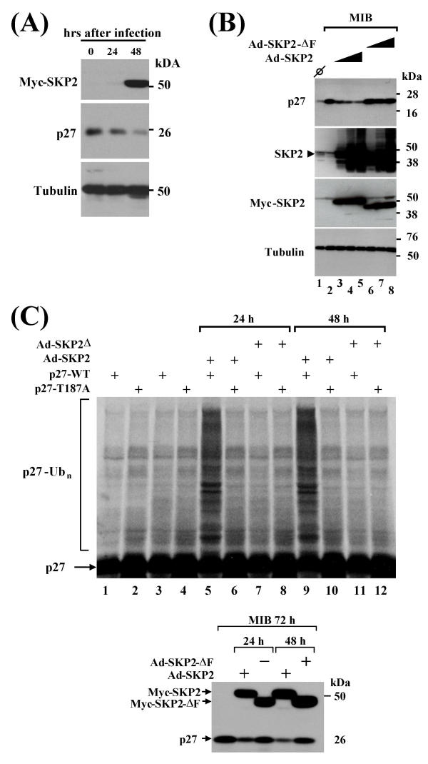
Considering the inverse correlation between p27 and SKP2 protein levels, we asked whether ectopic SKP2 can forge p27 down-regulation in LNCaP cells. To test this, we generated recombinant adenoviruses driving the expression of SKP2 (Ad-SKP2) or a mutant of SKP2 lacking the F-box (Ad-SKP2-ΔF). Infection of asynchronous LNCaP cells with SKP2 virus led to time-dependent downregulation of p27 (Fig. 4A).
Figure 4.
Effect of SKP2 overexpression on p27 protein levels and ubiquitylation (A) Asynchronous LNCaP cells were infected with adenovirus driving the expression of Myc epitope-tagged SKP2 (Ad-SKP2). Cell lysates were prepared at the indicated times after infection and Myc-SKP2 and p27 expression were determined by immunoblotting with p27 and Myc antibodies. Tubulin is shown as a loading control. (B) LNCaP cells kept in the presence of 10 ng/ml MIB for 72 h were infected with Ad-SKP2 or Ad-SKP2-ΔF (lacking the F-box) for increasing periods (24, 48, and 72 h in lanes 3–5 and 6–8, respectively). Protein lysates were prepared and assayed for the expression of the indicated proteins by immunoblotting. Overexpression of SKP2 is demonstrated by blotting with SKP2 (second panel from top) and Myc (third panel from top) antibodies. The first lane contains lysate from untreated controls. Tubulin is shown as loading control. (C) Effect of SKP2 on in vitro p27 ubiquitylation activity reconstituted in LNCaP cell lysate. LNCaP cells were arrested with 10 ng/ml MIB for 72 h and infected with Ad-SKP2 or Ad-SKP2-ΔF for 24 or 48 h as indicated. Cell lysate was prepared and employed in ubiquitylation assays using wildtype or T187A mutant p27 as substrate. Polyubiquitylated reaction products are indicated (p27-Ubn). The lower panel shows an immunoblot of the lysates used for the ubiquitylation assays downregulation of p27 by SKP2, but not SKP2-ΔF in these lysates.
To determine whether SKP2 downregulation is a necessary step in MIB-induced p27 upregulation, LNCaP cells were blocked with MIB for 72 h, followed by infection with Ad-SKP2 or Ad-SKP2-ΔF for various periods. Immunoblotting revealed that SKP2 overexpression can efficiently downregulate p27 levels in the continuous presence of MIB (Fig. 4B). In contrast, overexpression of F-box deleted SKP2 did not result in a decline of p27 (Fig. 4B).
To determine whether SKP2 induces p27 ubiquitylation, cell lysate was prepared from MIB-exposed cells infected with Ad-SKP2 and Ad-SKP2-ΔF, and supplemented with phosphorylated p27, ATP, and ubiquitin. SKP overexpression for 24 or 48 h dramatically increased p27 ubiquitylation activity present in these cell lysates (Fig. 4C, lane 5 and 7). This increase was not observed with mutant p27T187A as substrate and upon overexpression of SKP-ΔF (Fig. 4C, lanes 9 and 11).
Overexpression of SKP2 is not sufficient to overcome androgen-mediated G1 arrest
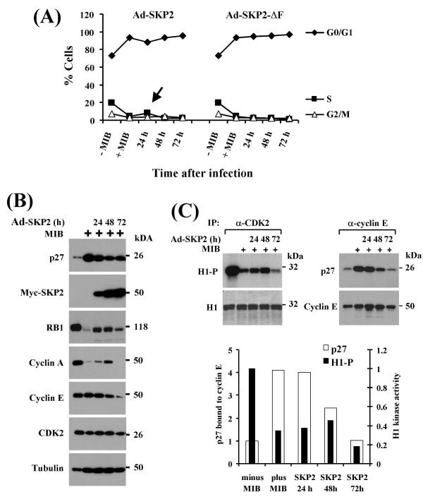
To test whether SKP2-mediated p27 degradation is sufficient to overcome the MIB-induced G1 cell cycle arrest, LNCaP cells were arrested with MIB for 72 h, followed by infection with SKP2 adenovirus. Cells were harvested and analyzed by flow cytometry after various periods. The mutant SKP2-ΔF was used as a negative control. Although, in three independent experiments, the fraction of cells in S phase was significantly (p = 0.009) higher in MIB-arrested cells overexpressing SKP2 (7.93%, +/-0.32) than in SKP2-ΔF overexpressing cells (4.02%, +/-1.41) at a time when p27 downregulation was already apparent (24 h), the majority of cells remained tightly arrested in G1 over the entire 72 h course of the experiment (Fig. 5A).
Figure 5.
Effect of SKP2 overexpression on cell cycle progression and CDK2 kinase activity (A) LNCaP cells were maintained in the absence or presence of 10 ng/ml MIB for 72 h. MIB-treated samples were subsequently infected with Ad-SKP2 or Ad-SKP2-ΔF for the indicated periods, after which cells were processed for flow cytometry. Percentage of cells in G1, S, and G2/M phases was blotted in a diagram. Note that SKP2 overexpression is not sufficient to override MIB-induced G1 arrest. The experiment was independently performed three times. (B) LNCaP cells were arrested with 10 ng/ml MIB for 72 h followed by infection with Ad-SKP2 for the indicated periods (24–72 h). Cell lysates were prepared and expression of the indicated proteins was determined by immunoblotting. (C) CDK2-dependent histone 1 (H1) in vitro kinase activity and cyclin E/p27 interaction were assessed in the same lysates used in (B). CDK2 immunoprecipitates were incubated with H1 in the presence of 32P-ATP and reaction products were separated by gel electrophoresis and detected by autoradiography (left panel). Parallel cyclin E immunoprecipitates were examined by immunoblotting for the amount of precipitated cyclin E and co-precipitated p27 (right panel). H1 kinase activity and the amount of p27 coprecipitated with cyclin E were quantitated using Totallab software (graph below blots). The input amount of H1 is shown in the lower panel on the left.
This finding is in contrast to serum starved rat fibroblasts and human U87 cells arrested in G1 by overexpression of PTEN, in which SKP2 overexpression can drive S phase entry. [29,30,32,33]. In both cases, SKP2 overexpression leads to induction of cyclin A and CDK2 kinase activity. We therefore asked whether SKP2-mediated p27 downregulation in MIB arrested LNCaP cells was able to actually bring about CDK2 activation.
We first determined the effect of SKP2 overexpression on MIB-induced RB1 downregulation and accumulation in the dephosphorylated state (see Fig. 2A), which is carried out by CDK2 in vivo[47]. Immunoblotting revealed that RB1 suppression in the hypophosphorylated state was efficiently reversed by SKP2 overexpression for 24 or 48 h (Fig. 5B). In addition, cyclin A expression was partially restored (Fig. 5B). However, both effects were largely annihilated after 72 h of SKP2 overexpression. In contrast, cyclin E levels, while not initially affected by MIB, were decreased after 72 h of SKP2 overexpression (Fig. 5B). In contrast, CDK2 and tubulin levels remained stable at all time points of SKP2 overexpression (Fig. 5B).
A parallel experiment, revealed that CDK2 kinase activity assayed in vitro using histone 1 as substrate was suppressed by MIB, but partially restored by overexpression of SKP2 for 24 and 48 h (Fig. 5C, left panel). This increase was paralleled by a decrease in the amount of p27 associated with cyclin E (Fig. 5C, right panel). While this decrease was maintained throughout the course of the experiment, CDK2 activation was attenuated again at 72 h of SKP2 overexpression. In summary, SKP2 overexpression caused transient upregulation of cyclin A expression and CDK2 activity, but was inefficient in promoting S phase entry of MIB-arrested LNCaP cells.
Discussion
Androgen control of LNCaP cell proliferation
We have used the cell line LNCaP as a model to examine the role of SKP2 in androgen control of p27 expression and cell cycle progression in human prostate cancer cells. LNCaP cells exhibit a complex, but characteristic biphasic response to androgens: (1.) Stimulation of proliferation at low androgen levels, and (2.) inhibition of proliferation at higher levels such as those used in this study [41]. This dichotomy was compared [43] to the situation in male rodents, where castration leads to atrophy of the prostate due to apoptosis. Re-administration of androgen induces transient epithelial cell proliferation, presumably mediated by proteolytic downregulation of p27, until pre-castration cell numbers have been restored [48,49]. No further proliferation ensues beyond this point, at which p27 levels are increased again, despite the continuous presence of androgen. This may reflect androgen activity in restoring the glandular structure by promoting postmitotic differentiation [48,49]. Consistent with this view, androgen administration to intact rats resulted in suppression of prostate epithelial cell proliferation and maintenance of morphological gland integrity [50]. Based on these findings, we propose that the effects of high doses of androgens on SKP2 and p27 in LNCaP cells described here reflect a differentiation and consolidation effect in vitro. If this is the case, LNCaP cells, despite being highly aneuploid derivatives of a metastatic lesion of an advanced cancer [39], may have retained some aspects of the normal biphasic mechanism of androgen control observed in castrated rodents. This mechanism may also be retained in other prostate cancers, thereby representing a potential target for intervention.
Role of SKP2 in androgen control of p27 stability
Our data indicate that SKP2 is an integral component of androgen control in LNCaP cells, as it is down-regulated in an AR-mediated manner (Fig. 2B and 2C). In contrast, all other known SCFSKP2 subunits examined were largely unresponsive to androgen (Fig. 2A). Importantly, SKP2 overexpression is sufficient to reverse androgen-mediated p27 accumulation and induce p27 ubiquitylation (Fig. 4B and 4D). Based on the F-box dependency of these processes (Fig. 4C), we conclude that SKP2 directly triggers the ubiquitylation and degradation of p27 in LNCaP cells, a process which is attenuated by androgen-mediated SKP2 downregulation. This interpretation is consistent with SKP2 being a proximal element in the previously identified androgen-mediated pathway of cell cycle inhibition in LNCaP cells. As a direct consequence of SKP2 downregulation, p27 is upregulated and cyclin A is downregulated. This results in inhibition of CDK2 activity, accumulation of hypophosphorylated RB1, inactivation of E2F1 [45], and cell cycle arrest.
While it may surprise that SKP2 overexpression alone is sufficient to mediate p27 degradation in LNCaP cells, given the known requirement for p27 phosphorylation [24], similar observations were made in rat fibroblasts, human U87 cells, and primary rat hepatocytes [29-32]. In rat fibroblasts, these findings correlate with SKP2-induced upregulation of cyclin A and CDK2 kinase activity [29,32], which could further augment p27 phosphorylation and degradation, ultimately stimulating cells to enter S phase.
Unlike in rat fibroblasts and human U87 cells [29,30,32], SKP2 overexpression is not sufficient to override androgen-induced G1 arrest in LNCaP cells, despite a profound effect on p27 levels (Fig. 5). Consistent with this finding, ectopic SKP2 expression was not sufficient to maintain high cyclin A levels, CDK2 activation, and RB1 phosphorylation for prolonged periods (Fig. 5B and 5C). The transient character of these responses may explain the failure to cause efficient S phase entry in the presence of MIB, in particular as co-expression of cyclins was previously shown to synergize with SKP2 in stimulating CDK2 activity and S phase entry [30,31]. In addition, cyclin A is frequently overexpressed in prostate cancers [51]. In future experiments, it will be interesting to determine the effect of combined overexpression of SKP2 and cyclin A and cyclin E on cell cycle progression of MIB-arrested cells.
Regardless of the outcome of such experiments, our data firmly suggest that SKP2 is a central mediator of the androgen response mechanism of LNCaP cells. However, we do not believe that SKP2 is a direct transcriptional target of the AR, as the kinetics of its downregulation were rather slow (Fig. 2A), suggesting several intermediate steps culminating in this event. Such a mediator could be AS3, a gene induced by growth inhibiting levels of androgen with kinetics that precede cell cycle arrest [52]. In addition, AS3 overexpression mimics androgen-mediated cell cycle arrest in MCF7 cells stably expressing exogenous AR, while anti-sense AS3 confers resistance to androgen-induced growth arrest [53]. The function of AS3 is still unknown, but the encoded protein has features of a potential transcription factor [53].
While our study suggests that SKP2 is an important player in p27 regulation in prostate cancer cells, recent studies have also implicated another protein, CSN5/JAB1, in p27 regulation [54,55]. CSN5 is a subunit of the COP9/signalosome (CSN) complex [56-59], but also forms distinct complexes apparently lacking CSN subunits [55,60-62]. Overexpression of CSN5 in NIH3T3 cells causes p27 export from the nucleus and ectopic degradation [54]. Notably, like SKP2 [33,36-38], CSN5 was found to be overexpressed in cancers devoid of p27 [63]. However, our studies did not reveal any effect of MIB on CSN5 expression. Future studies aimed at eliminating CSN5 activity will show whether it is involved in androgen-mediated p27 control.
Finally, a recent report demonstrated SKP2-independent proteolysis of p27 in lymphocytes derived from SKP2 knockout mice [64]. It is thus possible that p27 proteolysis is subject to tissue-specific regulation. In prostate cells, however, SKP2 seems to be a major determinant of p27 degradation, as it is down-regulated by androgen and its overexpression is sufficient to mediate p27 ubiquitylation and degradation. Recent studies involving androgen administration to castrated rats have come to similar conclusions [49].
Conclusions
Our data suggest that androgen-mediated SKP2 downregulation is responsible for decreased ubiquitylation and accumulation of p27 in LNCaP cells, an effect that is readily overcome by overexpression of SKP2. We propose that frequent p27 downregulation in prostate cancer may be caused by SKP2 overexpression thus enabling escape from normal androgen control. Finally, our in vitro studies validate the SKP2 pathway as a potential target for therapeutic intervention in cancers devoid of p27.
Materials and methods
Plasmids and reagents
Human SKP2 and p27 cDNAs were kindly provided by H. Zhang (Yale University) and P. Jackson (Stanford University). The genes were amplified by PCR and cloned into pcDNA3 enabling in vitro transcription. The SKP2 mutant lacking the F-box was constructed using the Quickchange kit purchased from Stratagene. Probes for Northern blotting were generated by PCR. The GAPDH probe was generated by reverse transcription and PCR from LNCaP m RNA.
The synthetic androgen mibolerone (7α-17α-dimethyl-19-nortestosterone) was purchased from Perkin Elmer Life Sciences. The antiandrogen cyproterone acetate was a gift from C. Sonnenschein (Tufts University). The reagents were dissolved in absolute ethanol.
Antibodies: SKP2 (Zymed 32–3300, 1:250 for immunoblotting), SKP2 (Santa Cruz sc-7164, 1:500 for immunofluorescence staining), p27 (Transduction Laboratories K25030, 1:5000 for immunoblotting, 1:1000 for immunofluorescence staining), cyclin A (Neomarkers Ab-6, MS-1061, 1:100 for immunoblotting), cyclin E (Neomarkers Ab-1, RB-012, 2 ug per immunoprecipitation) cyclin E (Neomarkers Ab-2, MS-870, 1:200 for immunoblotting), HRT1 (affinity-purified rabbit polyclonal, gift from R. Deshaies, 1:1000 for immunoblotting), CUL1 (Zymed 71–8700, 1:250 for immunoblotting), SKP1 (Transduction laboratories 610530, 1:5000 for immunoblotting), JAB1/CSN5 (GeneTex MS-Jab11-PX1, 1:1000 for immunoblotting), RB1 (Pharmingen 554136, 1:500 for immunoblotting), CDK2 (Santo Cruz Biotechnology sc-163, 2 ug per immunoprecipitation, 1:500 for immunoblotting), Tubulin (Sigma T5168, 1:2000 for immunoblotting).
Tissue culture
LNCaP-FGC cells were obtained from ATCC (order number CRL-10995) and maintained in RPMI supplemented with 10% FBS as described [46]. We noticed that LNCaP cells at low density grow best on 15 cm Falcon 3025 tissue culture dishes and that the effects of SKP2 overexpression were most pronounced when cells were cultured on these dishes (data not shown).
Adenoviruses
For production of adenoviruses, the ADEASY system was used (generously provided by B. Vogelstein). The complete SKP2 cDNA or a mutant lacking the F-box (SKP2-ΔF) were cloned into a modified pcDNA3 plasmid providing an N-terminal Myc epitope tag and the human lamin B leader sequence. The lamin B-SKP2 cassette was removed and cloned into the pADTRACK1 shuttle vector. The resulting plasmid was transformed into BJ-ADEASY cells by electroporation. Adenoviral DNA generated by recombination in BJ-ADEASY cells was isolated. Recombinant adenoviral DNA was transfected into 293 cells using standard calcium phosphate procedures. Virus was harvested from cells and amplified by infection of 293 cells. Amplified virus was purified by cesium chloride gradient centrifugation and tittered. An approximate multiplicity of infection of 100 was used throughout.
Immunological techniques
Cell lysates for immunoblotting were prepared by scraping cells into heated 2 × SDS sample buffer. Samples were boiled for 10 min, centrifuged, and separated by SDS gel electrophoresis. Proteins were transferred onto Immobilon P membrane (Millipore), blocked in 5 % nonfat dry milk dissolved in tris-buffered saline / 0.1 % Tween and probed with the various antibodies.
Cell lysates for immunoprecipitation were prepared in IP-LB (25 mM Tris, pH 7.4, 150 mM NaCl, 0.5 % Triton X100, 10 μg/ml leupeptin, 10 μg/ml pepstatin, 1 mM PMSF, 17 μg/ml aprotinin). Cells were scraped into IP-LB and incubated on ice for 30 min. Cell lysate was cleared by centrifugation and the supernatant was incubated with various antibodies (see plasmid and reagents section above). Immunocomplexes were collected on protein A or G beads, washed five times in IP-LB for 5 minutes each, dissolved in 2 × SDS sample buffer, and separated by SDS gel electrophoresis.
For immunofluorescence staining, LNCaP cells were plated onto poly-lysine coated glass coverslips. Cells were fixed in 4 % para-formaldehyde in PBS and permeabilized by washing in PBS / 0.1 % Triton X100. Coverslips were blocked in PBS / 0.1 % Triton X100 containing 5 % nonfat dry milk for 45 min. Incubation with primary and secondary antibodies was in the same buffer for 45 min each. After antibody incubations, coverslips were washed five times in PBS / 0.1 % Triton X100, followed by mounting on microscopy slides. Micrographs were taken with a Spot CCD camera mounted on a Nikon E600 epifluorescence microscope. Brightness and contrast of the images was adjusted in Adobe Photoshop 5.0.
Northern blotting
Isolation of total cellular RNA using a urea/LiCl protocol and gel electrophoresis was performed as described previously [65]. Gels were transferred onto Amersham Hybond N+ nylon membranes in 20 × SSC and prehybridized in 4 × SSC, 1 × Denhardt's solution, 0.5% SDS for 2 h at 68°C. Complementary DNA fragments for SKP2, p27, and GAPDH were amplified by PCR and radiolabeled by random priming. Filters were hybridized for 16 h at 68°C followed by washing in 2 × SSC at 68°C. Membranes were exposed to X-ray film for 2 to 12 h and scanned with a phosphoimager for signal quantitation.
H1 kinase assay
Histone 1 kinase assays with cyclin E or CDK2 immunoprecipitates were performed as described [65].
In vitro ubiquitylation assay
Cell lysates for ubiquitylation assays were prepared by hypotonic lysis on ice in 20 mM Hepes pH 7.5, 15 mM MgCl2, 5 mM KCl, 1 mM DTT, 10 μg/ml leupeptin, 10 μg/ml pepstatin, 1 mM PMSF, 17 μg/ml aprotinin as described previously [30]. Lysates were cleared by centrifugation and used without prior freezing. Radiolabeled wildtype and T178A point mutant p27 were produced by combined in vitro transcription and translation in rabbit reticulocyte lysate containing 35S-methionine. The in vitro translated proteins were incubated 2 h at 30°C in the presence of 1 mM ATP with recombinant cyclin E / CDK2 complexes produced from baculoviruses in insect cells. Aliquots of phosphorylated proteins were added to a reaction containing 120 μg cell lysate, recombinant human E1, 0.25 mg/ml ubiquitin, 1 μM ubiquitin aldehyde, 1 μM okadaic acid, 20 μM MG132, and ATP regenerating system (20 mM ATP, 10 mM creatine phosphate, 0.1 μg/ml creatine kinase). After 90 min, the reactions were stopped by addition of SDS sample buffer, and reaction products were separated by SDS gel electrophoresis and detected by autoradiography.
Abbreviations
SCF = SKP1/Cullin/F-box protein complex
UBC = Ubiquitin conjugating enzyme
MIB = Mibolerone (7α-17α-dimethyl-19-nortestosterone)
SKP2 = S phase kinase associated protein 2
CDK2 = cyclin-dependent kinase 2
Authors' contributions
All experiments reported in this study were performed by L-F. L. with the following exceptions: H.S. prepared the SKP2-ΔF mutant, SKP2 adenoviruses, and provided initial evidence for p27 upregulation by MIB. D.A.W. performed the experiments shown in Fig. 5B and 5C, conceived the study, and participated in its design and coordination. All authors read and approved the final manuscript.
Acknowledgments
Acknowledgments
We thank H. Zhang for a human SKP2 plasmid, P. Jackson for a human p27 cDNA, S. Reed and D. Morgan for cyclin E and CDK2 baculoviruses, C. Sonnenschein for antiandrogens, and B. Vogelstein for adenoviral vectors. We are grateful to C. Sonnenschein for careful review of this manuscript and for discussion. This work was funded by grant DAMD17-00-1-0021 from the Department of Defense Prostate Cancer Research Program (managed by the U.S. Army Medical Research and Materiel Command), by grant MDPH41411159033 form the Massachusetts Department of Public Health Prostate Cancer Research Program to D.A.W., and by the NIEHS Kresge Center Grant ES-00002.
Contributor Information
Lifang Lu, Email: lflu@hsph.harvard.edu.
Holger Schulz, Email: hschulz@hsph.harvard.edu.
Dieter A Wolf, Email: dwolf@hsph.harvard.edu.
References
- De Marzo AM, Meeker AK, Epstein JI, Coffey DS. Prostate stem cell compartments: expression of the cell cycle inhibitor p27Kip1 in normal, hyperplastic, and neoplastic cells. Am J Pathol. 1998;153:911–9. doi: 10.1016/S0002-9440(10)65632-5. [DOI] [PMC free article] [PubMed] [Google Scholar]
- Cheville JC, Lloyd RV, Sebo TJ, Cheng L, Erickson L, Bostwick DG, Lohse CM, Wollan P. Expression of p27kip1 in prostatic adenocarcinoma. Mod Pathol. 1998;11:324–8. [PubMed] [Google Scholar]
- Tsihlias J, Kapusta LR, DeBoer G, Morava-Protzner I, Zbieranowski I, Bhattacharya N, Catzavelos GC, Klotz LH, Slingerland JM. Loss of cyclin-dependent kinase inhibitor p27Kip1 is a novel prognostic factor in localized human prostate adenocarcinoma. Cancer Res. 1998;58:542–8. [PubMed] [Google Scholar]
- Cote RJ, Shi Y, Groshen S, Feng AC, Cordon-Cardo C, Skinner D, Lieskovosky G. Association of p27Kip1 levels with recurrence and survival in patients with stage C prostate carcinoma. J Natl Cancer Inst. 1998;90:916–20. doi: 10.1093/jnci/90.12.916. [DOI] [PubMed] [Google Scholar]
- Mal A, Poon RY, Howe PH, Toyoshima H, Hunter T, Harter ML. Inactivation of p27Kip1 by the viral E1A oncoprotein in TGFbeta-treated cells. Nature. 1996;380:262–5. doi: 10.1038/380262a0. [DOI] [PubMed] [Google Scholar]
- Zerfass-Thome K, Zwerschke W, Mannhardt B, Tindle R, Botz JW, Jansen-Durr P. Inactivation of the cdk inhibitor p27KIP1 by the human papillomavirus type 16 E7 oncoprotein. Oncogene. 1996;13:2323–30. [PubMed] [Google Scholar]
- Kawamata N, Morosetti R, Miller CW, Park D, Spirin KS, Nakamaki T, Takeuchi S, Hatta Y, Simpson J, Wilcyznski S, et al. Molecular analysis of the cyclin-dependent kinase inhibitor gene p27/Kip1 in human malignancies. Cancer Res. 1995;55:2266–9. [PubMed] [Google Scholar]
- Ponce-Castaneda MV, Lee MH, Latres E, Polyak K, Lacombe L, Montgomery K, Mathew S, Krauter K, Sheinfeld J, Massague J, et al. p27Kip1: chromosomal mapping to 12p12-12p13.1 and absence of mutations in human tumors. Cancer Res. 1995;55:1211–4. [PubMed] [Google Scholar]
- Pietenpol JA, Bohlander SK, Sato Y, Papadopoulos N, Liu B, Friedman C, Trask BJ, Roberts JM, Kinzler KW, Rowley JD, et al. Assignment of the human p27Kip1 gene to 12p13 and its analysis in leukemias. Cancer Res. 1995;55:1206–10. [PubMed] [Google Scholar]
- Koff A, Polyak K. p27KIP1, an inhibitor of cyclin-dependent kinases. Prog Cell Cycle Res. 1995;1:141–7. doi: 10.1007/978-1-4615-1809-9_11. [DOI] [PubMed] [Google Scholar]
- Sherr CJ, Roberts JM. Inhibitors of mammalian G1 cyclin-dependent kinases. Genes Dev. 1995;9:1149–63. doi: 10.1101/gad.9.10.1149. [DOI] [PubMed] [Google Scholar]
- Coats S, Flanagan WM, Nourse J, Roberts JM. Requirement of p27Kip1 for restriction point control of the fibroblast cell cycle. Science. 1996;272:877–80. doi: 10.1126/science.272.5263.877. [DOI] [PubMed] [Google Scholar]
- Hengst L, Reed SI. Translational control of p27Kip1 accumulation during the cell cycle. Science. 1996;271:1861–4. doi: 10.1126/science.271.5257.1861. [DOI] [PubMed] [Google Scholar]
- Cordon-Cardo C, Koff A, Drobnjak M, Capodieci P, Osman I, Millard SS, Gaudin PB, Fazzari M, Zhang ZF, Massague J, Scher HI. Distinct altered patterns of p27KIP1 gene expression in benign prostatic hyperplasia and prostatic carcinoma. J Natl Cancer Inst. 1998;90:1284–91. doi: 10.1093/jnci/90.17.1284. [DOI] [PubMed] [Google Scholar]
- Loda M, Cukor B, Tam SW, Lavin P, Fiorentino M, Draetta GF, Jessup JM, Pagano M. Increased proteasome-dependent degradation of the cyclin-dependent kinase inhibitor p27 in aggressive colorectal carcinomas. Nat Med. 1997;3:231–4. doi: 10.1038/nm0297-231. [DOI] [PubMed] [Google Scholar]
- Esposito V, Baldi A, De Luca A, Groger AM, Loda M, Giordano GG, Caputi M, Baldi F, Pagano M, Giordano A. Prognostic role of the cyclin-dependent kinase inhibitor p27 in non-small cell lung cancer. Cancer Res. 1997;57:3381–5. [PubMed] [Google Scholar]
- Chiarle R, Budel LM, Skolnik J, Frizzera G, Chilosi M, Corato A, Pizzolo G, Magidson J, Montagnoli A, Pagano M, Maes B, De Wolf-Peeters C, Inghirami G. Increased proteasome degradation of cyclin-dependent kinase inhibitor p27 is associated with a decreased overall survival in mantle cell lymphoma. Blood. 2000;95:619–26. [PubMed] [Google Scholar]
- Piva R, Cancelli I, Cavalla P, Bortolotto S, Dominguez J, Draetta GF, Schiffer D. Proteasome-dependent degradation of p27/kip1 in gliomas. J Neuropathol Exp Neurol. 1999;58:691–6. doi: 10.1097/00005072-199907000-00002. [DOI] [PubMed] [Google Scholar]
- Hershko A, Ciechanover A. The ubiquitin system. Annu Rev Biochem. 1998;67:425–79. doi: 10.1146/annurev.biochem.67.1.425. [DOI] [PubMed] [Google Scholar]
- Carrano AC, Eytan E, Hershko A, Pagano M. SKP2 is required for ubiquitin-mediated degradation of the CDK inhibitor p27. Nat Cell Biol. 1999;1:193–199. doi: 10.1038/12013. [DOI] [PubMed] [Google Scholar]
- Tsvetkov LM, Yeh KH, Lee SJ, Sun H, Zhang H. p27(Kip1) ubiquitination and degradation is regulated by the SCF(Skp2) complex through phosphorylated Thr187 in p27. Curr Biol. 1999;9:661–4. doi: 10.1016/S0960-9822(99)80290-5. [DOI] [PubMed] [Google Scholar]
- Spruck C, Strohmaier H, Watson M, Smith AP, Ryan A, Krek TW, Reed SI. A CDK-independent function of mammalian Cks1: targeting of SCF(Skp2) to the CDK inhibitor p27Kip1. Mol Cell. 2001;7:639–50. doi: 10.1016/s1097-2765(01)00210-6. [DOI] [PubMed] [Google Scholar]
- Ganoth D, Bornstein G, Ko TK, Larsen B, Tyers M, Pagano M, Hershko A. The cell-cycle regulatory protein Cks1 is required for SCF(Skp2)-mediated ubiquitinylation of p27. Nat Cell Biol. 2001;3:321–4. doi: 10.1038/35060126. [DOI] [PubMed] [Google Scholar]
- Sheaff RJ, Groudine M, Gordon M, Roberts JM, Clurman BE. Cyclin E-CDK2 is a regulator of p27Kip1. Genes Dev. 1997;11:1464–78. doi: 10.1101/gad.11.11.1464. [DOI] [PubMed] [Google Scholar]
- Vlach J, Hennecke S, Amati B. Phosphorylation-dependent degradation of the cyclin-dependent kinase inhibitor p27. Embo J. 1997;16:5334–44. doi: 10.1093/emboj/16.17.5334. [DOI] [PMC free article] [PubMed] [Google Scholar]
- Montagnoli A, Fiore F, Eytan E, Carrano AC, Draetta GF, Hershko A, Pagano M. Ubiquitination of p27 is regulated by Cdk-dependent phosphorylation and trimeric complex formation. Genes Dev. 1999;13:1181–9. doi: 10.1101/gad.13.9.1181. [DOI] [PMC free article] [PubMed] [Google Scholar]
- Michel JJ, Xiong Y. Human CUL-1, but not other cullin family members, selectively interacts with SKP1 to form a complex with SKP2 and cyclin A. Cell Growth Differ. 1998;9:435–49. [PubMed] [Google Scholar]
- Lisztwan J, Marti A, Sutterluty H, Gstaiger M, Wirbelauer C, Krek W. Association of human CUL-1 and ubiquitin-conjugating enzyme CDC34 with the F-box protein p45(SKP2): evidence for evolutionary conservation in the subunit composition of the CDC34-SCF pathway. Embo J. 1998;17:368–83. doi: 10.1093/emboj/17.2.368. [DOI] [PMC free article] [PubMed] [Google Scholar]
- Sutterluty H, Chatelain E, Marti A, Wirbelauer C, Senften M, Muller U, Krek W. p45Skp2 promotes p27Kip1 degradation and induces S phase in quiescent cells. Nat Cell Biol. 1999;1:207–214. doi: 10.1038/12027. [DOI] [PubMed] [Google Scholar]
- Mamillapalli R, Gavrilova N, Mihaylova VT, Tsvetkov LM, Wu H, Zhang H, Sun H. PTEN regulates the ubiquitin-dependent degradation of the CDK inhibitor p27(KIP1) through the ubiquitin E3 ligase SCF(SKP2). Curr Biol. 2001;11:263–7. doi: 10.1016/S0960-9822(01)00065-3. [DOI] [PubMed] [Google Scholar]
- Nelsen CJ, Hansen LK, Rickheim DG, Chen C, Stanley MW, Krek W, Albrecht JH. Induction of hepatocyte proliferation and liver hyperplasia by the targeted expression of cyclin E and skp2. Oncogene. 2001;20:1825–31. doi: 10.1038/sj.onc.1204248. [DOI] [PubMed] [Google Scholar]
- Carrano AC, Pagano M. Role of the F-box protein Skp2 in adhesion-dependent cell cycle progression. J Cell Biol. 2001;153:1381–90. doi: 10.1083/jcb.153.7.1381. [DOI] [PMC free article] [PubMed] [Google Scholar]
- Gstaiger M, Jordan R, Lim M, Catzavelos C, Mestan J, Slingerland J, Krek W. Skp2 is oncogenic and overexpressed in human cancers. Proc Natl Acad Sci U S A. 2001;98:5043–8. doi: 10.1073/pnas.081474898. [DOI] [PMC free article] [PubMed] [Google Scholar]
- Latres E, Chiarle R, Schulman BA, Pavletich NP, Pellicer A, Inghirami G, Pagano M. Role of the F-box protein Skp2 in lymphomagenesis. Proc Natl Acad Sci U S A. 2001;98:2515–20. doi: 10.1073/pnas.041475098. [DOI] [PMC free article] [PubMed] [Google Scholar]
- Zhang H, Kobayashi R, Galaktionov K, Beach D. p19Skp1 and p45Skp2 are essential elements of the cyclin A-CDK2 S phase kinase. Cell. 1995;82:915–25. doi: 10.1016/0092-8674(95)90271-6. [DOI] [PubMed] [Google Scholar]
- Chao Y, Shih YL, Chiu JH, Chau GY, Lui WY, Yang WK, Lee SD, Huang TS. Overexpression of cyclin A but not Skp 2 correlates with the tumor relapse of human hepatocellular carcinoma. Cancer Res. 1998;58:985–90. [PubMed] [Google Scholar]
- Hershko D, Bornstein G, Ben-Izhak O, Carrano A, Pagano M, Krausz MM, Hershko A. Inverse relation between levels of p27(Kip1) and of its ubiquitin ligase subunit Skp2 in colorectal carcinomas. Cancer. 2001;91:1745–51. doi: 10.1002/1097-0142(20010501)91:9<1745::AID-CNCR1193>3.0.CO;2-H. [DOI] [PubMed] [Google Scholar]
- Kudo Y, Kitajima S, Sato S, Miyauchi M, Ogawa I, Takata T. High expression of S-phase kinase-interacting protein 2, human F-box protein, correlates with poor prognosis in oral squamous cell carcinomas. Cancer Res. 2001;61:7044–7. [PubMed] [Google Scholar]
- Horoszewicz JS, Leong SS, Chu TM, Wajsman ZL, Friedman M, Papsidero L, Kim U, Chai LS, Kakati S, Arya SK, Sandberg AA. The LNCaP cell line – a new model for studies on human prostatic carcinoma. Prog Clin Biol Res. 1980;37:115–32. [PubMed] [Google Scholar]
- Soto AM, Lin TM, Sakabe K, Olea N, Damassa DA, Sonnenschein C. Variants of the human prostate LNCaP cell line as tools to study discrete components of the androgen-mediated proliferative response. Oncol Res. 1995;7:545–58. [PubMed] [Google Scholar]
- Sonnenschein C, Olea N, Pasanen ME, Soto AM. Negative controls of cell proliferation: human prostate cancer cells and androgens. Cancer Res. 1989;49:3474–81. [PubMed] [Google Scholar]
- Kokontis JM, Hay N, Liao S. Progression of LNCaP prostate tumor cells during androgen deprivation: hormone-independent growth, repression of proliferation by androgen, and role for p27Kip1 in androgen-induced cell cycle arrest. Mol Endocrinol. 1998;12:941–53. doi: 10.1210/mend.12.7.0136. [DOI] [PubMed] [Google Scholar]
- Tsihlias J, Zhang W, Bhattacharya N, Flanagan M, Klotz L, Slingerland J. Involvement of p27Kip1 in G1 arrest by high dose 5 alpha-dihydrotestosterone in LNCaP human prostate cancer cells. Oncogene. 2000;19:670–9. doi: 10.1038/sj.onc.1203369. [DOI] [PubMed] [Google Scholar]
- Wolf DA, Kohlhuber F, Schulz P, Fittler F, Eick D. Transcriptional down-regulation of c-myc in human prostate carcinoma cells by the synthetic androgen mibolerone. Br J Cancer. 1992;65:376–82. doi: 10.1038/bjc.1992.76. [DOI] [PMC free article] [PubMed] [Google Scholar]
- Hofman K, Swinnen JV, Verhoeven G, Heyns W. E2F activity is biphasically regulated by androgens in LNCaP cells. Biochem Biophys Res Commun. 2001;283:97–101. doi: 10.1006/bbrc.2001.4738. [DOI] [PubMed] [Google Scholar]
- Wolf DA, Schulz P, Fittler F. Synthetic androgens suppress the transformed phenotype in the human prostate carcinoma cell line LNCaP. Br J Cancer. 1991;64:47–53. doi: 10.1038/bjc.1991.237. [DOI] [PMC free article] [PubMed] [Google Scholar]
- Hinds PW, Mittnacht S, Dulic V, Arnold A, Reed SI, Weinberg RA. Regulation of retinoblastoma protein functions by ectopic expression of human cyclins. Cell. 1992;70:993–1006. doi: 10.1016/0092-8674(92)90249-c. [DOI] [PubMed] [Google Scholar]
- Chen Y, Robles AI, Martinez LA, Liu F, Gimenez-Conti IB, Conti CJ. Expression of G1 cyclins, cyclin-dependent kinases, and cyclin-dependent kinase inhibitors in androgen-induced prostate proliferation in castrated rats. Cell Growth Differ. 1996;7:1571–8. [PubMed] [Google Scholar]
- Waltregny D, Leav I, Signoretti S, Soung P, Lin D, Merk F, Adams JY, Bhattacharya N, Cirenei N, Loda M. Androgen-driven prostate epithelial cell proliferation and differentiation in vivo involve the regulation of p27. Mol Endocrinol. 2001;15:765–82. doi: 10.1210/mend.15.5.0640. [DOI] [PubMed] [Google Scholar]
- Leav I, Merk FB, Kwan PW, Ho SM. Androgen-supported estrogen-enhanced epithelial proliferation in the prostates of intact Noble rats. Prostate. 1989;15:23–40. doi: 10.1002/pros.2990150104. [DOI] [PubMed] [Google Scholar]
- Mashal RD, Lester S, Corless C, Richie JP, Chandra R, Propert KJ, Dutta A. Expression of cell cycle-regulated proteins in prostate cancer. Cancer Res. 1996;56:4159–63. [PubMed] [Google Scholar]
- Geck P, Szelei J, Jimenez J, Lin TM, Sonnenschein C, Soto AM. Expression of novel genes linked to the androgen-induced, proliferative shutoff in prostate cancer cells. J Steroid Biochem Mol Biol. 1997;63:211–8. doi: 10.1016/S0960-0760(97)00122-2. [DOI] [PubMed] [Google Scholar]
- Geck P, Maffini MV, Szelei J, Sonnenschein C, Soto AM. Androgen-induced proliferative quiescence in prostate cancer cells: the role of AS3 as its mediator. Proc Natl Acad Sci U S A. 2000;97:10185–90. doi: 10.1073/pnas.97.18.10185. [DOI] [PMC free article] [PubMed] [Google Scholar]
- Tomoda K, Y. K, J.-Y. K. Degradation of the cyclin-dependent-kinase inhibitor p27Kip1 is instigated by Jab1. Nature. 1999;398:160–5. doi: 10.1038/18230. [DOI] [PubMed] [Google Scholar]
- Tomoda K, Kubota Y, Arata Y, Mori S, Maeda M, Tanaka T, Yoshida M, Yoneda-Kato N, Kato JY. The cytoplasmic shuttling and subsequent degradation of p27Kip1 mediated by Jab1/CSN5 and the COP9 signalosome complex. J Biol Chem. 2002;277:2302–10. doi: 10.1074/jbc.M104431200. [DOI] [PubMed] [Google Scholar]
- Chamovitz DA, Wei N, Osterlund MT, von Arnim AG, Staub JM, Matsui M, Deng XW. The COP9 complex, a novel multisubunit nuclear regulator involved in light control of a plant developmental switch. Cell. 1996;86:115–21. doi: 10.1016/s0092-8674(00)80082-3. [DOI] [PubMed] [Google Scholar]
- Glickman MH, Rubin DM, Coux O, Wefes I, Pfeifer G, Cjeka Z, Baumeister W, Fried VA, Finley D. A subcomplex of the proteasome regulatory particle required for ubiquitin-conjugate degradation and related to the COP9-signalosome and eIF3. Cell. 1998;94:615–23. doi: 10.1016/s0092-8674(00)81603-7. [DOI] [PubMed] [Google Scholar]
- Seeger M, Kraft R, Ferrell K, Bech-Otschir D, Dumdey R, Schade R, Gordon C, Naumann M, Dubiel W. A novel protein complex involved in signal transduction possessing similarities to 26S proteasome subunits. Faseb J. 1998;12:469–78. [PubMed] [Google Scholar]
- Wei N, Chamovitz DA, Deng XW. Arabidopsis COP9 is a component of a novel signaling complex mediating light control of development. Cell. 1994;78:117–24. doi: 10.1016/0092-8674(94)90578-9. [DOI] [PubMed] [Google Scholar]
- Zhou C, Seibert V, Geyer R, Rhee E, Lyapina S, Cope G, Deshaies RJ, Wolf DA. The fission yeast COP9/signalosome is involved in cullin modification by ubiquitin-related Ned8p. BMC Biochemistry. 2001;2:7. doi: 10.1186/1472-2091-2-7. [DOI] [PMC free article] [PubMed] [Google Scholar]
- Mundt KE, Liu C, Carr AM. Deletion mutants in COP9/signalosome subunits in fission yeast Schizosaccharomyces pombe display distinct phenotypes. Mol Biol Cell. 2002;13:493–502. doi: 10.1091/mbc.01-10-0521. [DOI] [PMC free article] [PubMed] [Google Scholar]
- Kwok SF, Solano R, Tsuge T, Chamovitz DA, Ecker JR, Matsui M, Deng XW. Arabidopsis homologs of a c-Jun coactivator are present both in monomeric form and in the COP9 complex, and their abundance is differentially affected by the pleiotropic cop/det/fus mutations. Plant Cell. 1998;10:1779–90. doi: 10.1105/tpc.10.11.1779. [DOI] [PMC free article] [PubMed] [Google Scholar]
- Sui L, Dong Y, Ohno M, Watanabe Y, Sugimoto K, Tai Y, Tokuda M. Jab1 Expression Is Associated with Inverse Expression of p27(kip1) and Poor Prognosis in Epithelial Ovarian Tumors. Clin Cancer Res. 2001;7:4130–5. [PubMed] [Google Scholar]
- Hara T, Kamura T, Nakayama K, Oshikawa K, Hatakeyama S. Degradation of p27(Kip1) at the G(0)-G(1) transition mediated by a Skp2-independent ubiquitination pathway. J Biol Chem. 2001;276:48937–43. doi: 10.1074/jbc.M107274200. [DOI] [PubMed] [Google Scholar]
- Wolf DA, Hermeking H, Albert T, Herzinger T, Kind P, Eick D. A complex between E2F and the pRb-related protein p130 is specifically targeted by the simian virus 40 large T antigen during cell transformation. Oncogene. 1995;10:2067–78. [PubMed] [Google Scholar]


