

Abstract
Introduction
Pyodermatitis-pyostomatitis vegetans (PPV) is a rare inflammatory mucocutaneous dermatosis of unknown etiology. It is characterized by the appearance of vesicles, pustules, vegetating plaques, and erythematous lesions, often associated with underlying inflammatory bowel diseases such as Crohn’s disease.
Objective
To report a case of vegetative pyostomatitis in a patient with Crohn’s disease, focusing on the diagnostic process and therapeutic approach.
Case Report
A 47-year-old female patient, identified as AMSP, with a known diagnosis of Crohn’s disease and a history of colostomy, presented with complaints of lesions affecting both the skin and oral mucosa. Dermatological examination revealed a well-defined, flat, round lesion with darkened borders in the right axillary region. Intraoral examination showed erythematous, net-like plaques along the lateral border of the tongue, accompanied by ulcerations and vesicles. An incisional biopsy of the tongue was performed. Histopathological analysis revealed a predominantly eosinophilic inflammatory infiltrate in the connective tissue and epithelial acantholysis. The clinical presentation, patient history, and histopathological findings led to the diagnosis of pyodermatitis-pyostomatitis vegetans. The patient was treated with topical 0.1% tacrolimus ointment applied twice daily for 15 days. Following this intervention, there was complete resolution of the lesions.
Conclusion
Pyodermatitis-pyostomatitis vegetans is an uncommon condition that may serve as an oral and cutaneous manifestation of Crohn’s disease. The presence of vegetating plaques on the skin and vesiculopustular lesions in the oral cavity should raise clinical suspicion. Histopathological examination via biopsy remains the gold standard for definitive diagnosis. Treatment is typically straightforward, with an excellent prognosis when managed appropriately.
Keywords: Crohn’s disease, Oral manifestations, Inflammatory bowel disease, Pyostomatitis vegetans, Pyodermatitis
Introduction
Pyodermatitis-pyostomatitis vegetans (PPV) is a rare, idiopathic inflammatory dermatosis with mucocutaneous involvement. The oral mucosa is frequently affected and may serve as the first site of manifestation for this condition [1–3]. Oral lesions are typically characterized by yellowish-white pustules, most commonly affecting the labial and buccal mucosa, although other sites such as the palate, tongue, floor of the mouth, and vermilion border of the lips may also be involved [4, 5]. PPV tends to develop towards the end of the third decade of life and exhibits a male predominance with a reported male-to-female ratio of approximately 3:1 [6, 7].
Cutaneous manifestations of PPV present as vesiculopustular, exudative, and vegetating plaques, commonly involving the scalp, face, axillae, and genital regions [8, 9]. The terminology varies according to the site of involvement: “vegetating pyodermatitis” is used for cutaneous lesions, “vegetating pyostomatitis” for oral mucosal lesions, and “vegetating pyodermatitis-pyostomatitis” when both mucosal and cutaneous sites are affected concurrently [10].
Although its exact etiology remains unknown, PPV has been closely associated with inflammatory bowel diseases (IBD), particularly ulcerative colitis and Crohn’s disease [1, 11]. In some instances, oral pustular lesions may precede gastrointestinal symptoms and even serve as the initial clinical indicator of an underlying chronic inflammatory bowel condition [3, 11].
The differential diagnosis of PPV includes several other bullous and vesiculopustular disorders with oral involvement, such as pemphigus vegetans, pemphigus vulgaris, bullous pemphigoid, dermatitis herpetiformis, herpes simplex, syphilis, erythema multiforme, epidermolysis bullosa acquisita, bullous drug eruptions, pyoderma gangrenosum, and Behçet’s disease [6, 12].
Crohn’s disease is a chronic, incurable inflammatory disorder marked by periods of remission and exacerbation. While it may affect any part of the gastrointestinal tract, the ileocolonic region is most frequently involved. The disease is associated with a range of complications that contribute to its debilitating nature. Although the etiology is not fully elucidated, there is a recognized genetic component. Mutations in the NOD2 gene—which encodes a microbial recognition receptor expressed in monocytes, macrophages, dendritic cells, and Paneth cells—are associated with altered immune responses to the intestinal microbiota and have been implicated in Crohn’s pathogenesis [11].
Early recognition of PPV is critical due to its nonspecific presentation, which may mimic other inflammatory or autoimmune conditions [13]. This underscores the importance of a multidisciplinary approach in the diagnosis and management of mucocutaneous manifestations, which can significantly affect patient quality of life. From a pathophysiological perspective, PPV exemplifies the systemic-dermatologic interface, wherein the oral mucosa may serve as a “mirror” reflecting underlying systemic inflammatory diseases [14, 15].
Given the diagnostic complexity and rarity of vegetative pyodermatitis-pyostomatitis, particularly in association with immunologically mediated disorders, this study aims to report the diagnostic process and therapeutic management of PPV in a patient with Crohn’s disease.
Case report
A 47-year-old woman with a diagnosis of Crohn’s disease and a history of colostomy presented to the Stomatology Outpatient Clinic of a hospital in João Pessoa in February 2022. She reported the onset of lesions affecting both her skin and oral mucosa.
Dermatological examination revealed vegetating plaques with darkened borders located in the infrahyoid region (Fig. 1A) and right axilla (Fig. 1B). Additionally, generalized facial and trapezoid region edema was observed, likely associated with pharmacologic treatment for Crohn’s disease.
Fig. 1.
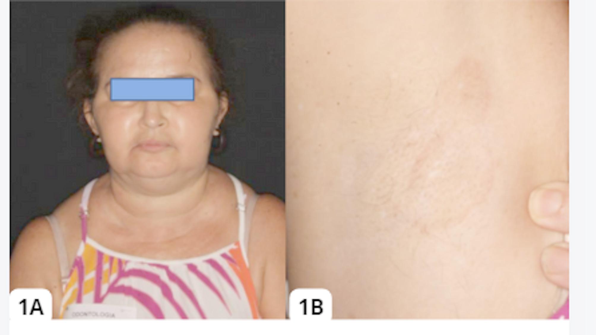
(A) Generalized edema in the infrahyoid region, extending superiorly to the cheeks, with visible vegetative plaques in the infracervical area. (B) Flat lesion with slightly pigmented borders in the right axillary region.
Source: Case files
Oroscopic examination revealed erythematous lesions, some exhibiting a leukoplakic surface, along with vesicles and ulcerations located on the lateral border and ventral surface of the tongue. These lesions were distributed in a characteristic net-like (reticular) pattern (Fig. 2A and B, and 2C).
Fig. 2.

A and B display a diffuse erythematous area on the lateral and ventral surfaces of the tongue, interspersed with leukoplakic regions. Multiple ruptured vesicles are evident, resulting in small ulcerations. C shows reticular erythematous plaques extending from the posterior region toward the apex of the tongue, with a distinct rounded ulcerated area visible more anteriorly. Source: Case files
An incisional biopsy of the lateral border of the tongue was performed for diagnostic clarification. Histopathological analysis revealed a predominantly eosinophilic inflammatory infiltrate within the underlying connective tissue, along with areas of epithelial acantholysis (Fig. 3). Instestinal histopathology was also obtained before and displayed on Fig. 4, associated with eosinophilic colitis.
Fig. 3.

Histopathological sections showing a subepithelial cleft and areas of acantholysis (A and B), accompanied by a dense eosinophilic infiltrate in the connective tissue (C and D). Hematoxylin and eosin (H&E) staining; original magnifications: 100× and 400×. Source: Case files
Fig. 4.

Histopathological examination of the large intestine revealing inflammation characterized by predominantly eosinophilic periglandular infiltration (Fig. 6A). Dense eosinophilic infiltrate is highlighted in Fig. 6C and D. Hematoxylin and eosin (H&E) staining; original magnifications: 100× and 400×. While not directly caused by Crohn’s disease, eosinophilic colitis can sometimes overlap with Crohn’s disease, particularly in cases of Crohn’s colitis. Source: Case files
The patient was prescribed topical tacrolimus 0.1% ointment, an immunosuppressive agent, to be applied three times daily for 15 days. She returned for follow-up evaluations at 15 and 30 days post-initiation of therapy.
At the first follow-up (Day 15), the patient reported only mild improvement in symptoms. However, at the second follow-up (Day 30), she described a marked clinical improvement. This was confirmed upon physical examination, which showed complete remission of the axillary lesion (Fig. 5), as well as full resolution of the oral lesions on the tongue (Fig. 6A and B).
Fig. 5.

Clinical remission of the right axillary lesion following topical immunosuppressive therapy. Source: Case files
Fig. 6.

Complete remission of oral lesions: (A) lateral border of the tongue; (B) ventral surface. Source: Case files
Discussion
Vegetative pyostomatitis is a rare dermatological condition that is often associated with inflammatory bowel diseases, with ulcerative colitis being the most commonly reported [1, 4]. However, Crohn’s disease is reported in 11% of patients [4]. Oral manifestations of intestinal diseases can be considered specific and non-specific. Specific manifestations include diffuse edema of the lips and mouth, deep, linear ulcers associated with hyperplastic folds of the mucosa in the vestibular fold, linear, serpiginous ulcers and mucogingivitis [11, 16].
Pyostomatitis is considered a non-specific manifestation, but when present it is a strong indicator of inflammatory bowel disease [11]. In the case reported, the rarity of the diagnosis is due to the association with Crohn’s disease, which is epidemiologically less frequently associated, and also because the patient was female, while the prevalence in men is approximately 3 times higher. In addition, the location of the lesions predominantly on the tongue is also a rare finding, since the most commonly affected sites are the labial and jugal mucosa [17].
The diagnosis of PPV is based on clinical findings (yellowish-white pustules with the appearance of snail trails), association with inflammatory bowel disease and histopathological findings of intraepithelial and/or subepithelial abscesses with an eosinophilic infiltrate [1, 11]. The differential diagnosis of PPV is necessary, since this condition can have clinical similarities with other bullous mucocutaneous disorders, such as pemphigus vulgaris [1, 4, 18].
However, the association with inflammatory bowel disease is an important factor in the patient’s medical history for the diagnosis of pyostomatitis. In immune-mediated diseases such as pemphigus, the bullous aspect of the lesions is important and can lead to the appearance of similar lesions on the skin as the disease progresses. However, histopathological findings include a suprabasal cleft, a row of tombstones, acantholysis and the presence of Tzank cells. Another essential complementary test for differentiating between these two diseases is immunofluorescence for IgA, which is positive for pemphigus and negative in the case of pyostomatitis-pyodermatitis [14, 18].
Direct immunofluorescence was not performed due to unavailability in our setting. Treatment for vegetative pyostomatitis-pyoderma is usually achieved by remission of the symptoms of the associated intestinal disease. Therefore, the use of systemic corticosteroids and immunosuppressants are reported in the literature [1, 4, 6, 18, 19]. In cases of oral manifestations without concomitant skin involvement, there are reports of topical treatments using corticosteroid rinses and ointments [20].
In the present case, due to the involvement of the oral mucosa and also skin lesions, associated with Crohn’s disease, the medication protocol was systemic through the use of immunosuppressants. Although systemic immunosuppressive therapy is commonly recommended for cases of pyodermatitis-pyostomatitis vegetans with mucocutaneous involvement, topical tacrolimus 0.1% was chosen in this case due to the limited extent of the lesions and the patient’s stable Crohn’s disease under ongoing systemic treatment. The topical approach was selected to provide localized control without altering the existing therapeutic regimen, and was supported by a multidisciplinary evaluation. Clinical response was initially modest, but complete remission was achieved within 30 days. Although less conventional, this strategy proved effective in the present case. Limitations include the absence of direct immunofluorescence and skin biopsy, due to resource constraint.
Conclusion
Dermatoses can be associated with systemic disorders, leading to the appearance of oral lesions. Knowledge of these associations allows dental surgeons, whether specialists or general practitioners, to make an early diagnosis and approach the pathology correctly, in order to improve the patient’s quality of life and prognosis.
Author contributions
Study design: JLC and IBAC; Data collection and case description: JLC, IBAC and IHGS; Histopathological and immunohistochemical analysis: PRFB, IHGS, JLC and IBAC; Interpretation of diagnostic findings: PRFB; Article writing: JLC, IBAC, IHGS and PRFB; Correction and final revision: PRFB; Approval of the study: All authors.
Funding
This article has not received any funding.
Data availability
No datasets were generated or analysed during the current study.
Declarations
Ethical statement
Write informed consent for publication was obtained from the patient.
Conflict of interest
The authors have no conflicts of interest to declare.
Study design
Case report.
Footnotes
Publisher’s note
Springer Nature remains neutral with regard to jurisdictional claims in published maps and institutional affiliations.
References
- 1.Atarbashi-Moghadam S, Lotfi A, Atarbashi-Moghadam F, Pyostomatitis Vegetans. A clue for diagnosis of silent crohn’s disease. J Clin Diagn Res. 2016;10(12):ZD12–3. 10.7860/JCDR/2016/22573.9032. Epub 2016 Dec 1. PMID: 28209014; PMCID: PMC5296587. [DOI] [PMC free article] [PubMed] [Google Scholar]
- 2.Katz TM, Katz AM. Idiopathic pyostomatitis-pyodermatitis vegetans with nasal obstruction: A case report. SAGE Open Med Case Rep. 2023;11. 10.1177/2050313X231160909. PMID: 36950050; PMCID: PMC10026124.:2050313X231160909. [DOI] [PMC free article] [PubMed]
- 3.Maruma F, Makuru H. Paediatric Pyodermatitis-Pyostomatitis vegetans without underlying inflammatory bowel disease: A case report of a 3-Year-Old African Girl. Clin Cosmet Investig Dermatol. 2022;15:2363–7. PMID: 36353092; PMCID: PMC9639368. [DOI] [PMC free article] [PubMed] [Google Scholar]
- 4.Matias F, de Rosa AT, de Carvalho DJ, de Castañon MTF. Bras Dermatol. 2011;86(4):137–40. 10.1590/S0365–05962011000700036. MCMN. Piodermatite-pioestomatite vegetante: relato de caso e revisão de literatura. [DOI]
- 5.Roldão J, de Saad A, de Campos VL, Leal GO. Arch Health. 2024;5(3):e2080. 10.46919/archv5n3espec–393. LS. Piodermatite-Pioestomatite Vegetante: relato de caso e revisão de literatura. [DOI]
- 6.Ko HC, Jung DS, Jwa SW, Cho HH, Kim BS, Kwon KS, Kim MB. Two cases of pyodermatitis-pyostomatitis vegetans. J Dermatol. 2009;36(5):293–7. 10.1111/j.1346–8138.2009.00641.x. PMID: 19383001. [DOI] [PubMed]
- 7.Bardasi G, Romagnoli A, Foschini MP, Mantovani A, Alvisi P. Pyostomatitis vegetans in a pediatric patient with ulcerative colitis: case report of a rare pediatric inflammatory bowel disease extraintestinal manifestation and review of the literature. Eur J Gastroenterol Hepatol. 2020;32(7):889–892. 10.1097/MEG.0000000000001723. PMID: 32282544. [DOI] [PubMed]
- 8.Femiano F, Lanza A, Buonaiuto C, Perillo L, Dell’Ermo A, Cirillo N. Pyostomatitis vegetans: a review of the literature. Med Oral Patol Oral Cir Bucal. 2009;14(3):E114–7. PMID: 19242389. [PubMed] [Google Scholar]
- 9.Carvalho S, Sanches M, Alves R, Selores M. Pyodermatitis vegetans of the vulva. Dermatol Online J. 2016;22(6):13030/qt3qr380c6.PMID: 27617602. [PubMed]
- 10.Clark LG, Tolkachjov SN, Bridges AG, Camilleri MJ. Pyostomatitis vegetans (PSV)-pyodermatitis vegetans (PDV): A clinicopathologic study of 7 cases at a tertiary referral center. J Am Acad Dermatol. 2016;75(3):578–84. 10.1016/j.jaad.2016.03.047. Epub 2016 Jun 24. PMID: 27349819. [DOI] [PubMed] [Google Scholar]
- 11.Pecci-Lloret MP, Ramirez-Santisteban E, Hergueta-Castillo A, Guerrero-Gironés J, Oñate-Sánchez RE. Oral manifestations of crohn’s disease: A systematic review. J Clin Med. 2023;12(20):6450. 10.3390/jcm12206450. PMID: 37892587; PMCID: PMC10607549. [DOI] [PMC free article] [PubMed] [Google Scholar]
- 12.Stagg B, Simpson A, Sidhu S. Similar but different: distinguishing between pemphigus vegetans and pyostomatitis-pyodermatitis vegetans. BMJ Case Rep. 2021;14(4):e242162. doi: 10.1136/bcr–2021–242162. PMID: 33879466; PMCID: PMC8061807. [DOI] [PMC free article] [PubMed]
- 13.Dupuis EC, Haber RM, Robertson LH. Pyoblepharitis vegetans in association with Pyodermatitis-Pyostomatitis vegetans: expanding the spectrum of a rare, multisystem disorder. J Cutan Med Surg. 2016 Mar-Apr;20(2):163–5. Epub 2015 Oct 30. PMID: 26519160. [DOI] [PubMed]
- 14.Antonelli E, Bassotti G, Tramontana M, Hansel K, Stingeni L, Ardizzone S, Genovese G, Marzano AV, Maconi G. Dermatological manifestations in inflammatory bowel diseases. J Clin Med. 2021;10(2):364. 10.3390/jcm10020364. PMID: 33477990; PMCID: PMC7835974. [DOI] [PMC free article] [PubMed] [Google Scholar]
- 15.Diaconescu S, Strat S, Balan GG, Anton C, Stefanescu G, Ioniuc I, Stanescu AMA. Dermatological manifestations in pediatric inflammatory bowel disease. Med (Kaunas). 2020;56(9):425. 10.3390/medicina56090425. PMID: 32842528; PMCID: PMC7559248. [DOI] [PMC free article] [PubMed] [Google Scholar]
- 16.Pereira MS, Munerato MC. Oral manifestations of inflammatory bowel diseases: two case reports. Clin Med Res. 2016;14(1):46–52. 10.3121/cmr.2015.1307. Epub 2016 Feb 10. PMID: 26864508; PMCID: PMC4851452. [DOI] [PMC free article] [PubMed] [Google Scholar]
- 17.Hegarty AM, Barrett AW, Scully C. Pyostomatitis vegetans. Clin Exp Dermatol. 2004; 29(1):1–7. 10.1111/j.1365–2230.2004.01438.x. PMID: 14723710. [DOI] [PubMed]
- 18.Abellaneda C, Mascaro JM Jr, Vazquez MG, Pablo IM, Iranzo P. All that glitters is not pemphigus: Pyodermatitis-pyostomatitis vegetans misdiagnosed as IgA pemphigus for 8 years. Am J Dermatopathol. 2011;33(1):e1–6. 10.1097/DAD.0b013e3181d81ecb. [DOI] [PubMed] [Google Scholar]
- 19.Antunes H, Patraquim C, Baptista V, Silva Monteiro L. Oral manifestations of Crohn’s disease. BMJ Case Rep. 2015;2015:bcr2015212300. doi: 10.1136/bcr–2015–212300. Erratum in: BMJ Case Rep. 2015;2015:bcr2015212300corr1. 10.1136/bcr–2015–212300corr1. PMID: 26511994; PMCID: PMC4636709. [DOI] [PMC free article] [PubMed]
- 20.Werchniak AE, Storm CA, Plunkett RW, Beutner EH, Dinulos JG. Treatment of pyostomatitis vegetans with topical tacrolimus. J Am Acad Dermatol. 2005;52(4):722–3. doi: 10.1016/j.jaad.2004.11.041. PMID: 15793543. [DOI] [PubMed]
Associated Data
This section collects any data citations, data availability statements, or supplementary materials included in this article.
Data Citations
- Matias F, de Rosa AT, de Carvalho DJ, de Castañon MTF. Bras Dermatol. 2011;86(4):137–40. 10.1590/S0365–05962011000700036. MCMN. Piodermatite-pioestomatite vegetante: relato de caso e revisão de literatura. [DOI]
- Roldão J, de Saad A, de Campos VL, Leal GO. Arch Health. 2024;5(3):e2080. 10.46919/archv5n3espec–393. LS. Piodermatite-Pioestomatite Vegetante: relato de caso e revisão de literatura. [DOI]
Data Availability Statement
No datasets were generated or analysed during the current study.