

Abstract
Purpose
Effective management of patients undergoing treatment with lutetium-177 labeled DOTA-ibandronic acid (177Lu-DOTA-IBA) necessitates the identification of radiological response biomarkers that can mitigate disease progression and facilitate patient stratification for subsequent treatment decisions. This study aims to evaluate the metabolic tumor volume (MTV) as a quantitative measure of radiological response in bone metastases using gallium-68 labeled DOTA-ibandronic acid (68Ga-DOTA-IBA) PET/CT.
Methods
In a single-center retrospective study, 68Ga-DOTA-IBA PET/CT scans of patients with bone metastases who received 177Lu-DOTA-IBA injections. Eligible patients had available PET/CT scans both prior to 177Lu-DOTA-IBA therapy and at treatment cessation. The Hermes system was employed to delineate regions of interest at baseline and at treatment cessation to measure the MTV of bone metastases. Spearman’s rank correlation coefficient was utilized to assess the correlation between MTV and the baseline covariate, alkaline phosphatase (ALP). The Cox proportional hazards model and Kaplan-Meier curves were used to evaluate the association between baseline covariates, their changes at treatment termination, and overall survival (OS). The C-index measured the predictive discrimination of covariates for OS.
Results
Baseline 68Ga-DOTA-IBA PET/CT images were available for 54 patients. Additionally, 30 patients underwent both baseline and post-treatment 68Ga-DOTA-IBA PET/CT scans. Baseline MTV demonstrated a moderate correlation with ALP. Among baseline covariates, MTV and ALP were significantly associated with OS. Following treatment discontinuation, MTV decreased in 57% of patients, while ALP decreased in 83%. As a continuous variable, the relative change in MTV after treatment compared to baseline was significantly associated with OS, with a C-index of 0.69. Patients exhibiting a decrease in both MTV and ALP had a significantly longer median OS compared to those with a decrease in ALP alone.
Conclusions
Both baseline MTV and its changes at treatment cessation were significant parameters associated with OS. The study warrants prospective validation of MTV as a quantitative imaging response biomarker for predicting OS in patients with BM treated with 177Lu-DOTA-IBA.
Keywords: Metabolic tumor volume, Imaging biomarker, Bone metastases, 177Lu-DOTA-ibandronic acid
Introduction
Bone is one of the most common sites for cancer metastasis. While many malignant tumors frequently metastasize to the bones, certain specific types of cancer show a predilection for bone metastasis (BM) (Davilaet al. 2015; Denget al. 2024). Once cancer metastasizes to the skeleton, the mortality rate among patients significantly increases, rendering the disease incurable. In clinical practice, advancements in therapeutic technologies for malignant tumors have notably extended survival times for many patients, leading to a corresponding increase in the incidence of BM.
Bisphosphonates are a well-known class of drugs used to treat bone diseases by inhibiting protein prenylation through the blockade of farnesyl pyrophosphate synthase in osteoclasts, ultimately leading to the inhibition of bone resorption (Kavanaghet al. 2006). Currently, radionuclide-labeled bisphosphonates have been extensively studied as effective diagnostic and therapeutic approaches for BM. Previous research has successfully synthesized the chelating ligand DOTA-ibandronic acid, complexed with 177Lu, for the treatment of BM, demonstrating favorable outcomes (Li et al. 2024; Wang et al. 2022; Yang et al. 2023). Preclinical studies (Qiuet al. 2023) indicate that 177Lu-DOTA-IBA possesses favorable biological properties, safety, rapid blood clearance, and a high target-to-background ratio. A preliminary clinical study involving 18 participants has shown that 177Lu-DOTA-IBA therapy is well-tolerated and safe in this population.
The extent of skeletal involvement is typically reported using qualitative or semi-quantitative indicators, such as the number of lesions. Metabolic Tumor Volume (MTV) is a valuable PET parameter for measuring the overall glucose metabolic activity of cancerous lesions (Bailey and Willowson 2013; Qiu et al. 2023). Numerous studies have demonstrated the prognostic significance of MTV in malignant tumors, with applications for risk stratification in esophageal cancer (Bailey and Willowson 2014), non-small cell lung cancer (Shimet al. 2014), and other malignancies (Tamandlet al. 2016). However, the optimal application of 177Lu-DOTA-IBA in the therapeutic toolkit for patients with BM remains to be determined. The use of 68Ga-DOTA-IBA PET/CT as an alternative to bone scintigraphy offers superior image resolution and enables absolute quantification, aligning with clinical application and management trends toward the integration of nuclear medicine and diagnostics (Deng et al. 2024). Herein, we report on the quantitative bone PET/CT analysis of bisphosphonate uptake in the central skeleton. We hypothesize that measurements of bone metabolism may serve as a prognostic indicator for patients undergoing 177Lu therapy. We conceived a retrospective clinical study aimed at evaluating MTV as a biomarker of radiographic response in patients with BM treated with 177Lu-DOTA-IBA.
Methods
Study design and patient population
We collected raw DICOM-format 68Ga-DOTA-IBA PET/CT images from patients with bone metastases who underwent 177Lu-DOTA-IBA injections every 6–8 weeks (from November 2021 to December 2022). This study received approval from the hospital’s Ethics Committee (approval no. KY2022114). It was registered as a clinical trial (registration no. ChiCTR2200064487). Patients with baseline PET/CT images in their original DICOM format and ALP levels were eligible for this study. Follow-up PET/CT scans and ALP values (when available) were also obtained within three months after the last 177Lu-DOTA-IBA injection. Ethical approval for the retrospective analysis of patients treated with 177Lu-DOTA-IBA was obtained.
68Ga-DOTA-IBA PET/CT imaging
Imaging was performed according to a previously described protocol (Qiu et al. 2023). No special preparation was required prior to the examination. 68Ga-DOTA-IBA was administered intravenously at a dosage of 1.85 MBq (0.05 mCi) per kilogram of body weight. PET/CT scanning was conducted 40–60 min post-injection, covering the entire body from head to feet, with a duration of 3 min per bed position. The acquired images were subjected to attenuation correction and iterative reconstruction to yield transverse, coronal, and sagittal PET/CT views.
Treatment
To the eligible participants, 370–2590 MBq of 177Lu-DOTA-IBA was administered intravenously, followed by a saline flush. 68Ga-DOTA-IBA PET/CT was performed to evaluate the imaging response 6–8 weeks post-177Lu-DOTA-IBA treatment. Subsequent treatments were administered based on the participant’s willingness and clinical conditions. Sex, age, primary tumor type, bone metastasis type, other accompanying metastases, dose and treatment times, survival status at the end of follow-up, and blood biomarkers were recorded.
Metabolic tumor volume
68Ga-DOTA-IBA PET/CT images were assessed by two experienced nuclear medicine physicians. Discrepancies among observers were resolved through discussion to reach a diagnostic consensus. MTV was automatically calculated from the retrospectively collected PET/CT images using OLINDA/EXM 2.0 software (Hermes Medical Solutions, Stockholm, Sweden), with diagnostic CT images serving as a reference.
Statistical analyses
As a retrospective study, no a priori assumptions were made regarding the clinical utility of MTV for power calculations. To assess MTV as a distinct independent variable, we employed the non-parametric Spearman’s rank correlation coefficient to determine the association between MTV and blood biomarkers. Univariate and multivariate Cox regression analyses were conducted to assess the correlation between baseline MTV, ALP levels, and OS. Additionally, Cox regression analysis was utilized to evaluate the relationship between MTV, ALP response (as continuous variables), and OS. Kaplan-Meier analysis was conducted to assess differences in median survival times associated with ALP levels and MTV response. A high concordance index indicates that the covariate is highly informative for predicting the relative mortality risk between any two patients at a specified time. The chi-square test was utilized to evaluate the relationship between the number of 177Lu-DOTA-IBA injections and the decrease in MTV and ALP levels. All statistical analyses were conducted using SPSS statistical software version 22.0 (SPSS Inc, Chicago, IL). The statistical significance level for each test was set at 0.05 (two-tailed).
Results
Of the total 108 screened patients,54 patients at baseline and 30 patients at treatment follow-up (with a median of 3 injections, ranging from 1 to 5) were deemed evaluable for retrospective analysis. Figure 1 presents a flowchart illustrating the total number of patients at baseline and the final number of patients eligible for analysis after treatment. Table 1 summarizes the demographic characteristics, prior treatment history, and baseline features of the evaluable patients with advanced BM.
Fig. 1.

Flow chart illustrating the evaluable patients
Table 1.
Patient characteristics
| Evaluable patients for baseline assessment (N = 54) | Evaluable patients for response assessment (N = 30) | |
|---|---|---|
| Baseline characteristics | Median (Range) | Median (Range) |
| Age | 60 (17–86) | 56 (17–86) |
| ALP (U/L) | 186.2 (39.3-971.9) | 134.25 (49–536) |
| MTV (cm3) | 269.52 (25.8-875.8) | 259.89 (25.8-861.2) |
| Type of primary tumor | N (%) | N (%) |
| Lung cancer | 21 (38.9) | 10 (33.3) |
| Prostate cancer | 14 (25.9) | 9 (30) |
| Breast cancer | 5 (9.3) | 5 (16.7) |
| Colorectal cancer | 8 (14.8) | 5 (16.7) |
| Other types | 6 (11.1) | 1 (3.3) |
| Type of bone metastasis | N (%) | N (%) |
| Osteoblastic metastasis | 28 (51.9) | 16 (53.3) |
| Osteolytic metastasis | 11 (20.4) | 6 (20) |
| Mixed metastasis | 15 (27.8) | 8 (26.7) |
| Accompanying other metastases | N (%) | N (%) |
| Liver | 9(17%) | 6(20%) |
| Lung | 9(17%) | 6(20%) |
| Lymph node | 27(50%) | 16(53%) |
| Other sites | 5(9%) | 3(10%) |
| Deaths | 29(54%) | 17(57%) |
| 177Lu Dosage | 2 (1–5) | 3 (1–5) |
The baseline MTV (median 259.89 cm3, IQR 173.49–517.06) showed a moderate correlation with baseline ALP levels (r = 0.37, p = 0.047). Details of the univariate and multivariate Cox regression analyses are presented in Table 2. In the univariate analysis, both MTV and baseline ALP levels were significantly associated with OS. In the bivariate model incorporating baseline ALP levels, no significant association was found between tumor metabolic volume and OS. The discriminatory ability of ALP in predicting OS (C-index 0.67) was significantly higher than that of MTV (C-index 0.47), with a p-value of 0.03.
Table 2.
Univariate and bivariate Cox regression analysis of MTV, and ALP level as continuous variable at baseline (n = 54)
| N = 54 | HR | 95%CI | P-value |
|---|---|---|---|
| Univariate | |||
| MTV baseline | 2.36 | 0.75–7.47 | 0.047 |
| ALP baseline | 0.23 | 0.06–0.89 | 0.017 |
| Bivariate | |||
| MTV baseline | 0.24 | 0.06–0.90 | 0.035 |
| ALP baseline | 1.3 | 0.24–7.03 | 0.022 |
Table 3 illustrates the changes in ALP and MTV relative to the number of 177Lu-DOTA-IBA injections. After the last administration of 177Lu-DOTA-IBA, 87% (26/30) of the patients experienced a decrease in ALP. Among the 26 patients with a decrease in ALP, 12 cases (46%) exhibited a reduction in ALP levels by more than 25%. The median interval for the first cycle of both 68Ga-DOTA-IBA and 177Lu-DOTA-IBA was 6 weeks, while the median time from the last treatment to the follow-up 68Ga-DOTA-IBA scan was 4 weeks. Overall, out of 30 patients, 17 (57%) exhibited a decrease in MTV, and among these 17 patients with a reduction in MTV, 11 (65%) had received three or more cycles of 177Lu-DOTA-IBA therapy.
Table 3.
The decline of ALP and MTV by number of 177Lu-DOTA-IBA injections
| N = 30 | ALP decline | MTV decline |
|---|---|---|
| 1–2 doses of Lu-177; N = 13 | 12/13(92%) | 6/13(46%) |
| 3–5 doses of Lu-177; N = 17 | 13/17(76%) | 11/17(65%) |
| Total | 25/30(83%) | 17/30(57%) |
It was observed that patients who received three or more injections of 177Lu-DOTA-IBA experienced a more frequent decrease in MTV compared to those who received fewer than three injections. No significant change was observed in the context of ALP levels. Figure 2 illustrates examples of MTV reduction and lesion regression in patient bone scans. Figure 3 demonstrates the relationship between the percentage changes in baseline ALP and MTV with the dosage of 177Lu-DOTA-IBA injections. After receiving 3 to 5 doses of 177Lu-DOTA-IBA treatment, the relative reductions in ALP levels (54%) and MTV (45%) were the most significant. Kaplan-Meier curves demonstrated that patients experiencing a decrease in MTV (median OS of 312 weeks) had a significantly longer median OS than patients with only a decrease in ALP levels (median OS of 145 weeks, p = 0.018), as shown in Fig. 4.
Fig. 2.

68Ga-DOTA-IBA PET MIP images of a 54-year-old man with lung cancer accompanied multiple metastases (including lymph node and bone metastases). MTV value decreased after Three treatment with 177Lu-DOTA-IBA. Utilize the Hermes system to delineate hotspots (shown as red) for MTV calculations
Fig. 3.

The Waterfall plot of the ALP (red) and MTV (green) change in each patients by number of 177Lu-DOTA-IBA injections
Fig. 4.
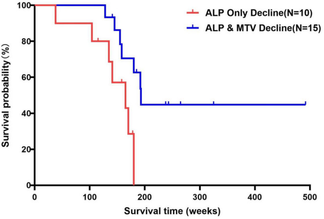
Kaplan Meier demonstrating the median overall survival of patients with 177Lu-DOTA-IBA treatment induced decline in levels of ALP (n = 10) in comparison to patients with both treatment induced decline in both MTV and ALP levels (n = 15)
In the Cox regression analysis, the change in MTV after treatment, as a continuous variable, was the only covariate significantly associated with OS, as shown in Table 4. The discriminatory power of MTV change in predicting OS after 177Lu-DOTA-IBA treatment (C-index 0.69) was significantly higher than that of ALP change and was not affected by visceral metastasis.
Table 4.
Cox regression analysis of change in MTV, ALP as continuous variables and presence or absence of visceral metastasis
| N = 30 | HR | 95%CI | P-value |
|---|---|---|---|
| On-Treatment change | |||
| MTV % difference | 0.26 | 0.08–0.77 | 0.016 |
| ALP % difference | 2.193 | 0.53–9.02 | 0.277 |
| visceral metastasis | 0.731 | 0.26–2.07 | 0.56 |
Discussion
Over the past decade, numerous radiopharmaceuticals, including 32P, 89Sr, 223Ra, 188Re-/186Re-HEDP, 153Sm-EDTMP, 177Lu-EDTMP, and 177Lu-BPAMD, have been used to treat BM (Chakraborty et al. 2008; Liepe et al. 2003; Parlak et al. 2015; Sun et al. 2016). While these agents have demonstrated significant potential for pain palliation, most cannot be used as theranostic pairs due to the lack of corresponding diagnostic analogs, with the exception of 68Ga-/177Lu-BPAMD. Compared to 99mTc MDP SPECT, 68Ga-DOTA-IBA PET has shown superior efficacy in detecting BM. 68Ga-DOTA-IBA PET/CT can be employed for diagnosis, staging, and dynamic evaluation of therapeutic responses, providing opportunities for personalized treatment with 177Lu-DOTA-IBA (Deng et al. 2024). The 68Ga-/177Lu-DOTA-IBA combination offers a promising therapeutic approach for BM.
Fernández et al. conducted a pivotal dosimetry and safety assessment of 177Lu-DOTA-ZOL in metastatic castration-resistant prostate cancer, establishing the red marrow as the dose-limiting organ with a median maximum tolerated activity of 6 GBq (Fernándezet al. 2021). While their work validated the therapeutic potential of zoledronate-based radiopharmaceuticals, our study advances the field by introducing 177Lu-DOTA-IBA, a third-generation bisphosphonate with distinct pharmacokinetic advantages, including rapid blood clearance and higher target-to-background ratios (Qiu et al. 2023). Critically, we leveraged 68Ga-DOTA-IBA PET/CT to quantify treatment response through MTV, a novel imaging biomarker absent in prior studies. This theranostic approach combining 68Ga/177Lu-DOTA-IBA with MTV monitoring provides an actionable framework for personalized therapy adjustment, addressing the unmet need for response-guided dosing highlighted by Fernández et al. Furthermore, compared to Fernández et al.‘s cohort (100% prostate cancer), our study included diverse malignancies (lung, breast, prostate, etc.), demonstrating broader applicability. The favorable safety profile of 177Lu-DOTA-IBA (grade 3–4 hematotoxicity: 0% in our cohort vs. 33% with 177Lu-DOTA-ZOL) and its efficacy in pain palliation (82%) underscore its clinical potential for individualized BM management.
The objective of this study is to assess whether changes in MTV induced by 177Lu-DOTA-IBA therapy could serve as a predictive biomarker for OS in patients with BM. The lack of reliable biomarkers in routine clinical management of patients undergoing 177Lu-DOTA-IBA therapy remains a challenge. Previous studies have also shown variability in alkaline phosphatase (ALP) levels during 177Lu-DOTA-IBA treatment. To date, no effective imaging biomarker is routinely available for assessing treatment response. MTV has been demonstrated as a prognostic biomarker in various studies (Chardin et al. 2020; Jemaa et al. 2022; Pfannkuchen et al. 2017), and as a quantitative tool in PET/CT, it shows potential in evaluating the efficacy of 177Lu-DOTA-IBA therapy. IBA, a third-generation aminobisphosphonate, possesses anti-resorptive and anti-hypercalcemic properties. We successfully synthesized the chelating ligand DOTA-IBA complexed with DOTA. 68Ga-DOTA-IBA PET/CT can be utilized for the dynamic diagnosis, staging, and evaluation of therapeutic efficacy, enabling personalized 177Lu-DOTA-IBA radionuclide therapy. Our baseline data confirmed the prognostic value of both MTV and ALP for OS. In the bivariate analysis, baseline ALP levels exhibited a stronger correlation with OS compared to baseline MTV values, showing greater discriminatory power for OS. However, post-treatment analysis indicated that changes in MTV were more closely associated with OS. The findings suggest that treatment-induced variations in MTV could serve as a biomarker for response to 177Lu therapy. Kaplan-Meier analysis demonstrated that MTV response adds predictive value over ALP response alone in determining treatment outcomes. In our multivariable Cox proportional hazards model adjusted for the presence of visceral metastasis as a key covariate, the results demonstrated that even after adjusting for this prognostic factor, the MTV of BM remained a significant and independent predictor of OS (HR = 0.731, 95% CI = 0.26–2.07, p = 0.56). This indicates that the metabolic response of BM inherently possesses unique and independent prognostic value beyond that provided by other lesions. Our data also suggest an association between the administered dose of 177Lu-DOTA-IBA and changes in MTV. Patients receiving three or more doses of 177Lu-DOTA-IBA showed a significantly greater reduction in MTV compared to those receiving fewer doses (p = 0.035). Patients who received a higher number of injections of 177Lu-DOTA-IBA tended to experience better outcomes, including a significantly reduced risk of mortality, along with decreased pain scores and ALP levels.
This is important to note that 68Ga-DOTA-IBA uptake demonstrates heterogeneous patterns between osteoblastic and osteolytic lesions. Our prior work has demonstrated that the SUVmax of osteoblastic lesions is significantly higher than that of osteolytic lesions (7.88 versus 6.28, P < 0.001) (Liet al. 2025). For osteoblastic lesions: due to high 68Ga-DOTA-IBA uptake and excellent background contrast (tumor-to-background ratio > 3:1), changes in MTV can more sensitively reflect treatment response. For osteolytic lesions, however, MTV changes must be interpreted with caution. If treatment induces “osteolysis-to-osteoblasia” (e.g., after 177Lu-DOTA-IBA therapy), 68Ga-DOTA-IBA uptake may increase due to new bone formation, and an increase in MTV does not indicate progression. Interestingly, our prior work have shown that there are no significant differences in imaging responses among different types of BM (Xuet al. 2025). Bone scintigraphy with hydroxyapatite reflects increased osteoblastic activity; post-nuclear therapy bone scans may demonstrate a temporary increase in lesion activity (Isenseeet al. 2018). As a quantitative parameter, MTV can capture delayed responses in bone scans and stratify patients, facilitating more precise management and informing subsequent treatment decisions.
Limitations
Retrospective design and selection bias
The inherent limitations of a retrospective analysis must be acknowledged. Data collection relied on pre-existing clinical records and imaging studies, which may introduce selection bias. For instance, only patients with both baseline and post-treatment 68Ga-DOTA-IBA PET/CT scans were included (n = 30/54), potentially excluding those with rapid progression or early death. This could skew results toward a cohort with better prognosis, overestimating the predictive value of MTV changes. Additionally, treatment schedules (1–5 cycles of 177Lu-DOTA-IBA) and follow-up intervals were not standardized, introducing heterogeneity in response assessment.
Limited sample size and tumor heterogeneity
The final cohort (n = 30) lacked statistical power for robust subgroup analyses. Patients had diverse primary malignancies (e.g., prostate, breast, lung), each with distinct biological behaviors and metastatic patterns. Notably, 20% had liver/lung metastases (Table 1), which independently impact OS but were not adjusted for in MTV-OS analyses. The small sample precluded stratification by tumor type or metastatic burden, limiting generalizability of MTV as a universal biomarker. Future multi-center studies with larger cohorts are needed to validate MTV’s prognostic role across tumor subtypes.
Technical variability in MTV quantification
MTV measurements using the Hermes platform (OLINDA/EXM 2.0) depend on threshold-based segmentation, which may vary between observers. Although consensus readings minimized inter-observer discordance, the lack of a standardized MTV calculation protocol (e.g., fixed SUV threshold vs. adaptive methods) could affect reproducibility. This is particularly relevant for sclerotic or heterogeneous lesions, where background uptake may confound MTV accuracy.
Nevertheless, the MTV response data are unique and merit prospective validation.
Conclusion
Quantitative, reproducible radiologic assessments such as MTV may enhance the clinical utility of existing methods for evaluating soft tissue metastases by CT and blood biomarkers, providing a more comprehensive understanding of disease status. This study demonstrated that baseline MTV and its changes following treatment are significantly correlated with OS, suggesting their potential as additional predictive factors in patients with advanced solid tumors undergoing 177Lu therapy. Future prospective studies are necessary to validate these findings and may lead to new strategies for personalized cancer treatment. As research continues, this therapeutic approach may further improve treatment efficacy, enhance patients’ quality of life, and positively influence prognosis.
Acknowledgement
We are grateful to the members of Department of Nuclear Medicine, The Affiliated Hospital, Southwest Medical University and Nuclear Medicine and Molecular Imaging Key Laboratory of Sichuan Province for their technical guidance, cooperation, and assistance in completing this study.
Author contributions
All authors contributed to the study conception and design. Material preparation, data collection and analysis were performed by Juan Yang and Lihan Zhang. Yue Chen was responsible for revising for important intellectual content. The first draft of the manuscript was written by Juan Yang and all authors commented on previous versions of the manuscript. All authors read and approved the final manuscript.
Funding
This work was supported by the Luzhou science and technology plan projects (2022-JYJ- 118, 2021LZXNYD-C02, and 2021LZXNYD-P03) and major science and technology project in Gansu Province (23ZDFA014).
Data availability
No datasets were generated or analysed during the current study.
Declarations
Competing interests
The authors declare no competing interests.
Ethics approval
The Affiliated Hospital of Southwest Medical University Review Board approved this study, and all patients received oral and written information on the routine tests performed in this study.
Footnotes
Publisher’s note
Springer Nature remains neutral with regard to jurisdictional claims in published maps and institutional affiliations.
Juan Yang and Lihan Zhang have contributed equally to this work.
References
- Bailey DL, Willowson KP (2013) An evidence-based review of quantitative SPECT imaging and potential clinical applications. J Nucl Med 54:83–89. 10.2967/jnumed.112.111476 [DOI] [PubMed] [Google Scholar]
- Bailey DL, Willowson KP (2014) Quantitative SPECT/CT: SPECT joins PET as a quantitative imaging modality. Eur J Nucl Med Mol Imaging 41(Suppl 1):S17–25. 10.1007/s00259-013-2542-4 [DOI] [PubMed] [Google Scholar]
- Chakraborty S, Das T, Banerjee S et al (2008) 177Lu-EDTMP: a viable bone pain palliative in skeletal metastasis. Cancer Biother Radiopharm 23:202–213. 10.1089/cbr.2007.374 [DOI] [PubMed] [Google Scholar]
- Chardin D, Paquet M, Schiappa R et al (2020) Baseline metabolic tumor volume as a strong predictive and prognostic biomarker in patients with non-small cell lung cancer treated with PD1 inhibitors: a prospective study. J Immunother Cancer 8. 10.1136/jitc-2020-000645 [DOI] [PMC free article] [PubMed]
- Davila D, Antoniou A, Chaudhry MA (2015) Evaluation of osseous metastasis in bone scintigraphy. Semin Nucl Med 45:3–15. 10.1053/j.semnuclmed.2014.07.004 [DOI] [PubMed] [Google Scholar]
- Deng J, Yang J, Wang Y et al (2024) Comparison of the relative diagnostic performance of (68)Ga-DOTA-IBA and (18)F-NaF for the detection of bone metastasis. Front Oncol 14:1364311. 10.3389/fonc.2024.1364311 [DOI] [PMC free article] [PubMed] [Google Scholar]
- Fernández R, Eppard E, Lehnert W et al (2021) Evaluation of safety and dosimetry of (177)Lu-DOTA-ZOL for therapy of bone metastases. J Nucl Med 62:1126–1132. 10.2967/jnumed.120.255851 [DOI] [PMC free article] [PubMed] [Google Scholar]
- Isensee G, Péporté A, Müller J et al (2018) Is there a flare phenomenon on bone scintigraphy in men with advanced prostate Cancer treated with Radium-223? Clin Genitourin Cancer 16:349–354. 10.1016/j.clgc.2018.04.002 [DOI] [PubMed] [Google Scholar]
- Jemaa S, Paulson JN, Hutchings M et al (2022) Full automation of total metabolic tumor volume from FDG-PET/CT in DLBCL for baseline risk assessments. Cancer Imaging 22:39. 10.1186/s40644-022-00476-0 [DOI] [PMC free article] [PubMed] [Google Scholar]
- Kavanagh KL, Guo K, Dunford JE et al (2006) The molecular mechanism of nitrogen-containing bisphosphonates as antiosteoporosis drugs. Proc Natl Acad Sci USA 103:7829–7834. 10.1073/pnas.0601643103 [DOI] [PMC free article] [PubMed] [Google Scholar]
- Li H, Pei W, Yang X et al (2024) Biodistribution and dosimetry of (177)Lu-DOTA-IBA for therapy of bone metastases. EJNMMI Res 14:30. 10.1186/s13550-024-01094-6 [DOI] [PMC free article] [PubMed] [Google Scholar]
- Li L, Chen L, Yang J et al (2025) Comparison of (18)F-FDG and (68)Ga-DOTA-IBA in detecting bone metastases: a lesion-basis study. Sci Rep 15:12766. 10.1038/s41598-025-97920-5 [DOI] [PMC free article] [PubMed] [Google Scholar]
- Liepe K, Hliscs R, Kropp J et al (2003) Dosimetry of 188Re-hydroxyethylidene diphosphonate in human prostate cancer skeletal metastases. J Nucl Med 44:953–960 [PubMed] [Google Scholar]
- Parlak Y, Gumuser G, Sayit E (2015) Samarium-153 therapy for prostate cancer: the evaluation of urine activity, staff exposure and dose rate from patients. Radiat Prot Dosimetry 163:468–472. 10.1093/rpd/ncu237 [DOI] [PubMed] [Google Scholar]
- Pfannkuchen N, Meckel M, Bergmann R et al (2017) Novel radiolabeled bisphosphonates for PET diagnosis and endoradiotherapy of bone metastases. Pharmaceuticals (Basel) 10. 10.3390/ph10020045 [DOI] [PMC free article] [PubMed]
- Qiu L, Wang Y, Liu H et al (2023) Safety and efficacy of 68 Ga- or 177 Lu-Labeled DOTA-IBA as a novel theranostic radiopharmaceutical for bone metastases: a phase 0/I study. Clin Nucl Med 48:489–496. 10.1097/rlu.0000000000004634 [DOI] [PubMed] [Google Scholar]
- Shim SH, Kim DY, Lee DY et al (2014) Metabolic tumour volume and total lesion glycolysis, measured using preoperative 18F-FDG PET/CT, predict the recurrence of endometrial cancer. BJOG 121:1097–1106 discussion 1106. 10.1111/1471-0528.12543 [DOI] [PubMed] [Google Scholar]
- Sun Y, Lu P, Yu L (2016) The volume-metabolic combined parameters from (18)F-FDG PET/CT May help predict the outcomes of cervical carcinoma. Acad Radiol 23:605–610. 10.1016/j.acra.2016.01.001 [DOI] [PubMed] [Google Scholar]
- Tamandl D, Ta J, Schmid R et al (2016) Prognostic value of volumetric PET parameters in unresectable and metastatic esophageal cancer. Eur J Radiol 85:540–545. 10.1016/j.ejrad.2016.01.002 [DOI] [PubMed] [Google Scholar]
- Wang Y, Wang Q, Chen Z et al (2022) Preparation, biological characterization and preliminary human imaging studies of (68)Ga-DOTA-IBA. Front Oncol 12:1027792. 10.3389/fonc.2022.1027792 [DOI] [PMC free article] [PubMed] [Google Scholar]
- Xu T, Wang Y, Liu G et al (2025) Efficacy and safety of 177Lu-DOTA-IBA in tumor bone metastasis treatment: a prospective clinical trial. Clin Nucl Med 50:119–124. 10.1097/rlu.0000000000005573 [DOI] [PubMed] [Google Scholar]
- Yang J, Deng J, Fan D et al (2023) Biodistribution and internal dosimetry of 68 Ga-DOTA-IBA PET imaging for patients with bone metastases. Clin Nucl Med 48:847–852. 10.1097/rlu.0000000000004757 [DOI] [PubMed] [Google Scholar]
Associated Data
This section collects any data citations, data availability statements, or supplementary materials included in this article.
Data Availability Statement
No datasets were generated or analysed during the current study.