

Abstract
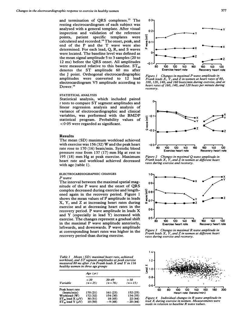
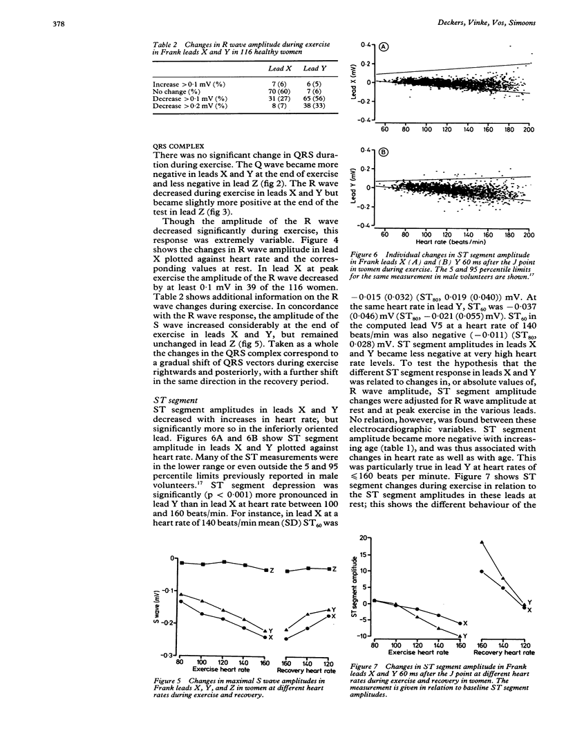
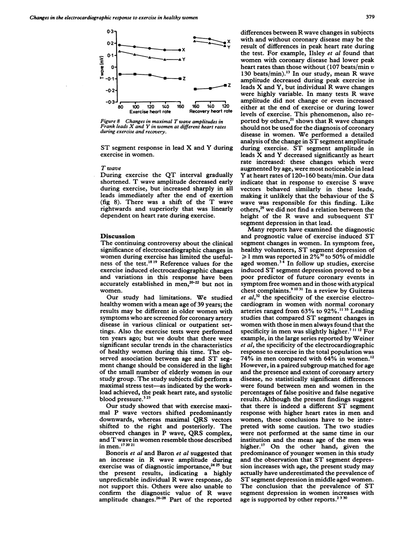
Changes in the P wave, QRS complex, ST segment, and T wave during and after maximal exercise were quantitatively analysed in 116 healthy women with a mean age of 39. The corrected orthogonal Frank lead electrocardiogram was continuously recorded and computer processed during bicycle ergometry. With exercise, maximal spatial P wave vectors shifted downward. The Q wave amplitude became more negative and the R wave amplitude diminished considerably in leads X and Y: the S wave amplitude decreased only slightly in these leads. The QRS vectors shifted towards right and posteriorly during exertion and a further shift in the same direction was seen in the recovery period. The ST segment amplitude 60 ms after the J point decreased with exertion and became negative at heart rates above 140 beats per minute, in particular in lead Y. ST segment depression increased with age. The T wave amplitude decreased during exercise and increased sharply in the recovery period. Though mean R wave amplitude in leads X and Y became more negative with exercise, this response was unpredictable in individual women. The exercise induced changes in QRS vectors in women resembled those described in men. Changes in the amplitude of the R wave should not be used for the diagnosis of coronary disease in women. ST segment depression was more pronounced in the inferiorly oriented lead Y than in lead X but it was unrelated to changes in the QRS vectors in these leads.
Full text
PDF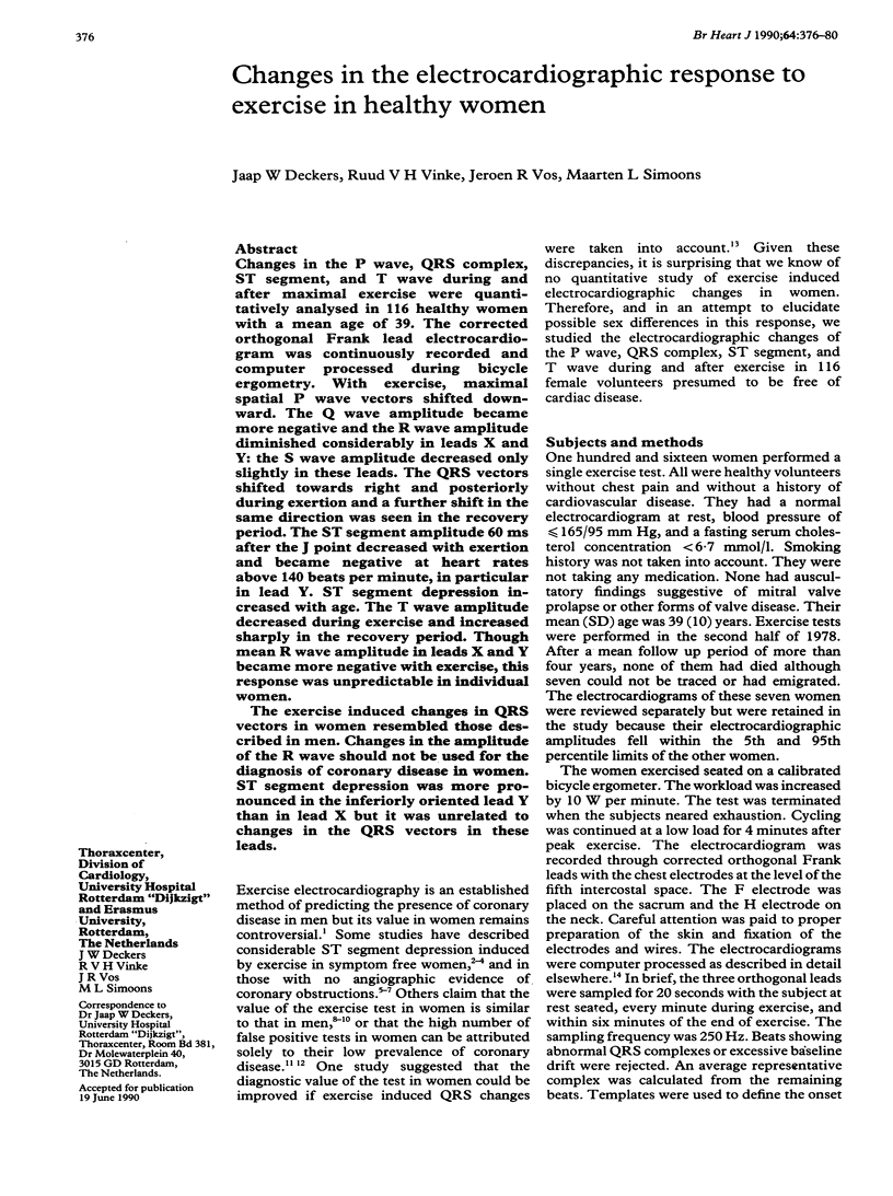
Selected References
These references are in PubMed. This may not be the complete list of references from this article.
- ASTRAND I. EXERCISE ELECTROCARDIOGRAMS RECORDED TWICE WITH AN 8-YEAR INTERVAL IN A GROUP OF 204 WOMEN AND MEN 48-63 YEARS OLD. Acta Med Scand. 1965 Jul;178:27–39. [PubMed] [Google Scholar]
- Allen W. H., Aronow W. S., De Cristofaro D. Treadmill exercise testing of mass screening for coronary risk factors. Cathet Cardiovasc Diagn. 1976;2(1):39–48. doi: 10.1002/ccd.1810020106. [DOI] [PubMed] [Google Scholar]
- Barolsky S. M., Gilbert C. A., Faruqui A., Nutter D. O., Schlant R. C. Differences in electrocardiographic response to exercise of women and men: a non-Bayesian factor. Circulation. 1979 Nov;60(5):1021–1027. doi: 10.1161/01.cir.60.5.1021. [DOI] [PubMed] [Google Scholar]
- Baron D. W., Ilsley C., Sheiban I., Poole-Wilson P. A., Rickards A. F. R wave amplitude during exercise. Relation to left ventricular function and coronary artery disease. Br Heart J. 1980 Nov;44(5):512–517. doi: 10.1136/hrt.44.5.512. [DOI] [PMC free article] [PubMed] [Google Scholar]
- Battler A., Froelicher V., Slutsky R., Ashburn W. Relationship of QRS amplitude changes during exercise to left ventricular function and volumes and the diagnosis of coronary artery disease. Circulation. 1979 Nov;60(5):1004–1013. doi: 10.1161/01.cir.60.5.1004. [DOI] [PubMed] [Google Scholar]
- Bengtsson C., Grimby G., Lindquist O., Noppa H., Sigurdsson J. A., Vedin J. A. Prognosis of women with exercise-induced ECG changes--results from a longitudinal population study. Cardiology. 1981;68 (Suppl 2):9–14. doi: 10.1159/000173311. [DOI] [PubMed] [Google Scholar]
- Bonoris P. E., Greenberg P. S., Castellanet M. J., Ellestad M. H. Significance of changes in R wave amplitude during treadmill stress testing: angiographic correlation. Am J Cardiol. 1978 May 1;41(5):846–851. doi: 10.1016/0002-9149(78)90723-3. [DOI] [PubMed] [Google Scholar]
- Bruce R. A. Editorial: Values and limitations of exercise electrocardiography. Circulation. 1974 Jul;50(1):1–3. doi: 10.1161/01.cir.50.1.1. [DOI] [PubMed] [Google Scholar]
- Cumming G. R., Dufresne C., Kich L., Samm J. Exercise electrocardiogram patterns in normal women. Br Heart J. 1973 Oct;35(10):1055–1061. doi: 10.1136/hrt.35.10.1055. [DOI] [PMC free article] [PubMed] [Google Scholar]
- Detrano R., Salcedo E., Passalacqua M., Friis R. Exercise electrocardiographic variables: a critical appraisal. J Am Coll Cardiol. 1986 Oct;8(4):836–847. doi: 10.1016/s0735-1097(86)80425-9. [DOI] [PubMed] [Google Scholar]
- Detry J. M., Kapita B. M., Cosyns J., Sottiaux B., Brasseur L. A., Rousseau M. F. Diagnostic value of history and maximal exercise electrocardiography in men and women suspected of coronary heart disease. Circulation. 1977 Nov;56(5):756–761. doi: 10.1161/01.cir.56.5.756. [DOI] [PubMed] [Google Scholar]
- Fox K., England D., Jonathan A., Selwyn A. Inability of exercise-induced R wave changes to predict coronary artery disease. Am J Cardiol. 1982 Mar;49(4):674–679. doi: 10.1016/0002-9149(82)91945-2. [DOI] [PubMed] [Google Scholar]
- Froelicher V. F., Wolthuis R., Fischer J., Uhl G. Variations in normal electrocardiographic response to treadmill testing. Am J Cardiol. 1981 May;47(5):1161–1167. doi: 10.1016/0002-9149(81)90229-0. [DOI] [PubMed] [Google Scholar]
- Guiteras P., Chaitman B. R., Waters D. D., Bourassa M. G., Scholl J. M., Ferguson R. J., Wagniart P. Diagnostic accuracy of exercise ECG lead systems in clinical subsets of women. Circulation. 1982 Jun;65(7):1465–1474. doi: 10.1161/01.cir.65.7.1465. [DOI] [PubMed] [Google Scholar]
- Hopkirk J. A., Leader S., Uhl G. S., Hickman J. R., Jr, Fischer J. Limitation of exercise-induced R wave amplitude changes in detecting coronary artery disease in asymptomatic men. J Am Coll Cardiol. 1984 Mar;3(3):821–826. doi: 10.1016/s0735-1097(84)80259-4. [DOI] [PubMed] [Google Scholar]
- Ilsley C., Canepa-Anson R., Westgate C., Webb S., Rickards A., Poole-Wilson P. Influence of R wave analysis upon diagnostic accuracy of exercise testing in women. Br Heart J. 1982 Aug;48(2):161–168. doi: 10.1136/hrt.48.2.161. [DOI] [PMC free article] [PubMed] [Google Scholar]
- Kasser I. S., Bruce R. A. Comparative effects of aging and coronary heart disease on submaximal and maximal exercise. Circulation. 1969 Jun;39(6):759–774. doi: 10.1161/01.cir.39.6.759. [DOI] [PubMed] [Google Scholar]
- Manca C., Dei Cas L., Albertini D., Baldi G., Visioli O. Different prognostic value of exercise electrocardiogram in men and women. Cardiology. 1978;63(5):312–319. doi: 10.1159/000169910. [DOI] [PubMed] [Google Scholar]
- Melin J. A., Wijns W., Vanbutsele R. J., Robert A., De Coster P., Brasseur L. A., Beckers C., Detry J. M. Alternative diagnostic strategies for coronary artery disease in women: demonstration of the usefulness and efficiency of probability analysis. Circulation. 1985 Mar;71(3):535–542. doi: 10.1161/01.cir.71.3.535. [DOI] [PubMed] [Google Scholar]
- Pratt C. M., Francis M. J., Divine G. W., Young J. B. Exercise testing in women with chest pain. Are there additional exercise characteristics that predict true positive test results? Chest. 1989 Jan;95(1):139–144. doi: 10.1378/chest.95.1.139. [DOI] [PubMed] [Google Scholar]
- Profant G. R., Early R. G., Nilson K. L., Kusumi F., Hofer V., Bruce R. A. Responses to maximal exercise in healthy middle-aged women. J Appl Physiol. 1972 Nov;33(5):595–599. doi: 10.1152/jappl.1972.33.5.595. [DOI] [PubMed] [Google Scholar]
- Simoons M. L., Boom H. B., Smallenburg E. On-line processing of orthogonal exercise electrocardiograms. Comput Biomed Res. 1975 Apr;8(2):105–117. doi: 10.1016/0010-4809(75)90032-4. [DOI] [PubMed] [Google Scholar]
- Simoons M. L., Hugenholtz P. G. Gradual changes of ECG waveform during and after exercise in normal subjects. Circulation. 1975 Oct;52(4):570–577. doi: 10.1161/01.cir.52.4.570. [DOI] [PubMed] [Google Scholar]
- Sketch M. H., Mohiuddin S. M., Lynch J. D., Zencka A. E., Runco V. Significant sex differences in the correlation of electrocardiographic exercise testing and coronary arteriograms. Am J Cardiol. 1975 Aug;36(2):169–173. doi: 10.1016/0002-9149(75)90521-4. [DOI] [PubMed] [Google Scholar]
- Weiner D. A., Ryan T. J., McCabe C. H., Kennedy J. W., Schloss M., Tristani F., Chaitman B. R., Fisher L. D. Exercise stress testing. Correlations among history of angina, ST-segment response and prevalence of coronary-artery disease in the Coronary Artery Surgery Study (CASS). N Engl J Med. 1979 Aug 2;301(5):230–235. doi: 10.1056/NEJM197908023010502. [DOI] [PubMed] [Google Scholar]
- Wolthuis R. A., Froelicher V. F., Hopkirk A., Fischer J. R., Keiser N. Normal electrocardiographic waveform characteristics during treadmill exercise testing. Circulation. 1979 Nov;60(5):1028–1035. doi: 10.1161/01.cir.60.5.1028. [DOI] [PubMed] [Google Scholar]
- van Bemmel J. H., Talmon J. L., Duisterhout J. S., Hengeveld S. J. Template waveform recognition applied to ECG-VCG analysis. Comput Biomed Res. 1973 Oct;6(5):430–441. doi: 10.1016/0010-4809(73)90076-1. [DOI] [PubMed] [Google Scholar]