

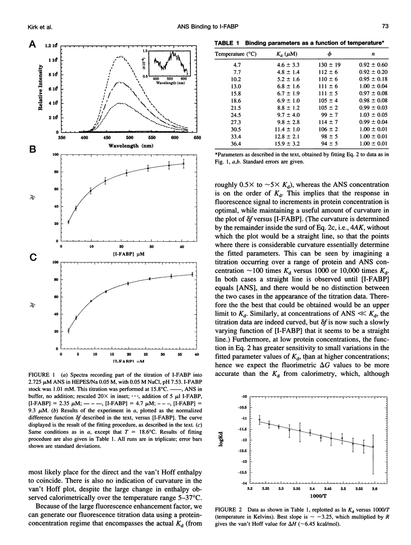
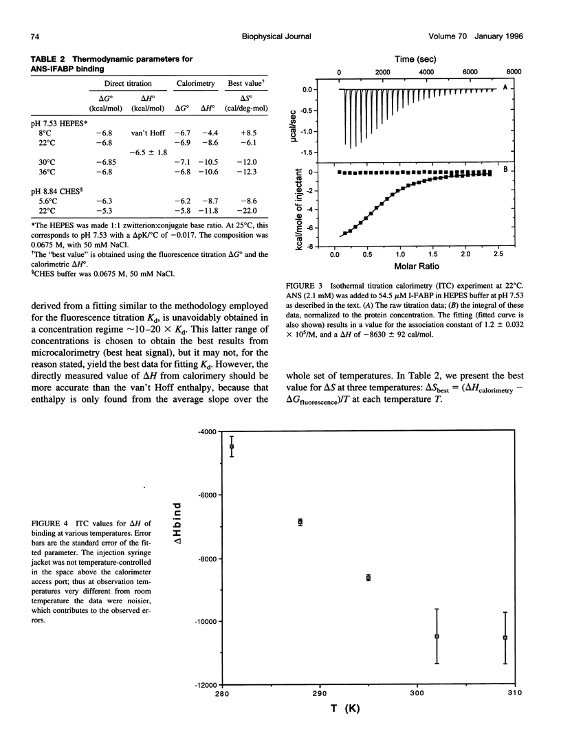
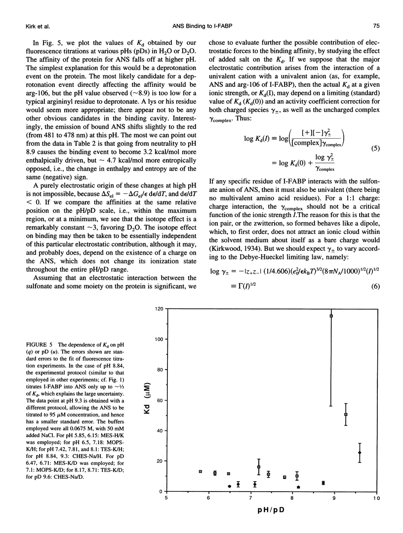
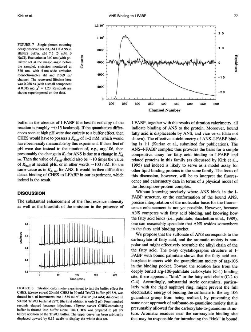
Abstract
1-Sulfonato-8-(1')anilinonaphthalene (1,8-ANS) was employed as a fluorescent probe of the fatty acid binding site of recombinant rat intestinal fatty acid binding protein (1-FABP). The enhancement of fluorescence upon binding allowed direct determination of binding affinity by fluorescence titration experiments, and measurement of the effects on that affinity of temperature, pH, and ionic strength. Solvent isotope effects were also determined. These data were compared to results from isothermal titration calorimetry. We obtained values for the enthalpy and entropy of this interaction at a variety of temperatures, and hence determined the change in heat capacity of the system consequent upon binding. The ANS-1-FABP is enthalpically driven; above approximately 14 degrees C it is entropically opposed, but below this temperature the entropy makes a positive contribution to the binding. The changes we observe in both enthalpy and entropy of binding with temperature can be derived from the change in heat capacity upon binding by integration, which demonstrates the internal consistency of our results. Bound ANS is displaced by fatty acids and can itself displace fatty acids bound to I-FABP. The binding site for ANS appears to be inside the solvent-containing cavity observed in the x-ray crystal structure, the same cavity occupied by fatty acid. From the fluorescence spectrum and from an inversion of the Debye-Hueckel formula for the activity coefficients as a function of added salt, we inferred that this cavity is fairly polar in character, which is in keeping with inferences drawn from the x-ray structure. The binding affinity of ANS is considered to be a consequence of both electrostatic and conditional hydrophobic effects. We speculate that the observed change in heat capacity is produced mainly by the displacement of strongly hydrogen-bonded waters from the protein cavity.
Full text
PDF

Selected References
These references are in PubMed. This may not be the complete list of references from this article.
- Banaszak L., Winter N., Xu Z., Bernlohr D. A., Cowan S., Jones T. A. Lipid-binding proteins: a family of fatty acid and retinoid transport proteins. Adv Protein Chem. 1994;45:89–151. doi: 10.1016/s0065-3233(08)60639-7. [DOI] [PubMed] [Google Scholar]
- Connelly P. R., Aldape R. A., Bruzzese F. J., Chambers S. P., Fitzgibbon M. J., Fleming M. A., Itoh S., Livingston D. J., Navia M. A., Thomson J. A. Enthalpy of hydrogen bond formation in a protein-ligand binding reaction. Proc Natl Acad Sci U S A. 1994 Mar 1;91(5):1964–1968. doi: 10.1073/pnas.91.5.1964. [DOI] [PMC free article] [PubMed] [Google Scholar]
- Dill K. A. The meaning of hydrophobicity. Science. 1990 Oct 12;250(4978):297–298. doi: 10.1126/science.2218535. [DOI] [PubMed] [Google Scholar]
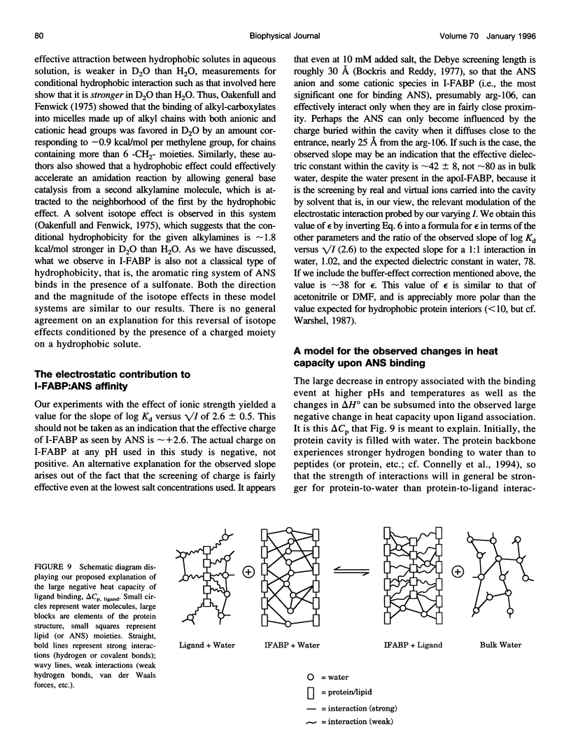
- Ernst J. A., Clubb R. T., Zhou H. X., Gronenborn A. M., Clore G. M. Demonstration of positionally disordered water within a protein hydrophobic cavity by NMR. Science. 1995 Mar 24;267(5205):1813–1817. doi: 10.1126/science.7892604. [DOI] [PubMed] [Google Scholar]
- Goldberg M. E., Semisotnov G. V., Friguet B., Kuwajima K., Ptitsyn O. B., Sugai S. An early immunoreactive folding intermediate of the tryptophan synthease beta 2 subunit is a 'molten globule'. FEBS Lett. 1990 Apr 9;263(1):51–56. doi: 10.1016/0014-5793(90)80703-l. [DOI] [PubMed] [Google Scholar]
- Jakoby M. G., Miller K. R., Toner J. J., Bauman A., Cheng L., Li E., Cistola D. P. Ligand-protein electrostatic interactions govern the specificity of retinol- and fatty acid-binding proteins. Biochemistry. 1993 Jan 26;32(3):872–878. doi: 10.1021/bi00054a019. [DOI] [PubMed] [Google Scholar]
- Jencks W. P. Binding energy, specificity, and enzymic catalysis: the circe effect. Adv Enzymol Relat Areas Mol Biol. 1975;43:219–410. doi: 10.1002/9780470122884.ch4. [DOI] [PubMed] [Google Scholar]
- Johnson J. D., El-Bayoumi M. A., Weber L. D., Tulinsky A. Interaction of alpha-chymotrypsin with the fluorescent probe 1-anilinonaphthalene-8-sulfonate in solution. Biochemistry. 1979 Apr 3;18(7):1292–1296. doi: 10.1021/bi00574a027. [DOI] [PubMed] [Google Scholar]
- Klotz I. M. Numbers of receptor sites from Scatchard graphs: facts and fantasies. Science. 1982 Sep 24;217(4566):1247–1249. doi: 10.1126/science.6287580. [DOI] [PubMed] [Google Scholar]
- LaLonde J. M., Levenson M. A., Roe J. J., Bernlohr D. A., Banaszak L. J. Adipocyte lipid-binding protein complexed with arachidonic acid. Titration calorimetry and X-ray crystallographic studies. J Biol Chem. 1994 Oct 14;269(41):25339–25347. doi: 10.2210/pdb1adl/pdb. [DOI] [PubMed] [Google Scholar]
- Levitt M., Park B. H. Water: now you see it, now you don't. Structure. 1993 Dec 15;1(4):223–226. doi: 10.1016/0969-2126(93)90011-5. [DOI] [PubMed] [Google Scholar]
- Ohyashiki T., Sekine T. Fluorometric studies on conformational changes in tropomyosin associated with depolymerization. J Biochem. 1979 Feb;85(2):575–580. doi: 10.1093/oxfordjournals.jbchem.a132366. [DOI] [PubMed] [Google Scholar]
- Parker C. W., Osterland C. K. Hydrophobic binding sites on immunoglobulins. Biochemistry. 1970 Mar 3;9(5):1074–1082. doi: 10.1021/bi00807a004. [DOI] [PubMed] [Google Scholar]
- Privalov P. L., Gill S. J. Stability of protein structure and hydrophobic interaction. Adv Protein Chem. 1988;39:191–234. doi: 10.1016/s0065-3233(08)60377-0. [DOI] [PubMed] [Google Scholar]
- Richieri G. V., Ogata R. T., Kleinfeld A. M. A fluorescently labeled intestinal fatty acid binding protein. Interactions with fatty acids and its use in monitoring free fatty acids. J Biol Chem. 1992 Nov 25;267(33):23495–23501. [PubMed] [Google Scholar]
- Sacchettini J. C., Banaszak L. J., Gordon J. I. Expression of rat intestinal fatty acid binding protein in E. coli and its subsequent structural analysis: a model system for studying the molecular details of fatty acid-protein interaction. 1990 Oct 15-Nov 8Mol Cell Biochem. 98(1-2):81–93. doi: 10.1007/BF00231371. [DOI] [PubMed] [Google Scholar]
- Sacchettini J. C., Gordon J. I., Banaszak L. J. Crystal structure of rat intestinal fatty-acid-binding protein. Refinement and analysis of the Escherichia coli-derived protein with bound palmitate. J Mol Biol. 1989 Jul 20;208(2):327–339. doi: 10.1016/0022-2836(89)90392-6. [DOI] [PubMed] [Google Scholar]
- Scapin G., Gordon J. I., Sacchettini J. C. Refinement of the structure of recombinant rat intestinal fatty acid-binding apoprotein at 1.2-A resolution. J Biol Chem. 1992 Feb 25;267(6):4253–4269. doi: 10.2210/pdb1ifc/pdb. [DOI] [PubMed] [Google Scholar]
- Sirangelo I., Bismuto E., Irace G. Solvent and thermal denaturation of the acidic compact state of apomyoglobin. FEBS Lett. 1994 Jan 24;338(1):11–15. doi: 10.1016/0014-5793(94)80107-x. [DOI] [PubMed] [Google Scholar]
- Slavík J. Anilinonaphthalene sulfonate as a probe of membrane composition and function. Biochim Biophys Acta. 1982 Aug 11;694(1):1–25. doi: 10.1016/0304-4157(82)90012-0. [DOI] [PubMed] [Google Scholar]
- Stryer L. The interaction of a naphthalene dye with apomyoglobin and apohemoglobin. A fluorescent probe of non-polar binding sites. J Mol Biol. 1965 Sep;13(2):482–495. doi: 10.1016/s0022-2836(65)80111-5. [DOI] [PubMed] [Google Scholar]
- WEBER G. Polarization of the fluorescence of macromolecules. II. Fluorescent conjugates of ovalbumin and bovine serum albumin. Biochem J. 1952 May;51(2):155–167. doi: 10.1042/bj0510155. [DOI] [PMC free article] [PubMed] [Google Scholar]
- Warshel A. What about protein polarity? Nature. 1987 Nov 5;330(6143):15–16. doi: 10.1038/330015a0. [DOI] [PubMed] [Google Scholar]
- Weber L. D., Tulinsky A., Johnson J. D., El-Bayoumi M. A. Expression of functionality of alpha-chymotrypsin. The structure of a fluorescent probe--alpha-chymotrypsin complex and the nature of its pH dependence. Biochemistry. 1979 Apr 3;18(7):1297–1303. doi: 10.1021/bi00574a028. [DOI] [PubMed] [Google Scholar]
- Xu Z. H., Buelt M. K., Banaszak L. J., Bernlohr D. A. Expression, purification, and crystallization of the adipocyte lipid binding protein. J Biol Chem. 1991 Aug 5;266(22):14367–14370. [PubMed] [Google Scholar]
- Yang M., Ghosh S. S., Millar D. P. Direct measurement of thermodynamic and kinetic parameters of DNA triple helix formation by fluorescence spectroscopy. Biochemistry. 1994 Dec 27;33(51):15329–15337. doi: 10.1021/bi00255a014. [DOI] [PubMed] [Google Scholar]