Abstract
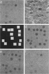
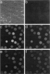
Scanning microphotolysis (Scamp), a recently developed photobleaching technique, was used to analyze the transport of two small organic anions and one inorganic cation through single pores formed in human erythrocyte membranes by the channel-forming toxin aerolysin secreted by Aeromonas species. The transport rate constants of erythrocyte ghosts carrying a single aerolysin pore were determined to be (1.83 +/- 0.43) x 10(-3) s-1 for Lucifer yellow, (0.33 +/- 0.10) x 10(-3) s-1 for carboxyfluorescein, and (8.20 +/- 2.30) x 10(-3) s-1 for Ca2+. The radius of the aerolysin pore was derived from the rate constants to be 19-23 A, taking steric hindrance and viscous drag into account. The size of the Ca2+ rate constant implies that at physiological extracellular Ca2+ concentrations (> 1 mM) the intracellular Ca2+ concentration would be elevated to the critical level of > 1 microM in much less than a second after formation of a single aerolysin pore in the plasma membrane. Thus changes in the levels of Ca2+ or other critical intracellular components may be more likely to cause cell death than osmotic imbalance.
Full text
PDF


Images in this article
Selected References
These references are in PubMed. This may not be the complete list of references from this article.
- Allbritton N. L., Meyer T., Stryer L. Range of messenger action of calcium ion and inositol 1,4,5-trisphosphate. Science. 1992 Dec 11;258(5089):1812–1815. doi: 10.1126/science.1465619. [DOI] [PubMed] [Google Scholar]
- Axelrod D., Koppel D. E., Schlessinger J., Elson E., Webb W. W. Mobility measurement by analysis of fluorescence photobleaching recovery kinetics. Biophys J. 1976 Sep;16(9):1055–1069. doi: 10.1016/S0006-3495(76)85755-4. [DOI] [PMC free article] [PubMed] [Google Scholar]
- Buckley J. T. Purification of cloned proaerolysin released by a low protease mutant of Aeromonas salmonicida. Biochem Cell Biol. 1990 Jan;68(1):221–224. doi: 10.1139/o90-029. [DOI] [PubMed] [Google Scholar]
- Dennert G., Podack E. R. Cytolysis by H-2-specific T killer cells. Assembly of tubular complexes on target membranes. J Exp Med. 1983 May 1;157(5):1483–1495. doi: 10.1084/jem.157.5.1483. [DOI] [PMC free article] [PubMed] [Google Scholar]
- Howard S. P., Buckley J. T. Membrane glycoprotein receptor and hole-forming properties of a cytolytic protein toxin. Biochemistry. 1982 Mar 30;21(7):1662–1667. doi: 10.1021/bi00536a029. [DOI] [PubMed] [Google Scholar]
- Jacobson K., Wu E., Poste G. Measurement of the translational mobility of concanavalin A in glycerol-saline solutions and on the cell surface by fluorescence recovery after photobleaching. Biochim Biophys Acta. 1976 Apr 16;433(1):215–222. doi: 10.1016/0005-2736(76)90189-9. [DOI] [PubMed] [Google Scholar]
- Kaplan J. H., Forbush B., 3rd, Hoffman J. F. Rapid photolytic release of adenosine 5'-triphosphate from a protected analogue: utilization by the Na:K pump of human red blood cell ghosts. Biochemistry. 1978 May 16;17(10):1929–1935. doi: 10.1021/bi00603a020. [DOI] [PubMed] [Google Scholar]
- Kubitscheck U., Pratsch L., Passow H., Peters R. Calcium pump kinetics determined in single erythrocyte ghosts by microphotolysis and confocal imaging. Biophys J. 1995 Jul;69(1):30–41. doi: 10.1016/S0006-3495(95)79875-7. [DOI] [PMC free article] [PubMed] [Google Scholar]
- Nicotera P., Zhivotovsky B., Orrenius S. Nuclear calcium transport and the role of calcium in apoptosis. Cell Calcium. 1994 Oct;16(4):279–288. doi: 10.1016/0143-4160(94)90091-4. [DOI] [PubMed] [Google Scholar]
- Orrenius S., McConkey D. J., Bellomo G., Nicotera P. Role of Ca2+ in toxic cell killing. Trends Pharmacol Sci. 1989 Jul;10(7):281–285. doi: 10.1016/0165-6147(89)90029-1. [DOI] [PubMed] [Google Scholar]
- PAPPENHEIMER J. R., RENKIN E. M., BORRERO L. M. Filtration, diffusion and molecular sieving through peripheral capillary membranes; a contribution to the pore theory of capillary permeability. Am J Physiol. 1951 Oct;167(1):13–46. doi: 10.1152/ajplegacy.1951.167.1.13. [DOI] [PubMed] [Google Scholar]
- Paine P. L., Scherr P. Drag coefficients for the movement of rigid spheres through liquid-filled cylindrical pores. Biophys J. 1975 Oct;15(10):1087–1091. doi: 10.1016/S0006-3495(75)85884-X. [DOI] [PMC free article] [PubMed] [Google Scholar]
- Parker M. W., Buckley J. T., Postma J. P., Tucker A. D., Leonard K., Pattus F., Tsernoglou D. Structure of the Aeromonas toxin proaerolysin in its water-soluble and membrane-channel states. Nature. 1994 Jan 20;367(6460):292–295. doi: 10.1038/367292a0. [DOI] [PubMed] [Google Scholar]
- Peters R. Nuclear envelope permeability measured by fluorescence microphotolysis of single liver cell nuclei. J Biol Chem. 1983 Oct 10;258(19):11427–11429. [PubMed] [Google Scholar]
- Peters R. Nucleo-cytoplasmic flux and intracellular mobility in single hepatocytes measured by fluorescence microphotolysis. EMBO J. 1984 Aug;3(8):1831–1836. doi: 10.1002/j.1460-2075.1984.tb02055.x. [DOI] [PMC free article] [PubMed] [Google Scholar]
- Peters R., Peters J., Tews K. H., Bähr W. A microfluorimetric study of translational diffusion in erythrocyte membranes. Biochim Biophys Acta. 1974 Nov 15;367(3):282–294. doi: 10.1016/0005-2736(74)90085-6. [DOI] [PubMed] [Google Scholar]
- Peters R., Sauer H., Tschopp J., Fritzsch G. Transients of perforin pore formation observed by fluorescence microscopic single channel recording. EMBO J. 1990 Aug;9(8):2447–2451. doi: 10.1002/j.1460-2075.1990.tb07421.x. [DOI] [PMC free article] [PubMed] [Google Scholar]
- Petersen O. H., Petersen C. C., Kasai H. Calcium and hormone action. Annu Rev Physiol. 1994;56:297–319. doi: 10.1146/annurev.ph.56.030194.001501. [DOI] [PubMed] [Google Scholar]
- RENKIN E. M. Filtration, diffusion, and molecular sieving through porous cellulose membranes. J Gen Physiol. 1954 Nov 20;38(2):225–243. [PMC free article] [PubMed] [Google Scholar]
- Sauer H., Pratsch L., Fritzsch G., Bhakdi S., Peters R. Complement pore genesis observed in erythrocyte membranes by fluorescence microscopic single-channel recording. Biochem J. 1991 Jun 1;276(Pt 2):395–399. doi: 10.1042/bj2760395. [DOI] [PMC free article] [PubMed] [Google Scholar]
- Schwoch G., Passow H. Preparation and properties of human erythrocyte ghosts. Mol Cell Biochem. 1973 Dec 15;2(2):197–218. doi: 10.1007/BF01795474. [DOI] [PubMed] [Google Scholar]
- Wedekind P., Kubitscheck U., Peters R. Scanning microphotolysis: a new photobleaching technique based on fast intensity modulation of a scanned laser beam and confocal imaging. J Microsc. 1994 Oct;176(Pt 1):23–33. doi: 10.1111/j.1365-2818.1994.tb03496.x. [DOI] [PubMed] [Google Scholar]
- Wilmsen H. U., Buckley J. T., Pattus F. Site-directed mutagenesis at histidines of aerolysin from Aeromonas hydrophila: a lipid planar bilayer study. Mol Microbiol. 1991 Nov;5(11):2745–2751. doi: 10.1111/j.1365-2958.1991.tb01983.x. [DOI] [PubMed] [Google Scholar]
- Wilmsen H. U., Leonard K. R., Tichelaar W., Buckley J. T., Pattus F. The aerolysin membrane channel is formed by heptamerization of the monomer. EMBO J. 1992 Jul;11(7):2457–2463. doi: 10.1002/j.1460-2075.1992.tb05310.x. [DOI] [PMC free article] [PubMed] [Google Scholar]
- Wilmsen H. U., Pattus F., Buckley J. T. Aerolysin, a hemolysin from Aeromonas hydrophila, forms voltage-gated channels in planar lipid bilayers. J Membr Biol. 1990 Apr;115(1):71–81. doi: 10.1007/BF01869107. [DOI] [PubMed] [Google Scholar]
- van der Goot F. G., Pattus F., Parker M., Buckley J. T. The cytolytic toxin aerolysin: from the soluble form to the transmembrane channel. Toxicology. 1994 Feb 28;87(1-3):19–28. doi: 10.1016/0300-483x(94)90152-x. [DOI] [PubMed] [Google Scholar]


