Abstract
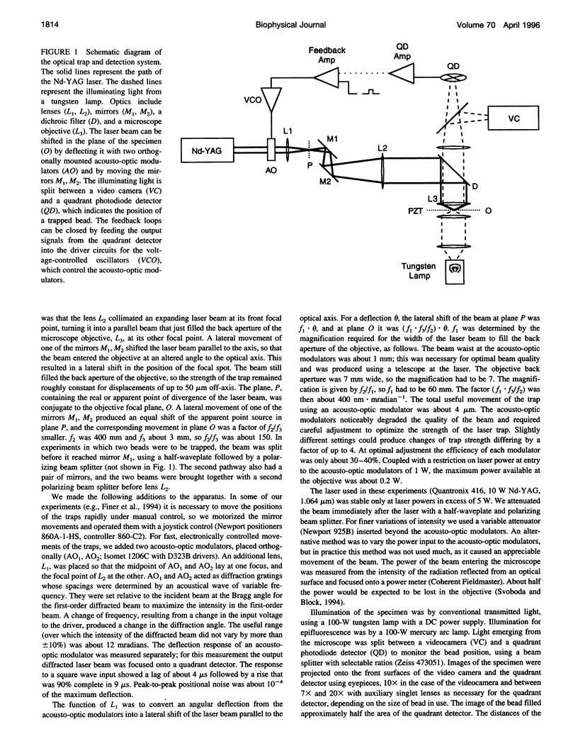
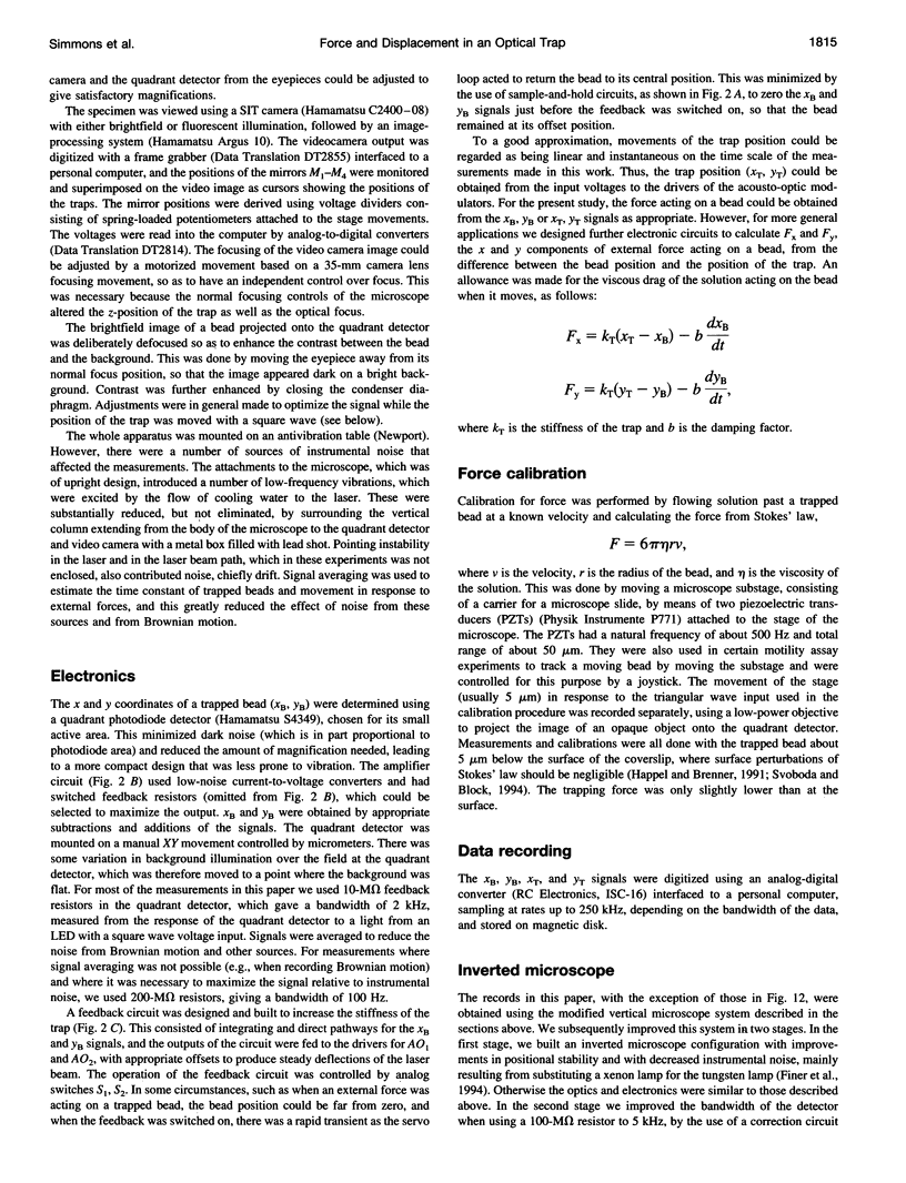
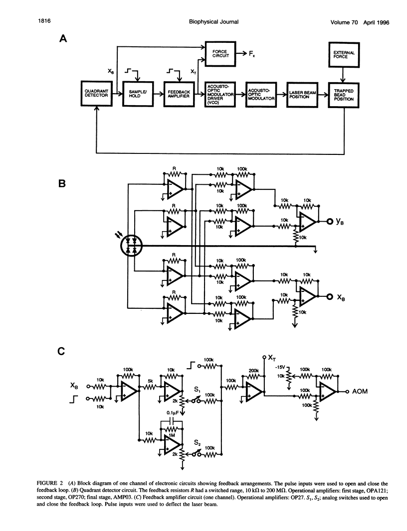
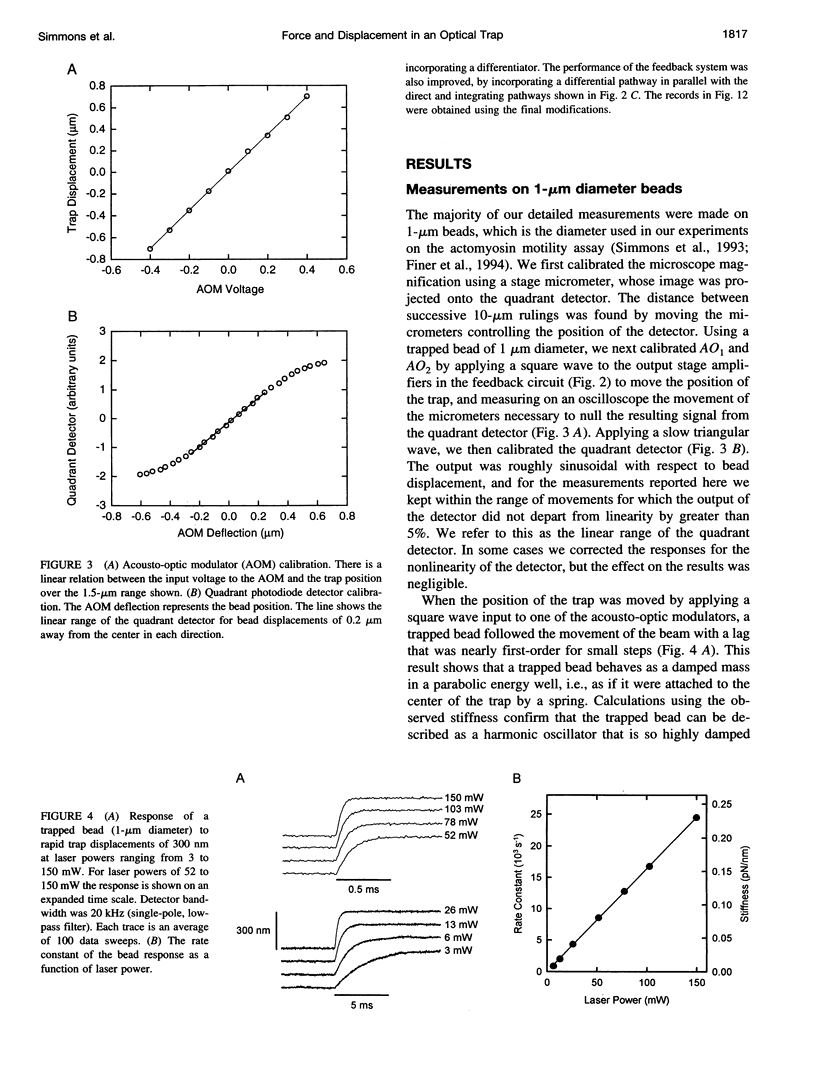
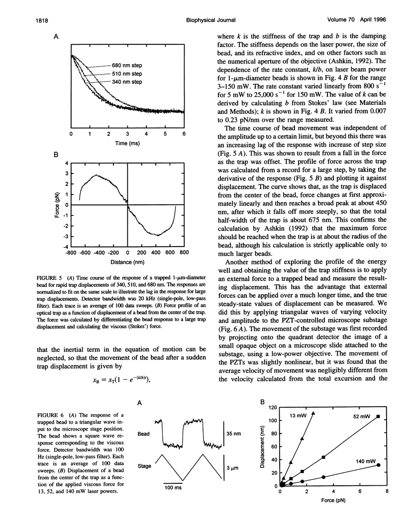
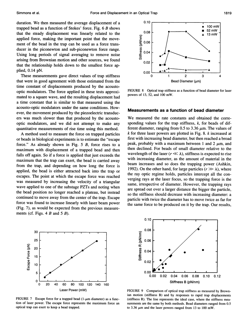
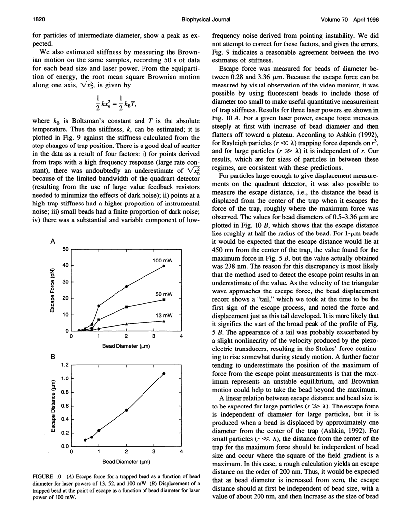
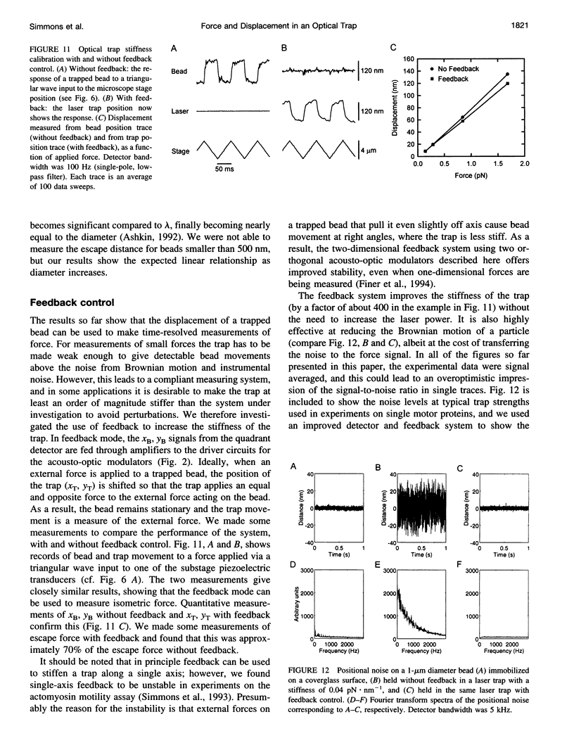
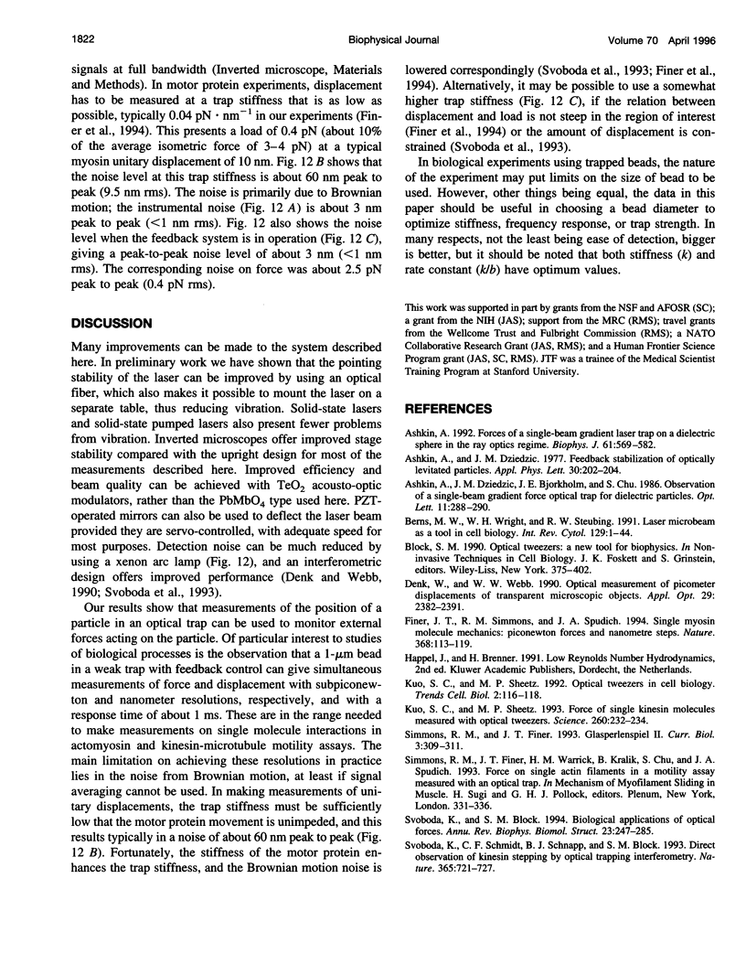
We combined a single-beam gradient optical trap with a high-resolution photodiode position detector to show that an optical trap can be used to make quantitative measurements of nanometer displacements and piconewton forces with millisecond resolution. When an external force is applied to a micron-sized bead held by an optical trap, the bead is displaced from the center of the trap by an amount proportional to the applied force. When the applied force is changed rapidly, the rise time of the displacement is on the millisecond time scale, and thus a trapped bead can be used as a force transducer. The performance can be enhanced by a feedback circuit so that the position of the trap moves by means of acousto-optic modulators to exert a force equal and opposite to the external force applied to the bead. In this case the position of the trap can be used to measure the applied force. We consider parameters of the trapped bead such as stiffness and response time as a function of bead diameter and laser beam power and compare the results with recent ray-optic calculations.
Full text
PDF
Selected References
These references are in PubMed. This may not be the complete list of references from this article.
- Ashkin A. Forces of a single-beam gradient laser trap on a dielectric sphere in the ray optics regime. Biophys J. 1992 Feb;61(2):569–582. doi: 10.1016/S0006-3495(92)81860-X. [DOI] [PMC free article] [PubMed] [Google Scholar]
- Berns M. W., Wright W. H., Wiegand Steubing R. Laser microbeam as a tool in cell biology. Int Rev Cytol. 1991;129:1–44. doi: 10.1016/s0074-7696(08)60507-0. [DOI] [PubMed] [Google Scholar]
- Finer J. T., Simmons R. M., Spudich J. A. Single myosin molecule mechanics: piconewton forces and nanometre steps. Nature. 1994 Mar 10;368(6467):113–119. doi: 10.1038/368113a0. [DOI] [PubMed] [Google Scholar]
- Kuo S. C., Sheetz M. P. Force of single kinesin molecules measured with optical tweezers. Science. 1993 Apr 9;260(5105):232–234. doi: 10.1126/science.8469975. [DOI] [PubMed] [Google Scholar]
- Kuo S. C., Sheetz M. P. Optical tweezers in cell biology. Trends Cell Biol. 1992 Apr;2(4):116–118. doi: 10.1016/0962-8924(92)90016-g. [DOI] [PubMed] [Google Scholar]
- Simmons R. M., Finer J. T. Optical tweezers: Glasperlenspiel II. Curr Biol. 1993 May 1;3(5):309–311. doi: 10.1016/0960-9822(93)90188-t. [DOI] [PubMed] [Google Scholar]
- Svoboda K., Block S. M. Biological applications of optical forces. Annu Rev Biophys Biomol Struct. 1994;23:247–285. doi: 10.1146/annurev.bb.23.060194.001335. [DOI] [PubMed] [Google Scholar]
- Svoboda K., Schmidt C. F., Schnapp B. J., Block S. M. Direct observation of kinesin stepping by optical trapping interferometry. Nature. 1993 Oct 21;365(6448):721–727. doi: 10.1038/365721a0. [DOI] [PubMed] [Google Scholar]


