Abstract
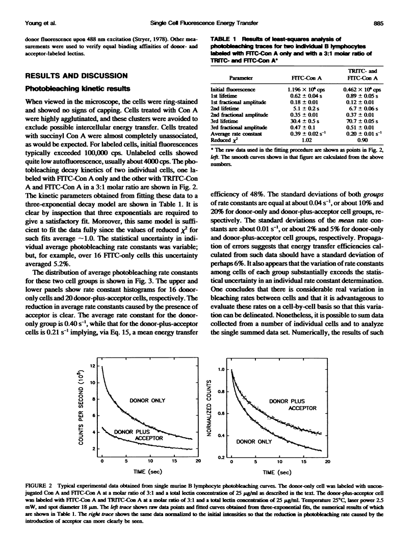
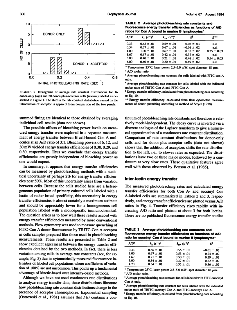
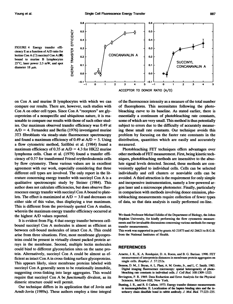
We describe practical aspects of photobleaching fluorescence energy transfer measurements on individual living cells. The method introduced by T. M. Jovin and co-workers (see, most recently, Kubitscheck et al. 1993. Biophys. J. 64:110) is based on the reduced rate of irreversible photobleaching of donor fluorophores when acceptor fluorophores are present. Measuring differences in donor photobleaching rates on cells labeled with donor only (fluorescein isothiocyanate-conjugated proteins) and with both donor and acceptor (tetramethylrhodamine-conjugated proteins) allows calculation of the fluorescence energy transfer efficiency. We assess possible methods of data analysis in light of the underlying processes of photobleaching and energy transfer and suggest optimum strategies for this purpose. Single murine B lymphocytes binding various ratios of donor and acceptor conjugates of tetravalent concanavalin A (Con A) and divalent succinyl Con A were examined for interlectin energy transfer by these methods. For Con A, a maximum transfer efficiency of 0.49 +/- 0.02 was observed. Under similar conditions flow cytometric measurements of donor quenching yielded a value of 0.54 +/- 0.03. For succinyl Con A, the maximum transfer efficiency was 0.36. To provide concrete examples of quantities arising in such energy transfer determinations, we present examples of individual cell data and kinetic analyses, population rate constant distributions, and error estimates for the various quantities involved.
Full text
PDF
Selected References
These references are in PubMed. This may not be the complete list of references from this article.
- Benson D. M., Bryan J., Plant A. L., Gotto A. M., Jr, Smith L. C. Digital imaging fluorescence microscopy: spatial heterogeneity of photobleaching rate constants in individual cells. J Cell Biol. 1985 Apr;100(4):1309–1323. doi: 10.1083/jcb.100.4.1309. [DOI] [PMC free article] [PubMed] [Google Scholar]
- Chan S. S., Arndt-Jovin D. J., Jovin T. M. Proximity of lectin receptors on the cell surface measured by fluorescence energy transfer in a flow system. J Histochem Cytochem. 1979 Jan;27(1):56–64. doi: 10.1177/27.1.374620. [DOI] [PubMed] [Google Scholar]
- Eisen H., Nasi S., Georgopoulos C. P., Arndt-Jovin D., Ostertag W. Surface changes in differentiating Friend erythroleukemic cells in culture. Cell. 1977 Apr;10(4):689–695. doi: 10.1016/0092-8674(77)90102-7. [DOI] [PubMed] [Google Scholar]
- Fairclough R. H., Cantor C. R. The use of singlet-singlet energy transfer to study macromolecular assemblies. Methods Enzymol. 1978;48:347–379. doi: 10.1016/s0076-6879(78)48019-x. [DOI] [PubMed] [Google Scholar]
- Fernandez S. M., Berlin R. D. Cell surface distribution of lectin receptors determined by resonance energy transfer. Nature. 1976 Dec 2;264(5585):411–415. doi: 10.1038/264411a0. [DOI] [PubMed] [Google Scholar]
- Fleming P. J., Koppel D. E., Lau A. L., Strittmatter P. Intramembrane position of the fluorescent tryptophanyl residue in membrane-bound cytochrome b5. Biochemistry. 1979 Nov 27;18(24):5458–5464. doi: 10.1021/bi00591a031. [DOI] [PubMed] [Google Scholar]
- Fung B. K., Stryer L. Surface density determination in membranes by fluorescence energy transfer. Biochemistry. 1978 Nov 28;17(24):5241–5248. doi: 10.1021/bi00617a025. [DOI] [PubMed] [Google Scholar]
- Herman B. Resonance energy transfer microscopy. Methods Cell Biol. 1989;30:219–243. doi: 10.1016/s0091-679x(08)60981-4. [DOI] [PubMed] [Google Scholar]
- Jongsma A. P., Hijmans W., Ploem J. S. Quantitative immunofluorescence. Standardization and calibration in microfluorometry. Histochemie. 1971;25(4):329–343. doi: 10.1007/BF00278226. [DOI] [PubMed] [Google Scholar]
- Jovin T. M., Arndt-Jovin D. J. Luminescence digital imaging microscopy. Annu Rev Biophys Biophys Chem. 1989;18:271–308. doi: 10.1146/annurev.bb.18.060189.001415. [DOI] [PubMed] [Google Scholar]
- Kubitscheck U., Kircheis M., Schweitzer-Stenner R., Dreybrodt W., Jovin T. M., Pecht I. Fluorescence resonance energy transfer on single living cells. Application to binding of monovalent haptens to cell-bound immunoglobulin E. Biophys J. 1991 Aug;60(2):307–318. doi: 10.1016/S0006-3495(91)82055-0. [DOI] [PMC free article] [PubMed] [Google Scholar]
- Kubitscheck U., Schweitzer-Stenner R., Arndt-Jovin D. J., Jovin T. M., Pecht I. Distribution of type I Fc epsilon-receptors on the surface of mast cells probed by fluorescence resonance energy transfer. Biophys J. 1993 Jan;64(1):110–120. doi: 10.1016/S0006-3495(93)81345-6. [DOI] [PMC free article] [PubMed] [Google Scholar]
- Leuther M. D., Peacock J. S., Krakauer H., Barisas B. G. Changes in lectin receptor lateral mobilities accompany lymphocyte stimulation. J Immunol. 1981 Sep;127(3):893–899. [PubMed] [Google Scholar]
- Steinkamp J. A., Kraemer P. M. Flow microfluorometric studies of lectin binding to mammalian cells. II. Estimation of the surface density of receptor sites by multiparameter analysis. J Cell Physiol. 1974 Oct;84(2):197–204. doi: 10.1002/jcp.1040840206. [DOI] [PubMed] [Google Scholar]
- Stryer L. Fluorescence energy transfer as a spectroscopic ruler. Annu Rev Biochem. 1978;47:819–846. doi: 10.1146/annurev.bi.47.070178.004131. [DOI] [PubMed] [Google Scholar]
- Szöllösi J., Trón L., Damjanovich S., Helliwell S. H., Arndt-Jovin D., Jovin T. M. Fluorescence energy transfer measurements on cell surfaces: a critical comparison of steady-state fluorimetric and flow cytometric methods. Cytometry. 1984 Mar;5(2):210–216. doi: 10.1002/cyto.990050216. [DOI] [PubMed] [Google Scholar]
- Trón L., Szöllósi J., Damjanovich S., Helliwell S. H., Arndt-Jovin D. J., Jovin T. M. Flow cytometric measurement of fluorescence resonance energy transfer on cell surfaces. Quantitative evaluation of the transfer efficiency on a cell-by-cell basis. Biophys J. 1984 May;45(5):939–946. doi: 10.1016/S0006-3495(84)84240-X. [DOI] [PMC free article] [PubMed] [Google Scholar]
- Uster P. S., Pagano R. E. Resonance energy transfer microscopy: observations of membrane-bound fluorescent probes in model membranes and in living cells. J Cell Biol. 1986 Oct;103(4):1221–1234. doi: 10.1083/jcb.103.4.1221. [DOI] [PMC free article] [PubMed] [Google Scholar]


