

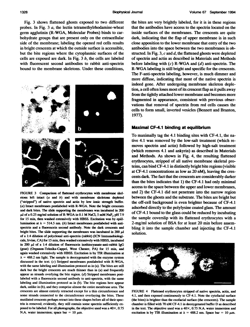
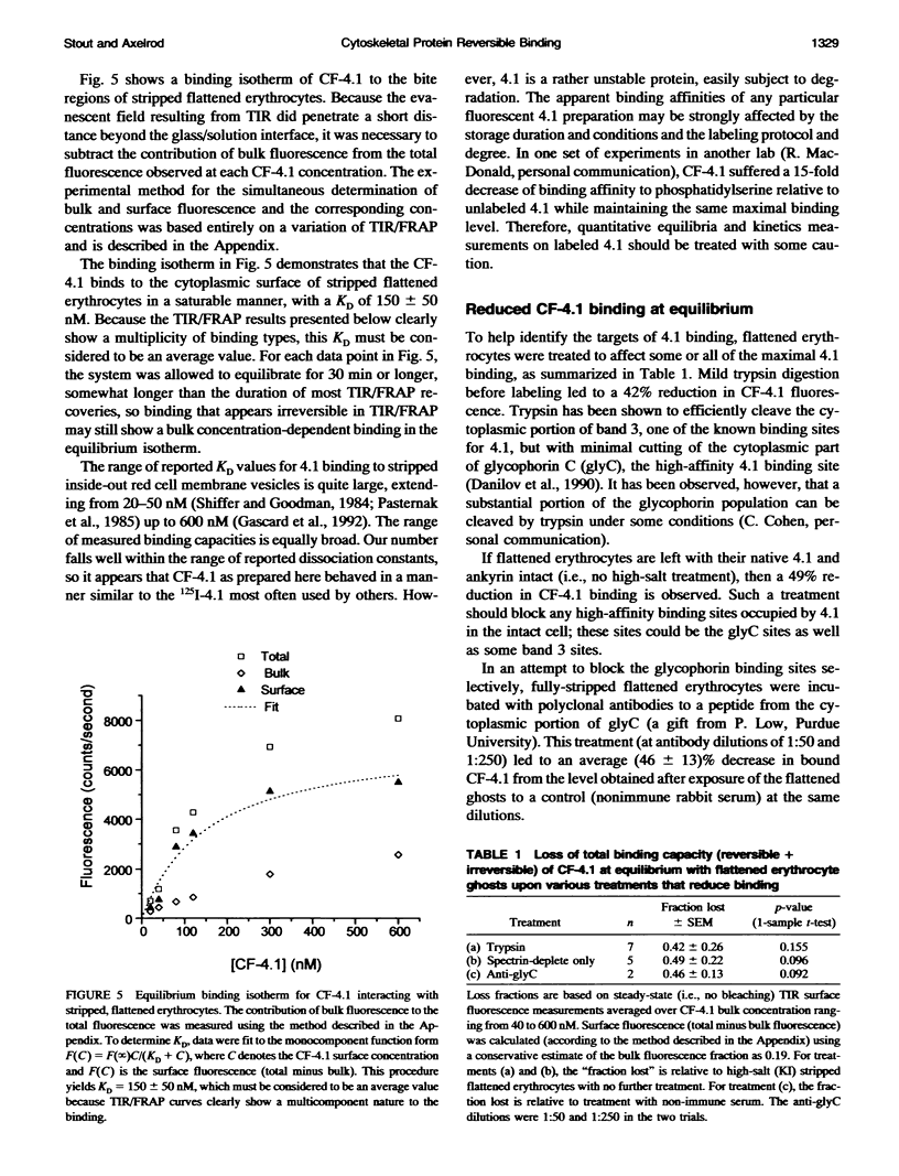
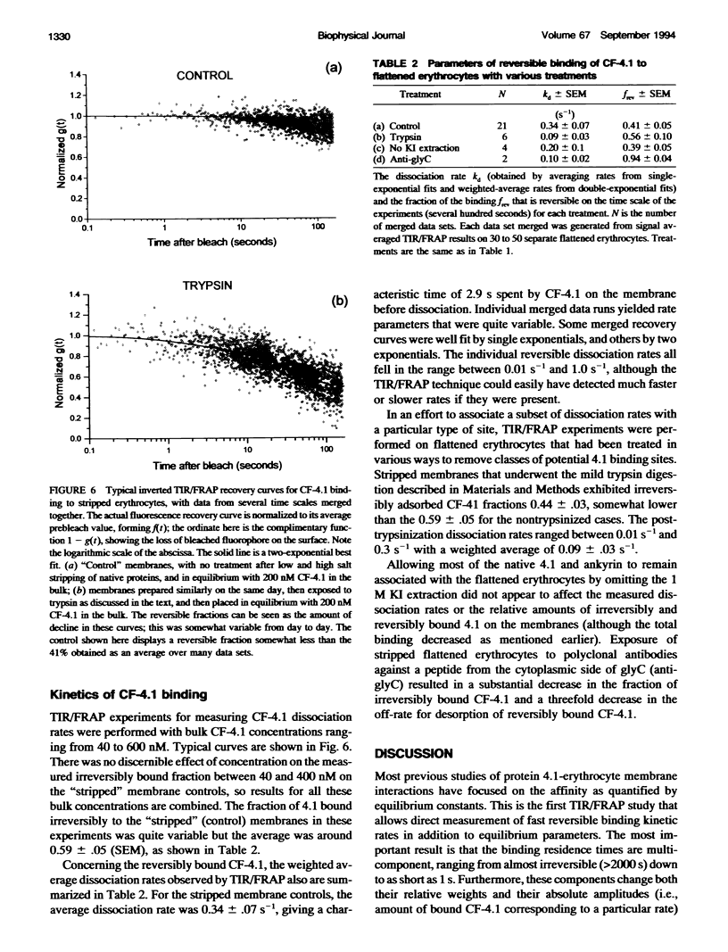
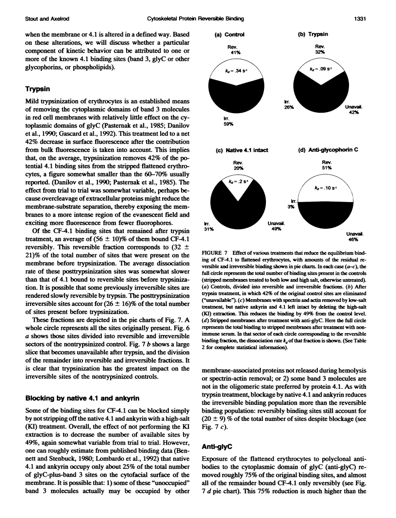
Abstract
Reversible binding among components of the cellular submembrane cytoskeleton and reversible binding of some of these components with the plasma membrane likely play a role in nonelastic morphological changes and mechanoplastic properties of cells. However, relatively few studies have been devoted to investigating directly the kinetic aspects of the interactions of individual components of the membrane skeleton with the membrane. The experiments described here investigated whether one component of the erythrocyte membrane cytoskeleton, protein 4.1, binds to its sites on the membrane reversibly and if so, whether the different 4.1-binding sites display distinct kinetic behavior. Protein 4.1 is known to stabilize the membrane and to mediate the attachment of spectrin filaments to the membrane. Protein 4.1 previously has been shown to bind to integral membrane proteins band 3, glycophorin C, and to negatively charged phospholipids. To examine the kinetic rates of dissociation of carboxymethyl fluorescein-labeled 4.1 (CF-4.1) to the cytofacial surface of erythrocyte membrane, a special preparation of hemolyzed erythrocyte ghosts was used, in which the ghosts became flattened on a glass surface and exposed their cytofacial surfaces to the solution through a membrane rip in a distinctive characteristic pattern. This preparation was examined by the microscopy technique of total internal reflection/fluorescence recovery after photobleaching (TIR/FRAP). Four different treatments were employed to help identify which membrane binding sites gave rise to the multiplicity of observed kinetic rates. The first treatment, the control, stripped off the native spectrin, actin, 4.1, and ankyrin. About 60% of the CF-4.1 bound to this control binded irreversibly (dissociation time > 20 min), but the remaining approximately 40% binded reversibly with a range of residency times averaging approximately 3 s. The second treatment subjected these stripped membranes to trypsin, which presumably removed most of the band 3. CF-4.1 binded significantly less to these trypsinized membranes and most of the decrease was a loss of the irreversibly binding sites. The third treatment simply preserved the native 4.1 and ankyrin. CF-4.1 binded less to this sample too, and the loss involved both the irreversible and reversible sites. The fourth treatment blocked the gycophorin C sites on the native 4.1-stripped membranes with an antibody. CF-4.1 again binded less to this sample than to a nonimmune serum control, and almost all of the decrease is a loss of irreversible sites. These rest suggest that 1) protein 4.1 binds to membrane or submembrane sites at least in part reversibly ; 2) the most reversible sites are probably not proteinaceous and not glycophorin C, but possibly are phospholipids (especially phosphatidylserine); and 3) TIWRFRAP can successfully examine the fast reversible dynamics of cytoskeletal components binding to biological membranes.
Full text
PDF
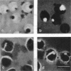
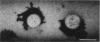
Images in this article
Selected References
These references are in PubMed. This may not be the complete list of references from this article.
- Bennett V., Branton D. Selective association of spectrin with the cytoplasmic surface of human erythrocyte plasma membranes. Quantitative determination with purified (32P)spectrin. J Biol Chem. 1977 Apr 25;252(8):2753–2763. [PubMed] [Google Scholar]
- Bennett V. Spectrin-based membrane skeleton: a multipotential adaptor between plasma membrane and cytoplasm. Physiol Rev. 1990 Oct;70(4):1029–1065. doi: 10.1152/physrev.1990.70.4.1029. [DOI] [PubMed] [Google Scholar]
- Bennett V., Stenbuck P. J. Association between ankyrin and the cytoplasmic domain of band 3 isolated from the human erythrocyte membrane. J Biol Chem. 1980 Jul 10;255(13):6424–6432. [PubMed] [Google Scholar]
- Berg H. C., Purcell E. M. Physics of chemoreception. Biophys J. 1977 Nov;20(2):193–219. doi: 10.1016/S0006-3495(77)85544-6. [DOI] [PMC free article] [PubMed] [Google Scholar]
- Brinkley M. A brief survey of methods for preparing protein conjugates with dyes, haptens, and cross-linking reagents. Bioconjug Chem. 1992 Jan-Feb;3(1):2–13. doi: 10.1021/bc00013a001. [DOI] [PubMed] [Google Scholar]
- Burghardt T. P., Axelrod D. Total internal reflection/fluorescence photobleaching recovery study of serum albumin adsorption dynamics. Biophys J. 1981 Mar;33(3):455–467. doi: 10.1016/S0006-3495(81)84906-5. [DOI] [PMC free article] [PubMed] [Google Scholar]
- Cohen A. M., Liu S. C., Lawler J., Derick L., Palek J. Identification of the protein 4.1 binding site to phosphatidylserine vesicles. Biochemistry. 1988 Jan 26;27(2):614–619. doi: 10.1021/bi00402a018. [DOI] [PubMed] [Google Scholar]
- Corbett J. D., Cho M. R., Golan D. E. Deoxygenation affects fluorescence photobleaching recovery measurements of red cell membrane protein lateral mobility. Biophys J. 1994 Jan;66(1):25–30. doi: 10.1016/S0006-3495(94)80760-X. [DOI] [PMC free article] [PubMed] [Google Scholar]
- Danilov Y. N., Fennell R., Ling E., Cohen C. M. Selective modulation of band 4.1 binding to erythrocyte membranes by protein kinase C. J Biol Chem. 1990 Feb 15;265(5):2556–2562. [PubMed] [Google Scholar]
- Englander S. W., Calhoun D. B., Englander J. J. Biochemistry without oxygen. Anal Biochem. 1987 Mar;161(2):300–306. doi: 10.1016/0003-2697(87)90454-4. [DOI] [PubMed] [Google Scholar]
- Gascard P., Pawelczyk T., Lowenstein J. M., Cohen C. M. The role of inositol phospholipids in the association of band 4.1 with the human erythrocyte membrane. Eur J Biochem. 1993 Feb 1;211(3):671–681. doi: 10.1111/j.1432-1033.1993.tb17595.x. [DOI] [PubMed] [Google Scholar]
- Jacobson B. S., Cronin J., Branton D. Coupling polylysine to glass beads for plasma membrane isolation. Biochim Biophys Acta. 1978 Jan 4;506(1):81–96. doi: 10.1016/0005-2736(78)90436-4. [DOI] [PubMed] [Google Scholar]
- Lerche D., Kozlov M. M., Meier W. Time-dependent elastic extensional RBC deformation by micropipette aspiration: redistribution of the spectrin network? Eur Biophys J. 1991;19(6):301–309. doi: 10.1007/BF00183319. [DOI] [PubMed] [Google Scholar]
- Leto T. L., Marchesi V. T. A structural model of human erythrocyte protein 4.1. J Biol Chem. 1984 Apr 10;259(7):4603–4608. [PubMed] [Google Scholar]
- Lombardo C. R., Willardson B. M., Low P. S. Localization of the protein 4.1-binding site on the cytoplasmic domain of erythrocyte membrane band 3. J Biol Chem. 1992 May 15;267(14):9540–9546. [PubMed] [Google Scholar]
- Ohanian V., Gratzer W. Preparation of red-cell-membrane cytoskeletal constituents and characterisation of protein 4.1. Eur J Biochem. 1984 Oct 15;144(2):375–379. doi: 10.1111/j.1432-1033.1984.tb08474.x. [DOI] [PubMed] [Google Scholar]
- Pasternack G. R., Anderson R. A., Leto T. L., Marchesi V. T. Interactions between protein 4.1 and band 3. An alternative binding site for an element of the membrane skeleton. J Biol Chem. 1985 Mar 25;260(6):3676–3683. [PubMed] [Google Scholar]
- Pearce K. H., Hiskey R. G., Thompson N. L. Surface binding kinetics of prothrombin fragment 1 on planar membranes measured by total internal reflection fluorescence microscopy. Biochemistry. 1992 Jul 7;31(26):5983–5995. doi: 10.1021/bi00141a005. [DOI] [PubMed] [Google Scholar]
- Peters R., Peters J., Tews K. H., Bähr W. A microfluorimetric study of translational diffusion in erythrocyte membranes. Biochim Biophys Acta. 1974 Nov 15;367(3):282–294. doi: 10.1016/0005-2736(74)90085-6. [DOI] [PubMed] [Google Scholar]
- Pisarchick M. L., Gesty D., Thompson N. L. Binding kinetics of an anti-dinitrophenyl monoclonal Fab on supported phospholipid monolayers measured by total internal reflection with fluorescence photobleaching recovery. Biophys J. 1992 Jul;63(1):215–223. doi: 10.1016/S0006-3495(92)81592-8. [DOI] [PMC free article] [PubMed] [Google Scholar]
- Takakuwa Y., Tchernia G., Rossi M., Benabadji M., Mohandas N. Restoration of normal membrane stability to unstable protein 4.1-deficient erythrocyte membranes by incorporation of purified protein 4.1. J Clin Invest. 1986 Jul;78(1):80–85. doi: 10.1172/JCI112577. [DOI] [PMC free article] [PubMed] [Google Scholar]
- Takeshita K., MacDonald R. I., MacDonald R. C. Band 4.1 enhances spectrin binding to phosphatidylserine vesicles. Biochem Biophys Res Commun. 1993 Feb 26;191(1):165–171. doi: 10.1006/bbrc.1993.1198. [DOI] [PubMed] [Google Scholar]
- Thevenin B. J., Low P. S. Kinetics and regulation of the ankyrin-band 3 interaction of the human red blood cell membrane. J Biol Chem. 1990 Sep 25;265(27):16166–16172. [PubMed] [Google Scholar]
- Thompson N. L., Burghardt T. P., Axelrod D. Measuring surface dynamics of biomolecules by total internal reflection fluorescence with photobleaching recovery or correlation spectroscopy. Biophys J. 1981 Mar;33(3):435–454. doi: 10.1016/S0006-3495(81)84905-3. [DOI] [PMC free article] [PubMed] [Google Scholar]
- Tilton R. D., Gast A. P., Robertson C. R. Surface diffusion of interacting proteins. Effect of concentration on the lateral mobility of adsorbed bovine serum albumin. Biophys J. 1990 Nov;58(5):1321–1326. doi: 10.1016/S0006-3495(90)82473-5. [DOI] [PMC free article] [PubMed] [Google Scholar]
- Tyler J. M., Reinhardt B. N., Branton D. Associations of erythrocyte membrane proteins. Binding of purified bands 2.1 and 4.1 to spectrin. J Biol Chem. 1980 Jul 25;255(14):7034–7039. [PubMed] [Google Scholar]
- Wang D., Gou S. Y., Axelrod D. Reaction rate enhancement by surface diffusion of adsorbates. Biophys Chem. 1992 Jun;43(2):117–137. doi: 10.1016/0301-4622(92)80027-3. [DOI] [PubMed] [Google Scholar]
- Whatmore J. L., Tang E. K., Hickman J. A. Cytoskeletal proteolysis during calcium-induced morphological transitions of human erythrocytes. Exp Cell Res. 1992 Jun;200(2):316–325. doi: 10.1016/0014-4827(92)90178-b. [DOI] [PubMed] [Google Scholar]