

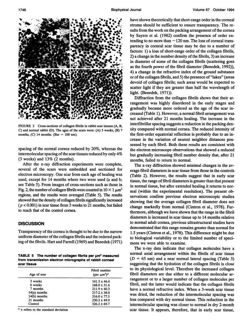
Abstract
Full-thickness corneal wounds (2 mm diameter) were produced in rabbits at the Schepens Eye Research Institute, Boston. These wounds were allowed to heal for periods ranging from 3 weeks to 21 months. The scar tissue was examined using low- and wide-angle x-ray diffraction from which average values were calculated for 1) the center-to-center collagen fibril spacing, 2) the fibril diameter, 3) the collagen axial periodicity D, and 4) the intermolecular spacing within the collagen fibrils. Selected samples were processed for transmission electron microscopy. The results showed that the average spacing between collagen fibrils within the healing tissue remained slightly elevated after 21 months and there was a small increase in the fibril diameter. The collagen D-periodicity was unchanged. There was a significant drop in the intermolecular spacing in the scar tissues up to 6 weeks, but thereafter the spacing returned to normal. The first-order equatorial reflection in the low-angle pattern was visible after 3 weeks and became sharper and more intense with time, suggesting that, as healing progressed, the number of nearest neighbor fibrils increased and the distribution of nearest neighbor spacings reduced. This corresponded to the fibrils becoming more ordered although, even after 21 months, normal packing was not achieved. Ultrastructural changes in collagen fibril density measured from electron micrographs were consistent with the increased order of fibril packing measured by x-ray diffraction. The results suggest that collagen molecules have a normal axial and lateral arrangement within the fibrils of scar tissue. The gradual reduction in the spread of interfibrillar spacings may be related to the progressive decrease in the light scattered from the tissue as the wound heals.
Full text
PDF


Images in this article

Selected References
These references are in PubMed. This may not be the complete list of references from this article.
- Borcherding M. S., Blacik L. J., Sittig R. A., Bizzell J. W., Breen M., Weinstein H. G. Proteoglycans and collagen fibre organization in human corneoscleral tissue. Exp Eye Res. 1975 Jul;21(1):59–70. doi: 10.1016/0014-4835(75)90057-3. [DOI] [PubMed] [Google Scholar]
- Cannon D. J., Cintron C. Collagen cross-linking in corneal scar formation. Biochim Biophys Acta. 1975 Nov 18;412(1):18–25. doi: 10.1016/0005-2795(75)90335-9. [DOI] [PubMed] [Google Scholar]
- Cintron C., Hassinger L. C., Kublin C. L., Cannon D. J. Biochemical and ultrastructural changes in collagen during corneal wound healing. J Ultrastruct Res. 1978 Oct;65(1):13–22. doi: 10.1016/s0022-5320(78)90017-5. [DOI] [PubMed] [Google Scholar]
- Cintron C., Hong B. S., Covington H. I., Macarak E. J. Heterogeneity of collagens in rabbit cornea: type III collagen. Invest Ophthalmol Vis Sci. 1988 May;29(5):767–775. [PubMed] [Google Scholar]
- Cintron C., Hong B. S., Kublin C. L. Quantitative analysis of collagen from normal developing corneas and corneal scars. Curr Eye Res. 1981;1(1):1–8. doi: 10.3109/02713688109019966. [DOI] [PubMed] [Google Scholar]
- Cintron C. Hydroxylysine glycosides in the collagen of normal and scarred rabbit corneas. Biochem Biophys Res Commun. 1974 Sep 9;60(1):288–294. doi: 10.1016/0006-291x(74)90203-4. [DOI] [PubMed] [Google Scholar]
- Cintron C., Kublin C. L. Regeneration of corneal tissue. Dev Biol. 1977 Dec;61(2):346–357. doi: 10.1016/0012-1606(77)90304-9. [DOI] [PubMed] [Google Scholar]
- Cintron C., Schneider H., Kublin C. Corneal scar formation. Exp Eye Res. 1973 Nov 11;17(3):251–259. doi: 10.1016/0014-4835(73)90176-0. [DOI] [PubMed] [Google Scholar]
- Fullwood N. J., Tuft S. J., Malik N. S., Meek K. M., Ridgway A. E., Harrison R. J. Synchrotron x-ray diffraction studies of keratoconus corneal stroma. Invest Ophthalmol Vis Sci. 1992 Apr;33(5):1734–1741. [PubMed] [Google Scholar]
- Goodfellow J. M., Elliott G. F., Woolgar A. E. X-ray diffraction studies of the corneal stroma. J Mol Biol. 1978 Feb 25;119(2):237–252. doi: 10.1016/0022-2836(78)90436-9. [DOI] [PubMed] [Google Scholar]
- HEDBLOM E. E. The role of polysaccharides in corneal swelling. Exp Eye Res. 1961 Sep;1:81–91. doi: 10.1016/s0014-4835(61)80012-2. [DOI] [PubMed] [Google Scholar]
- Hart R. W., Farrell R. A. Light scattering in the cornea. J Opt Soc Am. 1969 Jun;59(6):766–774. doi: 10.1364/josa.59.000766. [DOI] [PubMed] [Google Scholar]
- Hassell J. R., Cintron C., Kublin C., Newsome D. A. Proteoglycan changes during restoration of transparency in corneal scars. Arch Biochem Biophys. 1983 Apr 15;222(2):362–369. doi: 10.1016/0003-9861(83)90532-5. [DOI] [PubMed] [Google Scholar]
- Linsenmayer T. F., Gibney E., Igoe F., Gordon M. K., Fitch J. M., Fessler L. I., Birk D. E. Type V collagen: molecular structure and fibrillar organization of the chicken alpha 1(V) NH2-terminal domain, a putative regulator of corneal fibrillogenesis. J Cell Biol. 1993 Jun;121(5):1181–1189. doi: 10.1083/jcb.121.5.1181. [DOI] [PMC free article] [PubMed] [Google Scholar]
- MAURICE D. M. The structure and transparency of the cornea. J Physiol. 1957 Apr 30;136(2):263–286. doi: 10.1113/jphysiol.1957.sp005758. [DOI] [PMC free article] [PubMed] [Google Scholar]
- Marchini M., Morocutti M., Ruggeri A., Koch M. H., Bigi A., Roveri N. Differences in the fibril structure of corneal and tendon collagen. An electron microscopy and X-ray diffraction investigation. Connect Tissue Res. 1986;15(4):269–281. doi: 10.3109/03008208609001985. [DOI] [PubMed] [Google Scholar]
- Maroudas A., Wachtel E., Grushko G., Katz E. P., Weinberg P. The effect of osmotic and mechanical pressures on water partitioning in articular cartilage. Biochim Biophys Acta. 1991 Mar 4;1073(2):285–294. doi: 10.1016/0304-4165(91)90133-2. [DOI] [PubMed] [Google Scholar]
- Meek K. M., Elliott G. F., Sayers Z., Whitburn S. B., Koch M. H. Interpretation of the meridional x-ray diffraction pattern from collagen fibrils in corneal stroma. J Mol Biol. 1981 Jul 5;149(3):477–488. doi: 10.1016/0022-2836(81)90482-4. [DOI] [PubMed] [Google Scholar]
- Meek K. M., Fullwood N. J., Cooke P. H., Elliott G. F., Maurice D. M., Quantock A. J., Wall R. S., Worthington C. R. Synchrotron x-ray diffraction studies of the cornea, with implications for stromal hydration. Biophys J. 1991 Aug;60(2):467–474. doi: 10.1016/S0006-3495(91)82073-2. [DOI] [PMC free article] [PubMed] [Google Scholar]
- Meek K. M., Leonard D. W. Ultrastructure of the corneal stroma: a comparative study. Biophys J. 1993 Jan;64(1):273–280. doi: 10.1016/S0006-3495(93)81364-X. [DOI] [PMC free article] [PubMed] [Google Scholar]
- Oh S. P., Griffith C. M., Hay E. D., Olsen B. R. Tissue-specific expression of type XII collagen during mouse embryonic development. Dev Dyn. 1993 Jan;196(1):37–46. doi: 10.1002/aja.1001960105. [DOI] [PubMed] [Google Scholar]
- Quantock A. J., Meek K. M., Thonar E. J. Analysis of high-angle synchrotron x-ray diffraction patterns obtained from macular dystrophy corneas. Cornea. 1992 May;11(3):185–190. [PubMed] [Google Scholar]
- Sayers Z., Koch M. H., Whitburn S. B., Meek K. M., Elliott G. F., Harmsen A. Synchrotron x-ray diffraction study of corneal stroma. J Mol Biol. 1982 Oct 5;160(4):593–607. doi: 10.1016/0022-2836(82)90317-5. [DOI] [PubMed] [Google Scholar]
- Scott J. E. Proteoglycan histochemistry--a valuable tool for connective tissue biochemists. Coll Relat Res. 1985 Dec;5(6):541–575. doi: 10.1016/s0174-173x(85)80008-x. [DOI] [PubMed] [Google Scholar]
- Scott J. E. The periphery of the developing collagen fibril. Quantitative relationships with dermatan sulphate and other surface-associated species. Biochem J. 1984 Feb 15;218(1):229–233. doi: 10.1042/bj2180229. [DOI] [PMC free article] [PubMed] [Google Scholar]
- Takahashi T., Cho H. I., Kublin C. L., Cintron C. Keratan sulfate and dermatan sulfate proteoglycans associate with type VI collagen in fetal rabbit cornea. J Histochem Cytochem. 1993 Oct;41(10):1447–1457. doi: 10.1177/41.10.8245404. [DOI] [PubMed] [Google Scholar]