Abstract
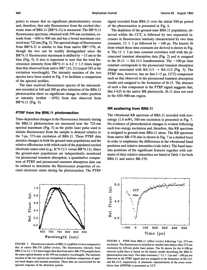
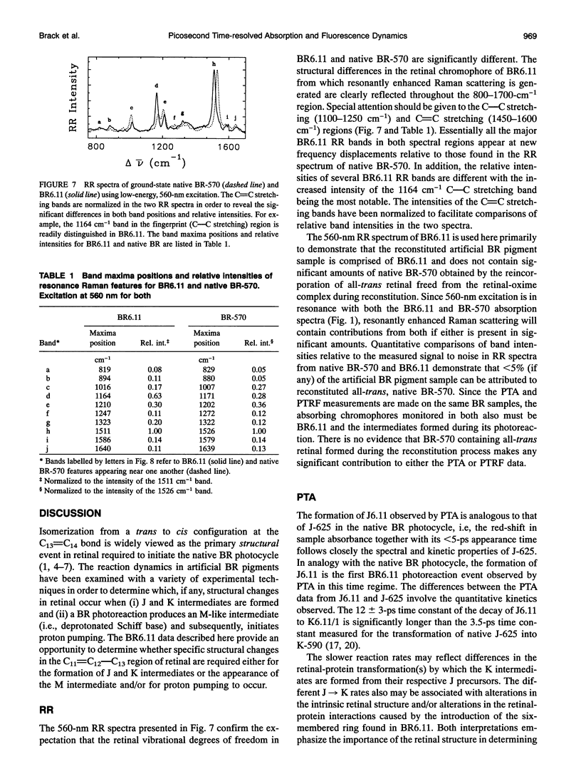
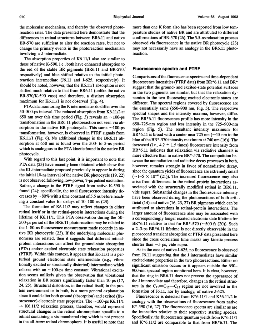
The picosecond molecular dynamics in an artificial bacteriorhodopsin (BR) pigment containing a structurally modified all-trans retinal chromphore with a six-membered ring bridging the C11=C12-C13 positions (BR6.11) are measured by picosecond transient absorption and picosecond time-resolved fluorescence spectroscopy. Time-dependent intensity and spectral changes in absorption in the 570-650-nm region are monitored for delays as long as 5 ns after the 7-ps, 573-nm excitation of BR6.11. Two intermediates, J6.11 and K6.11/1, both with enhanced absorption to the red (> 600 nm) of the BR6.11 spectrum are observed within approximately 50 ps. The J6.11 intermediate decays with a time constant of 12 +/- 3 ps to form K6.11/1. The K6.11/1 intermediate decays with an approximately 100-ps time constant to form a third intermediate, K6.11/2, which is observed through diminished 650-nm absorption (relative to that of K6.11/1). No other transient absorption changes are found during the remainder of the initial 5-ns period of the BR6.11 photoreaction. Fluorescence in the 650-900-nm region is observed from BR6.11, K6.11/1, and K6.11/2, but no emission assignable to J6.11 is found. The BR6.11 fluroescence spectrum has a approximately 725-nm maximum which is blue-shifted by approximately 15 nm relative to that of native BR-570 and is 4.2 +/- 1.5 times larger in intensity (same sample optical density). No differences in the profile of the fluorescence spectra of BR6.11 and the intermediates K6.11/1 and K6.11/2 are observed. Following ground-state depletion of the BR6.11 population, the time-resolved fluroescence intensity monitored at 725 nm increases with two time constants, 12 +/- 3 and approximately 100 ps, both of which correlate well with changes in the picosecond transient absorption data. The resonance Raman spectrum of ground-state BR6.11, measured with low-energy, 560-nm excitation, is significantly different from the spectrum of native BR-570, thus confirming that the picosecond transient absorption and picosecond time resolved fluorescence data are assignable to BR6.11 and its photoreaction alone and not to BR-570 reformed during there constitution process (<5% of the BR6.11 sample could be attributed to native BR-570).The J6.11 and K6.11 absorption and fluorescence data presented here are generally analogous to those measured for native J-625 and K-590, respectively, and therefore, the primary events in the BR6.11 photoreaction can be correlated with those in the native BR photocycle. The BR6.11 photoreaction, however, exhibits important differences including slower formation rates for J and K intermediates as well as the presence of a second K intermediate. These results demonstrate that the restricted motion in the C11=C12-C13 region of retinal found in BR6.11 does not greatly change the overall photoreaction mechanism,but does alter the rates at which processes occur.
Full text
PDF
Selected References
These references are in PubMed. This may not be the complete list of references from this article.
- Atkinson G. H., Blanchard D., Lemaire H., Brack T. L., Hayashi H. Picosecond time-resolved fluorescence spectroscopy of K-590 in the bacteriorhodopsin photocycle. Biophys J. 1989 Feb;55(2):263–274. doi: 10.1016/S0006-3495(89)82801-2. [DOI] [PMC free article] [PubMed] [Google Scholar]
- Birge R. R. Nature of the primary photochemical events in rhodopsin and bacteriorhodopsin. Biochim Biophys Acta. 1990 Apr 26;1016(3):293–327. doi: 10.1016/0005-2728(90)90163-x. [DOI] [PubMed] [Google Scholar]
- Birge R. R. Photophysics and molecular electronic applications of the rhodopsins. Annu Rev Phys Chem. 1990;41:683–733. doi: 10.1146/annurev.pc.41.100190.003343. [DOI] [PubMed] [Google Scholar]
- Harbison G. S., Smith S. O., Pardoen J. A., Courtin J. M., Lugtenburg J., Herzfeld J., Mathies R. A., Griffin R. G. Solid-state 13C NMR detection of a perturbed 6-s-trans chromophore in bacteriorhodopsin. Biochemistry. 1985 Nov 19;24(24):6955–6962. doi: 10.1021/bi00345a031. [DOI] [PubMed] [Google Scholar]
- Lewis A., Spoonhower J. P., Perreault G. J. Observation of light emission from a rhodopsin. Nature. 1976 Apr 22;260(5553):675–678. doi: 10.1038/260675a0. [DOI] [PubMed] [Google Scholar]
- Mathies R. A., Lin S. W., Ames J. B., Pollard W. T. From femtoseconds to biology: mechanism of bacteriorhodopsin's light-driven proton pump. Annu Rev Biophys Biophys Chem. 1991;20:491–518. doi: 10.1146/annurev.bb.20.060191.002423. [DOI] [PubMed] [Google Scholar]
- Milder S. J., Kliger D. S. A time-resolved spectral study of the K and KL intermediates of bacteriorhodopsin. Biophys J. 1988 Mar;53(3):465–468. doi: 10.1016/S0006-3495(88)83124-2. [DOI] [PMC free article] [PubMed] [Google Scholar]
- Oesterhelt D., Bräuchle C., Hampp N. Bacteriorhodopsin: a biological material for information processing. Q Rev Biophys. 1991 Nov;24(4):425–478. doi: 10.1017/s0033583500003863. [DOI] [PubMed] [Google Scholar]
- Ottolenghi M., Sheves M. Synthetic retinals as probes for the binding site and photoreactions in rhodopsins. J Membr Biol. 1989 Dec;112(3):193–212. doi: 10.1007/BF01870951. [DOI] [PubMed] [Google Scholar]
- Polland H. J., Franz M. A., Zinth W., Kaiser W., Kölling E., Oesterhelt D. Early picosecond events in the photocycle of bacteriorhodopsin. Biophys J. 1986 Mar;49(3):651–662. doi: 10.1016/S0006-3495(86)83692-X. [DOI] [PMC free article] [PubMed] [Google Scholar]


