Abstract
The retroviral Gag polyprotein directs budding from the plasma membrane of infected cells. Until now, it was believed that Gag proteins of type C retroviruses, including the prototypic oncoretrovirus Rous sarcoma virus, were synthesized on cytosolic ribosomes and targeted directly to the plasma membrane. Here we reveal a previously unknown step in the subcellular trafficking of the Gag protein, that of transient nuclear localization. We have identified a targeting signal within the N-terminal matrix domain that facilitates active nuclear import of the Gag polyprotein. We also found that Gag is transported out of the nucleus through the CRM1 nuclear export pathway, based on observations that treatment of virus-expressing cells with leptomycin B resulted in the redistribution of Gag proteins from the cytoplasm to the nucleus. Internal deletion of the C-terminal portion of the Gag p10 region resulted in the nuclear sequestration of Gag and markedly diminished budding, suggesting that the nuclear export signal might reside within p10. Finally, we observed that a previously described matrix mutant, Myr1E, was insensitive to the effects of leptomycin B, apparently bypassing the nuclear compartment during virus assembly. Myr1E has a defect in genomic RNA packaging, implying that nuclear localization of Gag might be involved in viral RNA interactions. Taken together, these findings provide evidence that nuclear entry and egress of the Gag polyprotein are intrinsic components of the Rous sarcoma virus assembly pathway.
Retroviruses must gain access to the nucleus to replicate. After receptor binding, entry, and reverse transcription, the integration-competent nucleoprotein complex (called the preintegration complex or PIC) enters the nucleus. For oncoretroviruses like Rous sarcoma virus (RSV) that primarily infect dividing cells, the PIC awaits breakdown of the nuclear envelope during mitosis for nuclear entry. Lentiviruses including HIV-1 infect nondividing cells, and PICs are transported through intact nuclear envelopes. HIV-1 nuclear entry is complex, and redundant signals have been identified in the viral matrix (MA), integrase, and Vpr proteins (reviewed in ref. 1). The recent report that RSV can replicate at low levels in quiescent cells does raise the possibility that a viral protein might mediate active nuclear targeting of the RSV PIC (2).
After nuclear entry of the PIC and proviral integration, viral RNA is transcribed, and unspliced genome-length viral mRNAs must exit the nucleus for translation into viral structural proteins and encapsidation into virions. The nuclear export of intron-containing mRNAs is normally inhibited by cellular mechanisms, so retroviruses must circumvent this obstacle. Lentiviruses encode trans-acting factors such as the HIV-1 Rev protein to mediate nuclear export of intron-containing viral RNAs (3). Oncoretroviruses including RSV lack Rev-like transport factors and instead have cis-acting constitutive transport elements to facilitate the export of unspliced viral RNA (4).
The regulation of nuclear transport is mediated by components of the nuclear pore complex (NPC) and the superfamily of nuclear transport receptors called importins. Although passive diffusion of small molecules can occur through NPCs, nucleocytoplasmic transport of nearly all proteins and RNAs is mediated by an active mechanism with directionality provided by the Ran-GTPase system (reviewed in refs. 3 and 5). Facilitated nuclear import of cytosolic proteins relies on specific interactions between importins and nuclear localization signals (NLSs). Classical examples of NLSs are the highly basic motifs of simian virus 40 T antigen and nucleoplasmin, although many additional nonclassical sequences have been shown to function as NLSs (1, 5, 6). The nuclear export of proteins and RNAs is also a signal-dependent process mediated by soluble receptors called exportins (3, 5). The best-characterized nuclear export signals (NESs) were initially identified in the HIV-1 Rev protein (7) and the protein kinase A inhibitor (8). These leucine-rich NESs consist of short peptide sequences with four closely spaced hydrophobic residues, a motif recognized by the CRM1 nuclear export receptor (9, 10). Leptomycin B (LMB) attaches to the central domain of CRM1 to disrupt its interaction with the NES, making LMB a specific tool for studying CRM1-mediated nuclear export (9, 11).
After nuclear export of full-length viral mRNAs, Gag and Gag-Pol proteins are synthesized on free ribosomes. Initial Gag–Gag contacts occur between I (interaction) domains found within the NC region (12–14). RNA is essential early in assembly as scaffolding for the assembly of Gag multimers (15, 16). Gag–RNA interactions also mediate selective encapsidation of the viral genome, a noncovalently linked RNA dimer, through association of NC and the cis-acting packaging element Ψ (17, 18). Assembly intermediates are directed to the plasma membrane via the M (membrane-binding) domain in the N-terminal MA sequence of Gag (19, 20). Because the RSV M domain has suboptimal membrane-binding activity, I domains provide cooperative protein–protein interactions to stabilize membrane association (14). Final release of the virus particle is controlled by the late (L) domain (21, 22). After budding, the RSV Gag polyprotein precursor is proteolytically cleaved into the structural proteins MA, p2a, p2b, p10, CA, NC, and PR.
In this report, we describe the unexpected finding that the RSV Gag protein enters the nucleus via a nuclear-targeting sequence in the MA domain. We show that Gag is subsequently transported into the cytoplasm by using a CRM1-mediated nuclear export pathway. Elimination of this nuclear step correlates with a defect in RNA packaging. These findings demonstrate a previously unknown step in the assembly pathway of RSV.
Materials and Methods
Viruses, Cells, and Plasmids.
Proviral constructs were derived from pRCV8 containing the RSV Prague C gag gene of pATV8 (23, 24). Plasmids pRC.Myr1E, p.RC.Myr1E−, pRC.Myr2.HB12, pMA-green fluorescent protein (GFP) (25), pGag-GFP (26), and pSV.Myr0.BgBs (12) have been described. QT6 cells, chemically transformed quail fibroblasts, were maintained as described (27, 28).
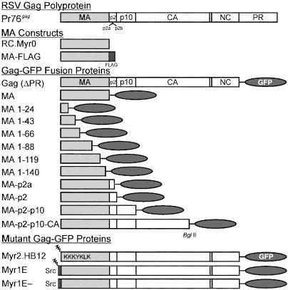
Plasmids encoding C-terminal truncations of the RSV Gag protein fused to GFP (Fig. 1) were made by PCR amplification of pRCV8 by using primer USP19.263 (25) and a set of downstream primers each containing an ApaI site, for cloning into SacI–ApaI sites of pEGFP.N2 (CLONTECH). MA truncations included amino acids 1–24, 1–43, 1–66, 1–88, 1–119, and 1–140. Gag truncations consisted of MA-p2a (amino acid 1–155), MA-p2 (amino acid 1–177), and MA-p2-p10 (amino acid 1–239). Deletions involving p10 were derived from pRC.Δp10.31, pRC.Δp10.52, and pRC.ΔQM1 [(29), kind gifts of Becky Craven and John Wills (Pennsylvania State University College of Medicine)] by PCR using primers USP19.263 and 5′-TCAGTATAGGGGCCCCGAGTCGGCAGGTGGCTCA.
Figure 1.
The RSV Gag protein and derivatives. The RSV Gag protein (Pr76) is depicted at the top, with cleavage proteins MA, p2a, p2b, p10, CA, NC, and PR indicated. RC.Myr0 is a derivative of pRCV8 and expresses the wild-type Prague C MA protein (45). Gag-GFP fusion proteins and truncations of MA fused to GFP are depicted below. Note that GFP replaces the PR sequence in Gag-GFP (ΔPR). Gag-GFP substitution mutants are shown in the bottom panel. The box labeled “Src” contains the N-terminal 10 residues of the Src oncoprotein. Myristic acid is shown as a zigzag line.
pMA-FLAG was made by ligating fragments from pMA-GFP (ApaI-Klenow/BglII) with pCMV.FLAG5b (Sigma) (EcoRV-Klenow/BglII). Mutations of the gag sequence (myr2.HB12, myr1e, and myr1e−) were cloned into pGag-GFP by SstI–BspEI fragment exchange.
The IKBα–GFP expression vector was a kind gift from Thomas Hope (University of Illinois, Chicago).
Confocal Microscopy.
QT6 cells were examined 18 h after transfection by using a Zeiss LSM 10 BioMed confocal microscope (25). In indicated experiments, cells were grown in medium augmented with LMB at a final concentration of 10 ng/ml (18 nM) for 2 h before imaging. For indirect immunofluorescence, cells were seeded in LabTek chamber slides (Nunc), transfected, and fixed in 2% paraformaldeyde or in 3:1 methanol:acetone at −20°C. Cells were rehydrated in PBS, blocked with 1.5% BSA, incubated with polyclonal anti-RSV or anti-MA (24, 27), washed, stained with goat anti-rabbit IgG-FITC (Sigma) or sheep anti-rabbit IgG-Cy3 (Sigma), mounted with SlowFade (Molecular Probes), and analyzed by confocal microscopy.
Viral Protein Detection and Budding Assay.
For immunoblotting, transfected cells were lysed in radioimmunoprecipitation assay buffer (50 mM Tris, pH 7.5/150 mM NaCl/1% Nonidet P-40/0.5% sodium deoxycholate/0.1% SDS), proteins were separated by electrophoresis, transferred to nitrocellulose, probed with anti-GFP Living Colors Peptide Ab (CLONTECH) and anti-mouse IgG-conjugated HRP (Sigma), and detected by chemiluminescence. Radioimmunoprecipitation assays were performed as described (30, 31). To calculate particle release, Gag protein bands were quantitated by using a PhosphoImager (Molecular Dynamics), and the amount of Gag protein in the medium was divided by the total Gag protein in lysates and medium. Statistical analysis was performed by using a two-sample t test.
Results
We have described mutants of the RSV MA sequence that interfere with viral RNA packaging and dimerization (24, 25). One such mutant, Myr1E (Fig. 1), contains the Src membrane-binding domain as an N-terminal extension of the Gag protein. Although Myr1E produces viral particles more efficiently than wild type, it has undetectable infectivity and packages only 40% of wild-type levels of viral RNA. The Myr1E.MA protein is mislocalized, demonstrating greatly enhanced plasma membrane accumulation compared to the wild-type MA protein (25). We hypothesized that the defect in genomic RNA incorporation results from transporting the Myr1E.Gag protein too quickly to the plasma membrane, omitting a cellular compartment or a trafficking pathway required for Gag–RNA interactions. This idea led us to further examine the subcellular localization of wild-type and mutant RSV MA and Gag proteins to dissect the targeting determinants within their sequences.
Subcellular Distribution of the MA Protein.
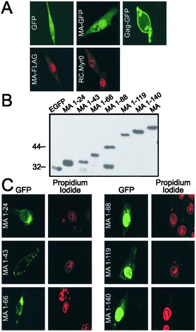
To study the subcellular localizations of MA and Gag in living cells, C-terminal fusions were made with GFP (Fig. 1) (25, 26). As analyzed by confocal microscopy, MA-GFP was observed throughout the cytosol with strong accumulation inside the nucleus (Fig. 2A) (25). In contrast, the distribution of unconjugated GFP was diffuse through the cell. To determine whether the presence of the foreign GFP protein altered the cellular distribution of MA, the FLAG epitope was fused to the C terminus of MA, and cells were examined by using indirect immunofluorescence with an anti-MA Ab. Similarly to MA-GFP, MA-FLAG also exhibited nuclear staining. Even a small sequence like FLAG could affect protein localization, so wild-type MA was expressed in the absence of other viral proteins by using a proviral vector, and cells were analyzed by indirect immunofluorescence. In these fixed cells, MA appeared within nuclei, confirming our previous results (Fig. 2A, RC.Myr0).
Figure 2.
Subcellular localization of the RSV MA protein. (A Upper) Subcellular localizations of GFP, MA-GFP, and Gag-GFP fusion proteins were analyzed in live cells using confocal microscopy 18 h after transfection. (Lower) Transfected QT6 cells were fixed and incubated with polyclonal anti-MA Ab and Cy3-conjugated secondary Ab. (B) Immunoblot analysis of intracellular expression levels of indicated proteins from lysates of transfected cells by using an anti-GFP Ab. Molecular weight markers are indicated to the left. (C) Mapping a nuclear-targeting sequence within the MA domain. Plasmids encoding truncations of the MA sequence fused to GFP were transfected and cells fixed, and epifluorescence was detected. The cells were stained with propidium iodide to visualize nuclear DNA, and identical fields are shown.
Because MA and MA-FLAG are small proteins, they could enter the nucleus by passive diffusion, because in theory proteins up to ≈40–60 kDa can diffuse through the NPC. In reality, few proteins actually traverse the NPC passively because the directional active transport system maintains strict compartmentalization (3, 5). However, even if MA and MA-FLAG entered by diffusion, they would be unlikely to accumulate in the nucleus under steady–state conditions. Furthermore, MA-GFP was concentrated in the nucleus despite its larger molecular weight (52 kDa) compared to GFP (33 kDa), which was distributed uniformly in the cell.
The nuclear concentration of MA-GFP is distinct from the localization of Gag-GFP, which excludes the nucleus and accumulates at focal patches along the plasma membrane (26) (Fig. 2A). A priori, we expected the MA protein to be localized at the plasma membrane given that MA and Gag share identical N-terminal sequences and therefore contain the same M domain. The unexpected difference in subcellular localization suggested that there must be additional targeting information located either within MA or Gag to account for their distinctive distributions. A conformational change resulting from cleavage of the Gag precursor during maturation might reveal new targeting information within MA, similar in principle to the myristyl-switch mechanism proposed to explain different conformations of the HIV-1 MA and Gag proteins (32–34). Alternatively, cleavage of the MA protein might remove downstream-targeting signals that are dominant over nuclear targeting in the context of the Gag polyprotein.
Nuclear Localization of MA Requires the Presence of the Four α-Helix Domain.
The localization of MA within the nucleus suggested the presence of an NLS, but sequences resembling a classical motif could not be identified by database searches (35, 36). However, MA is lysine- and arginine-rich, so perhaps the basic residues form an NLS in the three-dimensional conformation of the protein. To ascertain whether a simple nuclear-targeting sequence could be found, deletions were made based on the NMR structure of the N-terminal half of the MA protein, which consists of four overlapping α helices and a 310 helix joined by flexible loops (37). C-terminal truncations of MA were fused to GFP (Fig. 1). Immunoblot analysis revealed that each fusion protein was stably expressed, and there was no detectable free GFP except in the MA 1–88 construct (Fig. 2B). Examination by confocal microscopy revealed that addition of the first α helix (MA 1–24), the first two α helices (MA 1–43), or the first three α helices (MA 1–66) to GFP resulted in accumulation of the proteins in the cytoplasm (Fig. 2C). Fusion of the first 88 residues of MA (all four α helixes) to GFP restored nuclear concentration of the protein, and further extension into the second half of MA maintained nuclear localization (MA 1–119 and MA 1–140).
Identification of a CRM1-Dependent NES Within the Gag Sequence.
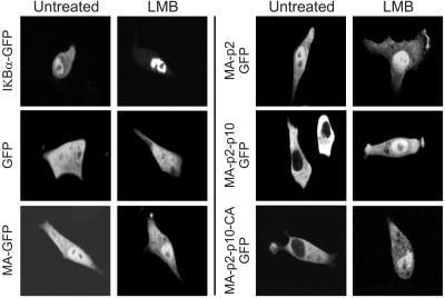
To identify the sequence required for the change from nuclear to plasma membrane localization, GFP fusion proteins including the p2a, p2 (p2a plus p2b), p10, and CA domains of Gag were examined (Fig. 1). MA-p2a-GFP (data not shown) and MA-p2-GFP fusion proteins displayed the same subcellular localization as MA-GFP with nuclear accumulation (Fig. 3). However, extending through the p10 and CA domains of Gag produced a dramatic alteration in localization: MA-p2-p10-GFP and MA-p2-p10-CA-GFP were present exclusively in the cytoplasm.
Figure 3.
Identification of an LMB-sensitive NES within the Gag protein. Live cells transfected with plasmids encoding C-terminal truncations of Gag fused to GFP were examined by confocal microscopy. LMB-treated cells were incubated with 10 ng/ml LMB for 2 h before imaging.
The nuclear exclusion of Gag-GFP fusion proteins including p10 and CA could be explained by either increased molecular weight, precluding passive nuclear entry, or by the presence of a nuclear export activity. To determine whether there is a CRM1-dependent NES in Gag, cells expressing GFP fusion proteins were treated with LMB (9, 11). As a control for LMB activity in QT6 cells, IκBα, a cellular protein containing a known NES (38), was expressed as a GFP fusion protein, and the minimal concentration of LMB (10 ng/ml) that blocked its nuclear export was used in subsequent experiments (Fig. 3). The distributions of GFP, MA-GFP, and MA-p2-GFP were unaffected by LMB. Strikingly, both GFP fusion proteins that contain p10 showed marked sensitivity to LMB, with retention of the MA-p2–p10-GFP and MA-p2-p10-CA-GFP proteins in the nucleus. Besides demonstrating the presence of a CRM1-dependent NES within the MA-p2-p10 region, this finding also confirmed that the nuclear-targeting activity within MA acts through an active process because it mediates nuclear entry of the ≈81 kDa MA-p2-p10-CA-GFP protein.
CRM1-dependent NESs are typically leucine-rich sequences, although other large hydrophobic amino acids may be substituted (39, 40). Two clusters of hydrophobic residues in p10 were potential candidates for an NES, as shown in Fig. 4A. To map the putative export signal, Gag-GFP mutants with deletions involving p10 were studied. Deletion of the N-terminal region of p10 did not alter the typical plasma membrane localization observed for Gag-GFP (ΔQM1, Fig. 4B). In contrast, an internal deletion that removes six hydrophobic residues was completely trapped in the nucleus, indicating that removal of this sequence interferes with nuclear export of Gag (Δp10.52). A smaller deletion that eliminates L219 and W222 from the C-terminal portion of p10 also abrogates nuclear export (Δp10.31). These results suggest that the NES is likely to be located in the second half of p10; although it remains possible that addition of the p10 sequence reveals a conformation-dependent NES within the MA or p2 sequences. Additionally, despite the existence of an LMB sensitive NES in this region of Gag, there might also be a cellular factor that mediates nuclear export of the protein.
Figure 4.
Effects of p10 deletions on subcellular localization. (A) Schematic diagram of the p10 sequence with hydrophobic residues that could function as an NES highlighted in red. Gag mutants with p10 deletions are shown below with heavy black lines indicating residues present in the mutant protein. (B) Confocal micrographs of cells expressing Gag-GFP p10 mutants reveal a putative NES. At 18 h after transfection, cells were fixed in paraformaldehyde. (Left) GFP epifluorescence. (Right) Propidium iodide-stained nuclei.
LMB Sensitivity of Gag and Mutants with Altered MA Sequences.
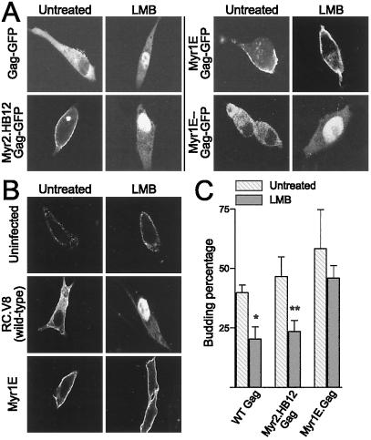
We next examined the effect of LMB on localization of the full-length Gag protein because previous constructs extended only through CA. We found that Gag was trapped in the nucleus after treatment with LMB, indicating that the trafficking pathway of Gag includes a nuclear phase (Fig. 5A). We reasoned that Gag mutants with strong membrane-binding domains might be targeted so rapidly to the plasma membrane that they fail to enter the nucleus. For example, Myr1E (Fig. 1) has the Src membrane-binding domain extended from the N terminus of Gag and efficiently associates with the plasma membrane, even without the contribution of I domains (25). In the presence of LMB, Myr1E.Gag remained localized to the plasma membrane with no evidence of nuclear retention (Fig. 5A). This is intriguing because Myr1E is noninfectious because of a defect in genomic RNA packaging and dimerization (24). In Myr1E, the Src membrane-targeting activity might override the NLS/NES activities normally found in Gag, bypassing the nuclear compartment. Thus, elimination of nuclear transport might explain the RNA packaging defect of Myr1E if nuclear localization of Gag is involved in genomic RNA incorporation. In support of this idea, we found that Myr1E−, which is identical to Myr1E except for a G2A substitution that eliminates Src myristylation, demonstrated nuclear localization in the presence of LMB. Myr1E− has normal infectivity and packages wild-type levels of RNA (24). A third mutant, Myr2.HB12, also remained in the nucleus with LMB treatment, and it packages nearly normal levels of genomic RNA (86% of wild-type levels, data not shown), although it is noninfectious. This mutant contains a myristic acid addition site at the Gag N terminus (designated Myr2) and a cluster of basic residues substituted for amino acids 12–18 in MA (25). Interestingly, Myr2.HB12 packages only monomers of RNA (25), suggesting that packaging and dimerization of viral RNA are not directly linked, and dimer formation might occur in a postnuclear compartment. Thus, there is a correlation between the nuclear localization of Gag proteins in response to LMB treatment and genomic RNA incorporation.
Figure 5.
LMB sensitivity of mutant Gag proteins with defects in RNA packaging and dimerization. (A) Live cells expressing the indicated Gag-GFP proteins were visualized without (Left) and with (Right) LMB treatment, revealing that wild-type Gag and mutants Myr2.HB12 and Myr1E− become trapped in the nucleus after treatment whereas Myr1E is insensitive to the effects of the drug. (B) Gag localization was analyzed in cells stably expressing wild-type or mutant RSV proviral genomes by indirect immunofluorescence in fixed cells either untreated or treated with LMB. Note that patterns of subcellular localization were the same as in A, indicating that GFP had not influenced the distribution of Gag. (C) Particle release in response to LMB treatment. The amount of immunoprecipitated Gag protein released from transfected cells during a 2.5-h-labeling period was divided by the total Gag protein detected in cells and medium to calculate budding percentage. Budding percentages from untreated (hatched bars) and treated (solid gray bars) cells were compared. Treated cells were incubated with LMB for 1–2 h followed by metabolic radiolabeling for 2.5 h in the presence of LMB. Each bar represents the average of three independent experiments with SDs indicated. Inhibition of particle assembly with LMB treatment was determined by a two-sample t test; *, P = 0.0049 and **, P = 0.014.
Effect of LMB Treatment on Gag Localization in Virus-Expressing Cells.
The subcellular trafficking of Gag proteins and their derivatives in response to LMB treatment was tested in cells expressing wild-type or mutant proviruses to determine whether GFP might have influenced localization. In cells expressing the wild-type RSV genome, Gag proteins were seen in the cytoplasm with focal concentration at the plasma membrane when analyzed by indirect immunofluorescence (Fig. 5B). When cells were incubated with LMB, Gag proteins became sequestered in the nucleus. Thus, during infection Gag undergoes nuclear entry and export independently of GFP and localization is not altered by the expression of other viral gene products. In cells expressing the myr1e genome, Myr1E. Gag was seen at the plasma membrane both in untreated and treated cells, indicating that the nuclear compartment was bypassed.
Particle Assembly in the Presence of LMB.
Because LMB treatment of infected cells resulted in nuclear accumulation of Gag proteins, we expected that fewer Gag molecules would be available for transport to the plasma membrane, and virus budding might be diminished. Cells were transfected with wild-type or mutant Gag-GFP constructs, pretreated with LMB for 1–2 h, metabolically labeled for 2.5 h in the presence of LMB, and RSV Gag proteins were immunoprecipitated from cell lysates and medium. Budding directed by Gag-GFP was significantly inhibited in the presence of LMB (P = 0.0049; mean of 49% reduction, 95% confidence interval 26.8–77.4; Fig. 5C). Myr2.HB12.Gag-GFP, which showed LMB-dependent nuclear localization, also had a significant reduction in extracellular particle release (P = 0.014; mean of 49% reduction, 95% confidence interval 17.7–83.4). In contrast, budding of Myr1E.Gag-GFP was not significantly altered with LMB treatment (P = 0.28).
Analysis of budding for the p10 deletion mutants revealed that ΔQM1.Gag-GFP, which had a subcellular localization similar to wild-type Gag, showed a trend toward reduction in budding because of LMB, but the effect was not statistically significant (P = 0.097, 45% reduction; data not shown). Mutants with deletions affecting hydrophobic residues in the C-terminal portion of p10 released particles at a much lower rate that wild-type (Δp10.31.Gag-GFP, 69% reduced and Δp10.52.Gag-GFP, 64% reduced, data not shown). Budding efficiency was not further diminished with LMB treatment (P = 0.22 and 0.43, respectively). Taken together, these results indicate that inhibition of nuclear export, either pharmacologically or by mutagenesis of p10, limits the amount of Gag in the cytoplasm thereby interfering with the normal trafficking pathway required for particle assembly.
Discussion
In this report, we describe the discovery of an unexpected step in the RSV assembly pathway. Previously, Gag proteins were believed to be located exclusively within the cytoplasm after synthesis, with subsequent targeting directly to the plasma membrane. Now we demonstrate that RSV Gag has a nuclear phase that is mediated by two targeting signals: one for nuclear import and the other for CRM1-dependent nuclear export.
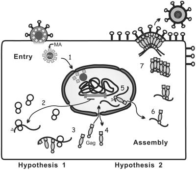
Although the reason for the transient nuclear localization of the RSV Gag protein is unknown, we propose two main hypotheses (Fig. 6). First, we suggest that the NES in Gag counteracts the nuclear import signal in MA to keep Gag out of the nucleus. In this scenario, RSV MA plays a role early in infection to facilitate nuclear entry of the viral PIC. Although a requirement for facilitated nuclear targeting of the PIC has not been demonstrated for RSV, the report that RSV can infect growth-arrested cells makes it possible that the nuclear-targeting sequence in MA might be involved (2). Because the nuclear import signal in MA also is present on the Gag polyprotein, Gag, too, would be transported into the nucleus but this would certainly be detrimental to virus assembly. To counteract nuclear import, the NES would return Gag to the cytoplasm for virus particle production. Besides import of the PIC, other roles could be imagined for the MA protein in the nucleus, including regulation of reverse transcription, integration, or viral transcription; however, mutants of MA that affect these processes have not yet been found. Whatever the function of MA might be in the nucleus, clearly its N-terminal α helical domain is sufficient for nuclear import of the MA and Gag proteins. The MA nuclear-targeting signal is large and complex compared to classical NLSs, and further experiments may reveal specific residues critical for interaction with nuclear import machinery or with a host factor that mediates the nuclear entry of MA and Gag.
Figure 6.
Model for the role of the nuclear localization of Gag during RSV replication. Hypothesis 1 (steps 1–4): The nuclear-targeting signal within MA delivers the cleaved MA protein to the nucleus during viral entry. Nuclear Gag proteins are exported via the CRM1-dependent NES so they are available in the cytoplasm to direct particle assembly. Hypothesis 2: (steps 5–7) The nuclear-targeting signal carries the Gag protein into the nucleus where Gag might interact with unspliced viral RNA to begin the packaging process. Cytoplasmic relocalization of Gag occurs via the NES; Gag–RNA complexes provide a nucleation point for Gag multimerization, and assembly intermediates are transported to the plasma membrane where budding occurs. Notably, hypotheses 1 and 2 are not mutually exclusive.
In the second model, we propose that Gag enters and exits the nucleus to fulfill a role during virion assembly (Fig. 6). After proviral integration, unspliced viral RNA transcripts are synthesized and exported out of the nucleus for translation into structural and enzymatic proteins. Export of unspliced viral transcripts must elude cellular mechanisms designed to retain intron-containing mRNA in the nucleus. For RSV, it was shown that cytoplasmic accumulation of unspliced viral RNA is mediated by cis-acting DR elements in a CRM1-independent fashion, although neither Gag proteins nor the RSV genome was present in those experiments (41, 42). It is possible that the fraction of unspliced RNA exported for translation is distinct from the fraction used for genome encapsidation. In our model, Gag proteins enter the nucleus where they might interact with unspliced viral RNA transcripts. The Gag-RNA nucleoprotein complex is then transported through the NPC via the CRM1 export pathway. In the cytoplasm, the Gag-RNA complex would be a nucleation point for the multimerization of additional Gag proteins that are then targeted to the plasma membrane. Importantly, these two models are not mutually exclusive, as MA might have a nuclear role early in infection and Gag might also enter the nucleus during assembly. It is also possible that Gag might have some other as yet unknown function in the nucleus unrelated to genomic RNA encapsidation.
Although the nuclear entry of oncoviral Gag proteins is unique, other viruses have well-described replication strategies that take advantage of nuclear machinery to transport viral genomes, structural proteins and regulatory proteins (reviewed in ref. 1). For example, HIV-1 uses multiple signals to deliver the PIC to the nucleus of nondividing cells (1), and a CRM1-dependent NES in MA influences the nuclear export of full-length viral RNAs (43). Alteration of the NES results in accumulation of HIV-1 Gag in the nucleus, although Gag itself was not shown to be sensitive to LMB treatment. Whether there are functional correlates between nuclear transport signals in RSV and HIV-1 Gag remains to be determined. As well, it is intriguing to consider possible parallels between the RSV Gag and influenza M1 proteins because both proteins are present in the nucleus and direct particle assembly at the plasma membrane. Influenza M1 associates with viral ribonucleoprotein complexes in the nucleus, and the viral genome is exported in a CRM1-dependent fashion (1, 44).
In this report, we demonstrated that the MA domain is involved in import of the Gag polyprotein into the nucleus and that an NES in Gag mediates its cytoplasmic relocalization to facilitate virus assembly. We found that Gag mutants that package normal amounts of genomic RNA also have nuclear entry and export phases (Myr2.HB12 and Myr1E−) whereas a mutant with a reduced level of viral RNA incorporation does not enter the nucleus (Myr1E). The observation that RSV replication includes transient nuclear localization of the Gag protein raises intriguing questions about early Gag–RNA interactions and the intracellular trafficking pathways taken by assembling virions.
Acknowledgments
We thank Becky Craven, Tina Cairns, Carol Wilson, and John Wills for reagents and for sharing unpublished results, and John Wills for thoughtful review of the manuscript. Tom Hope generously provided plasmids and helpful advice. We thank Dr. Vernon Chincilli (Pennsylvania State University College of Medicine) for help with statistical analysis. This work was supported by a grant from the National Institutes of Health to L.J.P (R01 CA76534) and a Graduate Student Fellowship from the National Science Foundation to L.Z.S.
Abbreviations
- RSV
Rous sarcoma virus
- LMB
leptomycin B
- PIC
preintegration complex
- NPC
nuclear pore complex
- NLS
nuclear localization signal
- NES
nuclear export signal
- GFP
green fluorescent protein
- MA
matrix
Footnotes
This paper was submitted directly (Track II) to the PNAS office.
References
- 1.Whittaker G R, Helenius A. Virology. 1998;246:1–23. doi: 10.1006/viro.1998.9165. [DOI] [PubMed] [Google Scholar]
- 2.Hatzoiioannou T G, Goff S P. J Virol. 2001;75:9526–9531. doi: 10.1128/JVI.75.19.9526-9531.2001. [DOI] [PMC free article] [PubMed] [Google Scholar]
- 3.Cullen B R. Mol Cell Biol. 2000;20:4181–4187. doi: 10.1128/mcb.20.12.4181-4187.2000. [DOI] [PMC free article] [PubMed] [Google Scholar]
- 4.Ogert R A, Beemon K L. J Virol. 1998;72:3407–3411. doi: 10.1128/jvi.72.4.3407-3411.1998. [DOI] [PMC free article] [PubMed] [Google Scholar]
- 5.Mattaj I W, Englmeier L. Annu Rev Biochem. 1998;67:265–306. doi: 10.1146/annurev.biochem.67.1.265. [DOI] [PubMed] [Google Scholar]
- 6.Christophe D, Christophe-Hobertus C, Pichon B. Cell Signalling. 2000;12:337–341. doi: 10.1016/s0898-6568(00)00077-2. [DOI] [PubMed] [Google Scholar]
- 7.Fischer U, Huber J, Boelens W C, Mattaj I M, Luhrmann R. Cell. 1995;82:475–483. doi: 10.1016/0092-8674(95)90436-0. [DOI] [PubMed] [Google Scholar]
- 8.Wen W, Meinkoth J L, Tsien R Y, Taylor S S. Cell. 1995;82:463–473. doi: 10.1016/0092-8674(95)90435-2. [DOI] [PubMed] [Google Scholar]
- 9.Fukuda M, Asano S, Nakamura T, Adachi M, Yoshida M, Yanagida M, Nishida E. Nature (London) 1997;390:308–311. doi: 10.1038/36894. [DOI] [PubMed] [Google Scholar]
- 10.Fornerod M, Ohno M, Yoshida M, Mattaj I W. Cell. 1997;90:1051–1060. doi: 10.1016/s0092-8674(00)80371-2. [DOI] [PubMed] [Google Scholar]
- 11.Kudo N, Matsumori N, Taoka H, Fujiwara D, Schreiner E P, Wolff B, Yoshida M, Horinouchi S. Proc Natl Acad Sci USA. 1999;96:9112–9117. doi: 10.1073/pnas.96.16.9112. [DOI] [PMC free article] [PubMed] [Google Scholar]
- 12.Weldon R A, Wills J W. J Virol. 1993;67:5550–5561. doi: 10.1128/jvi.67.9.5550-5561.1993. [DOI] [PMC free article] [PubMed] [Google Scholar]
- 13.Bowzard J B, Bennett R P, Krishna N K, Ernst S M, Rein A, Wills J W. J Virol. 1998;72:9034–9044. doi: 10.1128/jvi.72.11.9034-9044.1998. [DOI] [PMC free article] [PubMed] [Google Scholar]
- 14.Swanstrom R, Wills J W. In: Retroviruses. Coffin J M, Hughes S H, Varmus H E, editors. Plainview, NY: Cold Spring Harbor Lab. Press; 1997. pp. 263–334. [Google Scholar]
- 15.Campbell S, Vogt V M. J Virol. 1995;69:6487–6497. doi: 10.1128/jvi.69.10.6487-6497.1995. [DOI] [PMC free article] [PubMed] [Google Scholar]
- 16.Muriaux D, Mirro J, Harvin D, Rein A. Proc Natl Acad Sci USA. 2001;98:5246–5251. doi: 10.1073/pnas.091000398. [DOI] [PMC free article] [PubMed] [Google Scholar]
- 17.Shields A, Witte W N, Rothenberg E, Baltimore D. Cell. 1978;14:601–609. doi: 10.1016/0092-8674(78)90245-3. [DOI] [PubMed] [Google Scholar]
- 18.Vogt V M. In: Retroviruses. Coffin J M, Hugher S H, Varmus H E, editors. Plainview, NY: Cold Spring Harbor Lab. Press; 1997. pp. 27–69. [Google Scholar]
- 19.Nelle T D, Wills J W. J Virol. 1996;70:2269–2276. doi: 10.1128/jvi.70.4.2269-2276.1996. [DOI] [PMC free article] [PubMed] [Google Scholar]
- 20.Verderame M F, Nelle T D, Wills J W. J Virol. 1996;70:2664–2668. doi: 10.1128/jvi.70.4.2664-2668.1996. [DOI] [PMC free article] [PubMed] [Google Scholar]
- 21.Wills J W, Cameron C E, Wilson C B, Xiang Y, Bennett R P, Leis J. J Virol. 1994;68:6605–6618. doi: 10.1128/jvi.68.10.6605-6618.1994. [DOI] [PMC free article] [PubMed] [Google Scholar]
- 22.Parent L J, Bennett R P, Craven R C, Nelle T D, Krishna N K, Bowzard J B, Wilson C B, Puffer B A, Montelaro R C, Wills J W. J Virol. 1995;69:5455–5460. doi: 10.1128/jvi.69.9.5455-5460.1995. [DOI] [PMC free article] [PubMed] [Google Scholar]
- 23.Craven R C, Leure-duPree A E, Erdie C R, Wilson C B, Wills J W. J Virol. 1993;67:6246–6252. doi: 10.1128/jvi.67.10.6246-6252.1993. [DOI] [PMC free article] [PubMed] [Google Scholar]
- 24.Parent L J, Cairns T M, Albert J A, Wilson C B, Wills J W, Craven R C. J Virol. 2000;74:164–172. doi: 10.1128/jvi.74.1.164-172.2000. [DOI] [PMC free article] [PubMed] [Google Scholar]
- 25.Garbitt R A, Albert J A, Kessler M D, Parent L J. J Virol. 2001;75:260–268. doi: 10.1128/JVI.75.1.260-268.2001. [DOI] [PMC free article] [PubMed] [Google Scholar]
- 26.Callahan E M, Wills J W. J Virol. 2000;74:11222–11229. doi: 10.1128/jvi.74.23.11222-11229.2000. [DOI] [PMC free article] [PubMed] [Google Scholar]
- 27.Craven R C, Leure-duPree A E, Weldon R A, Jr, Wills J W. J Virol. 1995;69:4213–4227. doi: 10.1128/jvi.69.7.4213-4227.1995. [DOI] [PMC free article] [PubMed] [Google Scholar]
- 28.Moscovici C, Moscovici M G, Jimenez H, Lai M M C, Hayman M J, Vogt P K. Cell. 1977;11:95–103. doi: 10.1016/0092-8674(77)90320-8. [DOI] [PubMed] [Google Scholar]
- 29.Krishna N K, Campbell S, Vogt V M, Wills J W. J Virol. 1998;72:564–577. doi: 10.1128/jvi.72.1.564-577.1998. [DOI] [PMC free article] [PubMed] [Google Scholar]
- 30.Parent L J, Wilson C B, Resh M D, Wills J W. J Virol. 1996;70:1016–1026. doi: 10.1128/jvi.70.2.1016-1026.1996. [DOI] [PMC free article] [PubMed] [Google Scholar]
- 31.Weldon R A, Erdie C R, Oliver M G, Wills J W. J Virol. 1990;64:4169–4179. doi: 10.1128/jvi.64.9.4169-4179.1990. [DOI] [PMC free article] [PubMed] [Google Scholar]
- 32.Hermida-Matsumoto L, Resh M D. J Virol. 1999;73:1902–1908. doi: 10.1128/jvi.73.3.1902-1908.1999. [DOI] [PMC free article] [PubMed] [Google Scholar]
- 33.Paillart J C, Gottlinger H G. J Virol. 1999;73:2604–2612. doi: 10.1128/jvi.73.4.2604-2612.1999. [DOI] [PMC free article] [PubMed] [Google Scholar]
- 34.Spearman P, Horton R, Ratner L, Kuli-Zade I. J Virol. 1997;71:6582–6592. doi: 10.1128/jvi.71.9.6582-6592.1997. [DOI] [PMC free article] [PubMed] [Google Scholar]
- 35.Hofmann K, Bucher P, Falquet L, Bairoch A. Nucleic Acids Res. 1999;27:215–219. doi: 10.1093/nar/27.1.215. [DOI] [PMC free article] [PubMed] [Google Scholar]
- 36.Cokol M, Nair R, Rost B. EMBO Rep. 2000;1:411–415. doi: 10.1093/embo-reports/kvd092. [DOI] [PMC free article] [PubMed] [Google Scholar]
- 37.McDonnell J M, Fushman D, Cahill S M, Zhou W, Wolven A, Wilson C B, Nelle T D, Resh M D, Wills J, Cowburn D. J Mol Biol. 1998;279:921–928. doi: 10.1006/jmbi.1998.1788. [DOI] [PubMed] [Google Scholar]
- 38.Johnson C, Van Antwerp D, Hope T J. EMBO J. 1999;18:6682–6693. doi: 10.1093/emboj/18.23.6682. [DOI] [PMC free article] [PubMed] [Google Scholar]
- 39.Hope T J. Arch Biochem Biophys. 1999;365:186–191. doi: 10.1006/abbi.1999.1207. [DOI] [PubMed] [Google Scholar]
- 40.Gorlich D, Kutay U. Annu Rev Cell Dev Biol. 1999;15:607–660. doi: 10.1146/annurev.cellbio.15.1.607. [DOI] [PubMed] [Google Scholar]
- 41.Ogert R A, Lee L H, Beemon K L. J Virol. 1996;70:3834–3843. doi: 10.1128/jvi.70.6.3834-3843.1996. [DOI] [PMC free article] [PubMed] [Google Scholar]
- 42.Paca R E, Ogert R A, Hibbert C S, Izaurralde E, Beemon K L. J Virol. 2000;74:9507–9514. doi: 10.1128/jvi.74.20.9507-9514.2000. [DOI] [PMC free article] [PubMed] [Google Scholar]
- 43.Dupont S, Sharova N, DeHoratius C, Virbasius C-M A, Zhu X, Bukrinskaya A G, Stevenson M, Green M R. Nature (London) 1999;402:681–685. doi: 10.1038/45272. [DOI] [PubMed] [Google Scholar]
- 44.Ma K, Roy A M M, Whittaker G R. Virology. 2001;282:215–220. doi: 10.1006/viro.2001.0833. [DOI] [PubMed] [Google Scholar]
- 45.Wills J W, Craven R C, Weldon R A, Jr, Nelle T D, Erdie C R. J Virol. 1991;65:3804–3812. doi: 10.1128/jvi.65.7.3804-3812.1991. [DOI] [PMC free article] [PubMed] [Google Scholar]


