



Abstract
Background
Gingival crevicular fluid (GCF) volume can be used to evaluate periodontal diseases, along with matrix metalloproteinase-8 (MMP-8) and transforming growth factor-beta 1 (TGF-β1) found in GCF. Conversely, matrix metalloproteinase-20 (MMP-20) is implicated in amelogenesis and serves as a potential biomarker for assessing enamel mineralization. In this context, the present study aims to investigate the effect of molar incisor hypomineralization (MIH) on periodontal health and to evaluate the relationship between MIH and GCF volume, as well as GCF biomarkers such as MMP-8, MMP-20, and TGF-β1.
Methods
The study was conducted on a total of 40 pediatric patients diagnosed with MIH (Study Group/SG), and 40 pediatric patients healthy for MIH (Control Group/CG). Clinical periodontal measurements (plaque index (PI), gingival index (GI), probing pocket depth (PPD), and GCF volume were performed. Concentrations of MMP-8, MMP-20, and TGF-β1 in GCF were determined using the enzyme-linked immunosorbent assay (ELISA). Mann–Whitney U test and T-test were used for statistical comparisons between groups. Binary Logistic Regression Analysis was performed for independent risk factors affecting MIH status. Finally, the ROC curve was used to determine the optimal cut-off value for MMP-20.
Results
PI, GI, PPD, and GCF volume values were significantly higher in the SG than in CG (p < 0.001, p = 0.007, p = 0.001, p = 0.007, respectively). According to binary logistic regression analysis, a one-unit decrease in MMP-20 level increased the risk of MIH four-fold (p < 0.001). However, no significant differences between the groups in terms of MMP-8 and TGF-β1 were observed (p = 0.564, p = 0.088, respectively). Finally, a significant area under the curve (AUC) value was found in determining MIH status for the MMP-20 variable (AUC = 0.948; p < 0.001).
Conclusions
Along with the increased tendency for gingivitis in MIH, detecting low concentrations of MMP-20, one of the biomarkers known to be effective in enamel mineralization, in the GCF of patients with MIH is the first information to record in the dental literature.
Trial registration
The trial protocol was retrospectively registered under ID NCT06764043 (https://clinicaltrials.gov/); Jan 7, 2025.
Keywords: Gingival crevicular fluid, Matrix metalloproteinase 8, Matrix metalloproteinase 20, Molar incisor hypomineralization, Periodontal diseases, Transforming growth factor beta
Background
Molar incisor hypomineralization (MIH) is a common developmental defect characterized by hypomineralization of enamel that adversely affects at least one permanent first molar. MIH manifestations range from simple white-yellow opacities to severely dysplastic enamel that usually rapidly disintegrates after tooth eruption. In addition, clinical problems such as post-eruptive enamel breakdown, spontaneous or external stimuli hypersensitivity (air, water, brushing), and increased plaque accumulation are frequently observed in teeth affected by MIH [1, 2]. When the effect of MIH on periodontal health was evaluated, plaque accumulation was found to increase in children with MIH as compared to children with healthy teeth; similarly, it was found that gingival health is negatively affected with an increase of the severity of MIH [2].
Gingival crevicular fluid (GCF), defined as an inflammatory exudate originating from the gingival crevice or periodontal pocket, is formed as a result of the interaction of periodontal cells with the biofilm on the tooth surface [3]. Previous studies reported that GCF, which contains various cytokines and enzymes with tissue destruction capacity, can be used to aid in the diagnosis of periodontal diseases [4]. Matrix metalloproteinases (MMPs), which play an important role in the destruction of extracellular matrix (ECM) in connective tissue and in maintaining the balance between ECM formation and destruction, are a large family of enzymes controlling the activation, proliferation, differentiation and migration of cells by regulating the structure of the ECM [5, 6]. Available evidence suggests that matrix metalloproteinase-8 (MMP-8), considered the major MMP type in the gingiva, saliva, and GCF of periodontitis patients, changes from inactive to active as a result of periodontal inflammation [7]. Matrix metalloproteinase-20 (MMP-20) removes most of the enamel matrix proteins from the extracellular matrix to support the formation and growth of hydroxyapatite crystals during amelogenesis. In this way, the width and thickness of the hydroxyapatite crystals significantly increase, and thus the mineralization of the enamel is fully achieved [8]. Mineralization is not completed during amelogenesis in enamel with MIH lesions [1]. In addition, two different variants in the MMP-20 gene (rs1711399 and rs1711423) were preivously reported to have a significant association with MIH [9]. Transforming growth factor-beta (TGF-β) is a multifunctional cytokine that plays a role in the immune system, cell differentiation, apoptosis, and angiogenesis [10]. Concurrently, transforming growth factor-beta 1 (TGF-β1) is expressed in the early stages of tooth formation and later periods [11]. The rs10733708 polymorphism in the TGF-β R1 gene is known to be associated with severe MIH [12]. An examination of TGF-β1 levels in the GCF of individuals with periodontitis and gingivitis, as well as in periodontally healthy individuals, revealed that TGF-β1 levels were higher in the periodontitis group than in the healthy group [13].
However, to date, none of the previous studies has evaluated the relationship between MIH and biomarkers in GCF. There are also limited studies on the effects of MIH on periodontal health. To fill this gap in the literature, the present study aimed to measure the levels of MMP-8, MMP-20, and TGF-β1 in GCF together with the volume of GCF so as to investigate the effect of MIH on oral hygiene and gingival health, as well as to evaluate the relationship between MIH and various biomarkers.
Materials and methods
Ethical approval
Recep Tayyip Erdoğan University Faculty of Medicine Non-Interventional Clinical Research Ethics Committee (2023/121) approved this study. In full compliance with the principles described in the Declaration of Helsinki, the study protocol included all amendments and revisions.
Sample size
The results of power analysis performed to determine the number of volunteers to be included in the study revealed that the study should be conducted on a total of 76 samples, with at least 38 in each group, with a confidence level of 95% (1-α), a test power of 90% (1-β), and an effect size of d = 0.678 [14]. Accordingly, the present study was conducted on a total of 80 children based on this analysis.
Inclusion criteria
The study was conducted on children diagnosed with MIH in their upper permanent first molars (Study Group / SG) and children with healthy upper permanent first molars (Control Group / CG). Both groups included patients aged between 8 and 14 years old who scored 3 (positive) or 4 (definitely positive) on the Frankl Behavior Scale [15], had no mental or physical disabilities, no periodontal disease, did not need urgent treatment and/or were in pain, agreed to participate in the study with their parents, and signed the consent form. Children with systemic, chronic, and/or metabolic diseases, under active orthodontic treatment, with developmental defects of enamel (excluding MIH), or those born prematurely were excluded.
Study design
The participants were divided into two groups: 40 individuals with a MIH diagnosis in their upper permanent first molars (SG), and 40 individuals with healthy upper permanent first molars (CG). Periodontal measurements of the participants (plaque index (PI), gingival index (GI), and probable pocket depth (PPD) were performed and recorded. The collection of GCF and measurement of its volume were performed. GCF samples taken from the patients were stored at -80 °C following the manufacturer’s instructions until the day of biochemical analysis (Fig. 1).
Fig. 1.

Participant procedure steps
Clinical ındexes and measurements
PI, GI, and PPD measurements were performed to evaluate periodontal health of the children participating in the study. A millimetrically calibrated Williams probe (Hu-Friedy, Chicago, IL, USA) was used in the periodontal measurements. All measurements were performed by the same researcher.
Plaque Index (PI)
After isolating the relevant teeth with a cotton roll, the area was dried with an air–water spray. The presence of plaque on the teeth was determined with a periodontal probe and via visual examination. Measurements were made with a periodontal probe from each of the four surfaces of the index teeth (mesial, distal, buccal, palatal/lingual). According to the PI scoring, the PI values of the relevant teeth were recorded between 0 and 3 [14, 16]. These scores were as follows. 0: no plaque; 1: thin film of microbial plaque along the free gingival margin; 2: moderate accumulation with plaque in the sulcus; 3: large amount of plaque in sulcus or pocket along the free gingival margin.
Gingival Index (GI)
One of the methods used to evaluate inflammation in the gingiva is GI [16]. GI values obtained by taking measurements from four surfaces of the index teeth (mesial, distal, buccal, palatal/lingual) with a periodontal probe were recorded between 0 and 3. These scores were as follows. 0: normal gingiva; 1: mild inflammation, slight change in color, slight edema, no bleeding on probing; 2: moderate inflammation, redness, edema, and glazing, bleeding on probing; 3: severe inflammation, marked redness, and edema, ulceration, tendency to spontaneous bleeding. PI and GI were evaluated on Ramfjord Teeth (teeth numbered 16, 21, 24/64, 36, 41, 44/84) [14, 17].
Probing Pocket Depth (PPD)
To evaluate the presence of periodontal pockets, the periodontal probe was placed in the gingival groove with its weight, parallel to the tooth’s axis. Measurements were made from the following six surfaces of the teeth: mesiobuccal, distobuccal, midbuccal, midpalatinal, mesiopalatinal, and distopalatinal. The measured PPD values were recorded in millimeters (mm), and their averages were taken [14, 18].
Gingival Crevicular Fluid (GCF)
GCF was collected in the morning hours (between 09:00 and 11:00), a day after each patient’s clinical periodontal measurements were completed to prevent plaque and blood contamination or changes in the GCF composition. Furthermore, to prevent contamination of GCF with saliva, teeth located in the maxilla were preferred [14]. Accordingly, the mesial and distal surfaces of teeth 16 and 26 of SG and CG were selected to collect GCF samples. The supragingival plaque on the sampled teeth was removed from the relevant area using a sterile curette. The area was isolated from saliva with cotton rolls. The relevant tooth and gingiva were dried for 10 s by air–water spray without causing mechanical trauma. Special absorbent paper strips (Periopaper, Proflow, Inc., Amityville, NY) prepared in standard sizes were placed 1 mm into the sulcus (intracrevicular method) and maintained there for 30 s to collect GCF (see Fig. 2). Strips contaminated with blood and saliva were not included in the study, and the samples were taken from the relevant patient again [14]. Before collecting GCF, the Periotron device (Periotron 8010; Harco Electronics, Winnipeg, Canada) was calibrated. The numerical data obtained from the Periotron device during the measurements were converted to microliter (µl) values using the Periotron Professional 3.0 program provided by the manufacturer. After the measurement was completed, the absorbent paper strips were placed in a sterile Eppendorf tube containing a 500 phosphate buffered saline solution (7.4 pH). These tubes were stored at -80 °C (Celsius) (Thermo Fisher Scientific, Waltham, MA, USA) until the ELISA experiment was performed (MA). The collection of GCF samples was performed by the same researcher (BSB).
Fig. 2.

Presentation of clinical application. a. Patient with molar incisor hypomineralization, b. Absorbent paper strips to measure gingival crevicular fluid volume, c. Collection of gingival crevicular fluid sample from a healthy tooth
Evaluation of MMP-8, MMP-20 and TGF-β1 levels by ELISA method
The levels of MMP-8 (Elabscience® EL-H1450, Biotechnology Co., Ltd, Wuhan, China), MMP-20 (Bioassay Technology Laboratory, Cat. No E4139Hu), and TGF-β1 (Elabscience® EL-H0110, Biotechnology Co., Ltd, Wuhan, China) in the GCF samples were measured using ELISA kits. The collected samples were first brought to room temperature (18- 25 °C) before analysis. Then, the following procedures were performed for MMP-8 and TGF-β1, respectively. First, all samples were vortexed and then diluted tenfold as stated in the protocol, and vortexed again. Acid was added to activate TGF-β1, with the waiting time of 10 min. This activation process was not performed for MMP-8. Then, 100 μl of this prepared standard solution were pipetted into the wells, and incubated at 37 °C for 90 min. Then, the liquid was removed from the wells, and 100 μl of “Biotinylated detection Ab/Ag” working solution were pipetted onto the wells and incubated again at 37 °C for 60 min. After incubation, 100 μl of “HRP conjugate” were pipetted onto the wells that were washed three times, and incubated for another 30 min at 37 °C. After incubation, the wells were washed five times, and 90 μl of “Substrate reagent” were added for a final incubation at 37 °C for 15 min. Then, 50 μl of “Stop solution” were added, and the samples were read at 450 nm.
Similarly to the [procedure described above, the samples collected in MMP-20 were first brought to room temperature (18–25 °C) before analysis. The following procedures were then performed in order. First, 50 μl of the prepared standard solution were pipetted into the wells. At this stage, since the standard solution contains biotinylated antibodies, no antibodies were added to the standard well. After adding 40 μl of sample to the sample wells, 10 μl of “Human MMP-20 antibody” were added. Then, “streptavidin-HRP” was pipetted into all wells, and incubated at 37 °C for 60 min. After incubation, the samples were washed five times with awash buffer. Then, 50 μl of “Substrate A” solution, and 50 μl of “Substrate A” solution were added, which was followed by incubation at 37 °C for another 10 min. After incubation, 50 μl of “Stop solution” were added to the samples, and the reading was performed at 450 nm within 10 min.
Statistical analysis
The data obtained as a result of the study were evaluated using the SPSS statistical package program (SPSS V23.0, SPSS Inc, Chicago, IL, USA). The normality distribution of the data was evaluated using the Kolmogorov–Smirnov test. Accordingly, the normally distributed data belonging to the study groups were analyzed with the Independent Sample t-test, and those that were not normally distributed were analyzed with the Mann–Whitney U. Binary Logistic Regression Analysis was used to determine independent risk factors affecting MIH status. The cut-off value for the MMP-20 variable in determining MIH status was determined by ROC Analysis. The results were presented as mean ± standard deviation and median (minimum–maximum) for quantitative variables. The statistical significance level was set at p < 0.05.
To calibrate the researcher (BSB), additional PPD measurements were performed on 20 periodontally healthy children before the study began. Each measurement was taken twice: once during the initial appointment and again 10 days later at the follow-up appointment. Cohen’s κ was applied to evaluate the intra-observer agreement between the PPD values measured by the researcher. The analysis of the repeated measurements after 10 days, based on the weighted κ coefficient, showed a strong level of agreement between the measurements (κ = 0.823) [19].
Results
A total of 80 children aged between 8 and 14 years olf were included in this study. Of these, 51.25% (n = 41) were girls, 48.75% (n = 39) were boys, and their mean age was 10.16 ± 2.14.
The mean periodontal index values and GCF volumes of participants are summarized in Table 1. As can be seen in Table 1, the PI, GI, PPD, and GCF volumes in the SG were significantly higher than the CG (p = < 0.001, p = 0.007, p = 0.001, and p = 0.007, respectively).
Table 1.
Clinical periodontal measurements outcomes
| Study Group | Control Group | p | |||
|---|---|---|---|---|---|
| Mean ± SD | M (Min–Max) | Mean ± SD | M (Min–Max) | ||
| PI | 1.31 ± 0.50 | 1.33 (0.33- 2.33) | 0.81 ± 0.44 | 0.74 (0–2) | < 0.001+* |
| GI | 0.90 ± 0.52 | 1 (0–1.8) | 0.57 ± 0.43 | 0.66 (0–1.66) | 0.007+* |
| PPD (mm) | 1.27 ± 0.30 | 1.14 (1–2.12) | 1.08 ± 0.15 | 1 (0.75–1.50) | 0.001+* |
| GCF Volume (μL) | 0.24 ± 0.09 | 0.21 (0.12–0.53) | 0.19 ± 0.08 | 0.17 (0.10–0.61) | 0.007+* |
SD Standard deviation, M Median, Min Minimum, Max Maximum, PI Plaque index, GI Gingival index, PPD Probing pocket depth, GCF volume Gingival crevicular fluid volume, mm Millimeter, μL Microliter, +Mann–Whitney U test, Significance was determined as *p-values < 0.05
The biomarker parameters outcomes in GCF are reported in Table 2. When SG and CG were compared in terms of MMP-20 concentration, SG was significantly lower than CG (P < 0.001). No significant difference was found in MMP-8, and TGF-β1 concentrations between the groups (p = 0.564, p = 0.088, respectively).
Table 2.
Biochemical parameters outcomes
| Study Group | Control Group | p | |||
|---|---|---|---|---|---|
| Mean ± SD | M (Min–Max) | Mean ± SD | M (Min–Max) | ||
| MMP-8 (ng/mL) | 2.42 ± 0.52 | 2.32 (1.79- 4.18) | 2.30 ± 0.30 | 2.23 (1.87–3.49) | 0.5641 |
| MMP-20 (ng/mL) | 8.59 ± 1.56 | 8.59 (5.99-11.21) | 12.21 ± 1.53 | 12.59 (7.51-15.19) | < 0.0012* |
| TGF-β1 (pg/mL) | 151.38 ± 51.79 | 145 (91–295) | 172.08 ± 55.25 | 169 (91–289) | 0.0882 |
SD Standard deviation, M Median, Min Minimum, Max Maximum, MMP-8 Matrix metalloproteinase-8, MMP-20 Matrix metalloproteinase-20, TGF-β1 Transforming growth factor-beta 1, ng Nanogram, mL Milliliter, pg Picogram, 1Mann-Whitney U test, 2Independent Samples t-tests, Significance was determined as *p-values < 0.05
The results of Binary Logistic Regression Analysis examining whether the MMP-20 variable is an independent risk factor affecting the MIH status are summarized in Table 3. Accordingly, a one-unit decrease in the MMP-20 value was found to increase the risk of MIH (1/0.25) four-fold (p < 0.001).
Table 3.
Investigation of independent risk factor affecting MIH status using binary logistic regression analysis
| Groups | Univariate | |||
|---|---|---|---|---|
| Control Group | Study Group | OR (%95 CI) | p | |
| MMP-20 (ng/mL) | 12.21 ± 1.53 | 8.6 ± 1.56 | 0.25 (0.136 – 0.459) | < 0.001* |
OR Odds Ratio, Mean ± Standard deviation; *p-values < 0.05; ng Nanogram, mL Milliliter, CI Confidence interval
A significant area under the curve (AUC) value was found for the MMP-20 variable in determining the MIH status (AUC = 0.948; p < 0.001). A condition where the MMP-20 value was ≤ 10.57 indicated the presence of MIH. The sensitivity value was 87.50%, the specificity value was 90%, the positive predictive value (PPV) amounted to 89.74%, and the negative predictive value (NPV) was 87.80% (see Fig. 3, Table 4).
Fig. 3.
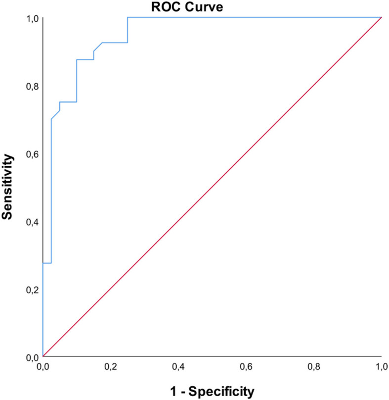
ROC curve for MMP-20 variable in determining MIH status
Table 4.
Determination of cut-off value for MMP-20 variable in determining MIH status
| Cut-off Value | AUC (%95 CI) | p | Sensitivity (%) | Specificity (%) | PPV (%) | NPV (%) | |
|---|---|---|---|---|---|---|---|
|
MMP-20 (ng/mL) |
≤ 10.57 | 0.948 (0.902 – 0.995) | < 0.001* | 87.50% | 90% | 89.74% | 87.80% |
AUC Area under the curve, PPV Positive predictive value, NPV Negative predictive value; *p-values < 0.05; ng Nanogram, mL Milliliter, CI confidence interval
Discussion
MIH, an enamel defect occurring due to insufficient removal of enamel proteins during enamel formation or phosphate and calcium deficiency during matrix formation, is associated with various factors such as hypoxia, premature birth, and antibiotic use [20, 21]. In the teeth affected by MIH clinical problems such as demarcated opacities, post-eruptive enamel breakdown, and difficulties in local anesthesia can be observed [1]. In addition, dental sensitivity can also be observed in MIH-affected teeth, and the inability of patients to brush their teeth sufficiently can increase the accumulation of microbial dental plaque [22]. The findings of this study revealed that periodontal parameters such as PI, GI, PPD, and GCF volume were significantly higher in the MIH group. This indicated that periodontal inflammation and tissue involvement were increased in individuals with MIH. Bycontrast, no significant difference was detected between the groups in terms of MMP-8 and TGF-β1 levels, which are biomarkers associated with periodontal disease. In addition, a significant reduction in MMP-20 levels was observed in individuals with MIH, suggesting a disruption in enamel-related protein activity.
In the diagnosis of periodontal disease, along with clinical and radiographic parameters, biomarkers in GCF are also used [4, 16]. PI is one of the clinical parameters that evaluates the presence and localization of plaque on the tooth [16]. Most previous studies evaluating the relationship between MIH and plaque reported that plaque in children with MIH was higher than in healthy children [2, 22, 23]. For instance, Ulusoy et al. found no statistically significant difference in terms of plaque accumulation between children with MIH and healthy children; however, plaque still tended to increase in the MIH group [24]. In the present study, PI was also found to be significantly higher in children with MIH as compared to the control group. Most previous studies reported that MIH was frequently observed in the patients who failrf to maintain oral hygiene: the increased amount of plaque on the irregular surfaces of the affected teeth and the increased sensitivity level to brushing made these patients prone to periodontal diseases.
Bleeding observed in the gingiva is one of the clinical findings of inflammation [16]. One relevant study found that MIH increased the level of gingival inflammation in children [2], while there were also studies reporting that MIH does not affect inflammation [23, 24]. In the present investigation, statistically significantly higher GI was detected in children with MIH as compared to the control group. These differences between the results of the studies may be due to the use of modified methods in the evaluation of bleeding and/or individual/environmental factors affecting oral hygiene habits.
Several previous studies evaluated the relationship between PPD and periodontal diseases [14, 25]. The studies comparing PPD in healthy and gingivitis children established that PPD in the gingivitis group was significantly higher [14, 25]. In the present study, PPD in children with MIH was evaluated for the first time, and statistically significantly higher PPD was detected in children with MIH as compared to healthy children. The fact that PI and GI were significantly higher in the MIH group as compared to the healthy group was interpreted as an indicator of increasing severity of periodontal disease, and the increase in PPD was thought to correlate with this situation.
GCF is used to evaluate the inflammation in periodontal tissues. Available evidence suggests that GCF volume increases with increasing inflammation severity [26]. Previous studies revealed that GCF volume is significantly higher in children with gingivitis as compared to periodontally healthy children [25, 26]. In this context, the present study is the first to measure GCF volume in individuals with MIH and to The higher GCF volume in the MIH group can be attributed to inflammation caused by increased plaque accumulation.
Furthermore, previous studies reported that high concentrations of MMP-8 secreted from inflammatory cells during periodontal disease are associated with tissue destruction and that the concentration of MMP-8 increases with the severity of periodontal disease [27]. Several previous studies evaluated the relationship between MMP-8 levels in children and various diseases [28]. For instance, Yamazaki‐Kubota et al. demonstrated that the MMP-8 level in GCF in children with Down syndrome was significantly higher than in the healthy child patient group, independent of periodontal inflammation [28]. The present study was the first to evaluate the MMP-8 levels in GCF of children with MIH, and although the MMP-8 level in GCF was found to be higher in the MIH group, this difference did not reach statistical significance. This outcome can be attributed to the fact that, while previous studies were predominantly conducted on adults with periodontitis, the present study did not include a pediatric population with advanced periodontal disease.
MMP-20, expressed by ameloblasts during the secretory phase of amelogenesis, is a type of proteinase that can be found in the enamel matrix. The primary function of MMP-20 is to degrade amelogenin, one of the enamel matrix proteins. In this way, a large portion of the enamel matrix proteins are removed from the ECM, and it is effective in the growth and development of hydroxyapatite crystals in the enamel [8]. Previous research reported that the amount of mineral in the defective enamel of teeth with MIH decreases [1], which was attributed to the inability to remove proteins from the ECM as much as necessary during enamel formation or due to the calcium-phosphate deficiency observed during the formation of the matrix [20]. For instance, in a study conducted by Shin et al., the authors found that MMP-20 expression must be in appropriate amounts for enamel formation to occur properly. Furthermore, there is also evidence to suggest that too little MMP-20 activity causes amelogenesis imperfecta, while too much results in a discontinuous ameloblast layer with impaired mineralization [29]. In an in vitro study, Mukhtar et al. Compared various protein amounts belonging to MIH-affected enamel and healthy enamel samples. The authors found that the amount of MMP-20 was higher in healthy enamel [30]. To the best of our knowledge, the present study is the first to evaluate the level of MMP-20 in GCF and to investigate its relationship with MIH. We found that the level of MMP-20 in GCF of MIH patients was statistically significantly lower as compared to healthy children. These findings are largely consistent with previous research reporting the effects of MMP-20 on amelogenesis and the finding that the low amount of the relevant enzyme causes high protein amounts in tooth enamel. In addition, according to the ROC analysis, it was determined that MMP-20 is a highly effective biomarker in the diagnosis of MIH and has a high success rate in both correctly identifying patients and distinguishing healthy individuals.
TGF-β1 is a cytokine with various functions such as cell proliferation, embryonic development, ECM synthesis, and regulation of the immune system [10]. In addition, the rs10733708 single nucleotide polymorphism in the TGF-β R1 gene was found to be associated with MIH [12]. TGF-β1 was also found to be effective in regulating collagen metabolism in diseases such as periodontitis, control the inflammatory response, and its level was found to increase with periodontal disease [13, 31]. Skalerič et al. reported that TGF-β1 concentrations in GCF increased in areas where pocket depth increased [32]. However, although the present study was the first to evaluate TGF-β1 levels in GCF of children with MIH and healthy children, no significant difference was found in GCF TGF-β1 levels between participants. Similarly to the findings on the MMP-8 biomarker, this finding can be attributed to the fact that the severity of periodontal disease is generally lower in children and the absence of participants with a direct diagnosis of periodontitis among the study groups does not create a significant difference in TGF-β1 levels between the groups.
The findings of this study revealed that the levels of MMP-20 in GCF are significantly lower in individuals with MIH compared to healthy controls. The ROC analysis further demonstrated that an MMP-20 cut-off value of ≤ 10.57 ng/mL could reliably predict the presence of MIH with high sensitivity (87.5%) and specificity (90%). These results highlight the potential of MMP-20 as a clinically useful biomarker for the non-invasive diagnosis of MIH. Early detection of MIH is crucial, particularly in mild or borderline cases that may otherwise go unnoticed during routine clinical examination. Detecting reduced MMP-20 levels in GCF may allow clinicians to effectively initiate preventive or minimally invasive interventions before substantial enamel breakdown, hypersensitivity, or caries occur. Therefore, to support early identification and management of MIH, MMP-20 testing should be integrated as a diagnostic adjunct in pediatric dental assessments.
The ultimate goal of this line of research would be to develop a biomolecular-based diagnostic tool that can complement conventional clinical evaluations. This approach may be particularly beneficial in populations with limited access to specialist dental care or in settings where early identification is key to preventing further oral health complications. Future studies should aim to validate these findings in larger, multi-centered, and ethnically diverse populations. In addition, longitudinal research is needed to evaluate the progression of MMP-20 levels over time and their association with MIH severity and treatment outcomes. Exploring the interaction between MMP-20 expression and genetic variations, as well as other enamel matrix proteins, may provide further important insights into the pathogenesis of MIH and guide the development of targeted preventive or therapeutic strategies.
The present study has several limitations. First, we did not evaluate individual and environmental factors affecting oral hygiene habits. Second, the number of patients included in the study was limited. Thirdly and finally, MIH could not be assessed by dividing cases into subgroups based on severity. All these limitations may have biased the results and prevented generalizability and depth of the findings, and should be carefully addressed in future research.
Conclusion
The present study is the first to evaluate the periodontal health of individuals with MIH, the volume of GCF, and the levels of MMP-8, MMP-20, and TGF-β1 in the GCF. Along with important clinical results, the detection of MMP-20, which plays an important role in enamel mineralization, in the GCF of teeth with MIH, is reported in the literature for the first time. Taken together, the present results demonstrate that MMP-20 may be a highly effective biomarker that allows approximately 90% of participants to be correctly diagnosed as MIH or healthy. This conclusion may guide future studies to better understand the effects of MMP-20 on enamel tissue.
Acknowledgements
Supported in part by Recep Tayyip Erdogan University, Department of Scientific Research Project with the ID number 1495.
This study was supported by the Recep Tayyip Erdoğan University Development Foundation (Grant number:02025002007284).
Authors’ contributions
B.S.B. performed the data collection. S.A. and D.N.G. participated in the writing and analysis of the article. B.S.B., M.A., I.A., O.K., and S.A. planned the study.
Funding
Supported in part by Recep Tayyip Erdogan University, Department of Scientific Research Project with the ID number 1495.
Data availability
The datasets used and/or analysed during the current study are available from the corresponding author on reasonable request.
Declarations
Ethics approval and consent to participate
Ethical approval was obtained from the Non-Invasive Clinical Research Ethics Committee of Recep Tayyip Erdoğan University (Decision no: 2023/121). The study was conducted in accordance with the Declaration of Helsinki of 1975, as revised in 2013. The purpose and content of the research were explained to all individuals included in the study and voluntary consent forms were signed.
Consent for publication
Not applicable.
Competing interests
The authors declare no competing interests.
Footnotes
Publisher’s Note
Springer Nature remains neutral with regard to jurisdictional claims in published maps and institutional affiliations.
References
- 1.Lygidakis NA, Garot E, Somani C, Taylor GD, Rouas P, Wong FSL. Best clinical practice guidance for clinicians dealing with children presenting with molar-incisor-hypomineralisation (MIH): an updated European academy of paediatric dentistry policy document. Eur Arch Paediatr Dent. 2022;23:3–21. 10.1007/s40368-021-00668-5. [DOI] [PMC free article] [PubMed] [Google Scholar]
- 2.Turkmen E, Ozukoc C. Impact of molar incisor hypomineralization on oral hygiene and gingival health in 8–15-years-old children. Aust Dent J. 2022;67:50–6. 10.1111/adj.12923. [DOI] [PubMed] [Google Scholar]
- 3.Alfano MC. The origin of gingival fluid. J Theor Biol. 1974;47:127–36. 10.1016/0022-5193(74)90103-9. [DOI] [PubMed] [Google Scholar]
- 4.Chapple IL. Periodontal diagnosis and treatment–where does the future lie? Periodontol. 2000;2009(51):9–24. 10.1111/j.1600-0757.2009.00319.x. [DOI] [PubMed] [Google Scholar]
- 5.Baker AH, Edwards DR, Murphy G. Metalloproteinase inhibitors: biological actions and therapeutic opportunities. J Cell Sci. 2002;115:3719–27. 10.1242/jcs.00063. [DOI] [PubMed] [Google Scholar]
- 6.Nagase H, Woessner JF Jr. Matrix metalloproteinases. J Biol Chem. 1999;30(274):21491–4. 10.1074/jbc.274.31.21491. [DOI] [PubMed] [Google Scholar]
- 7.Uitto VJ, Suomalainen K, Sorsa T. Salivary collagenase. Origin, characteristics and relationship to periodontal health. J Periodontal Res. 1990;25:135–42. 10.1111/j.1600-0765.1990.tb01035.x. [DOI] [PubMed] [Google Scholar]
- 8.Robinson C, Brookes SJ, Bonass WA, Shore RC, Kirkham J. Enamel maturation. Ciba Found Symp. 1997;205:156–70. 10.1002/9780470515303.ch11. [DOI] [PubMed] [Google Scholar]
- 9.Jeremias F, Pierri RA, Souza JF, Fragelli CM, Restrepo M, Finoti LS, et al. Family-based genetic association for molar-incisor hypomineralization. Caries Res. 2016;50:310–8. 10.1159/000445726. [DOI] [PubMed] [Google Scholar]
- 10.Momose M, Murata M, Kato Y, et al. Vascular endothelial growth factor and transforming growth factor-α and -β1 are released from human cultured gingival epithelial sheets. J Periodontol. 2002;73:748–53. 10.1902/jop.2002.73.7.748. [DOI] [PubMed] [Google Scholar]
- 11.SassáBenedete AP, Sobral AP, Lima DM, Kamibeppu L, Soares FA, Lourenço SV. Expression of transforming growth factor-beta 1, -beta 2, and -beta 3 in human developing teeth: immunolocalization according to the odontogenesis phases. Pediatr Dev Pathol. 2008;11:206–12. 10.2350/07-09-0333.1. [DOI] [PubMed] [Google Scholar]
- 12.Bussaneli DG, Restrepo M, Fragelli CMB, Santos-Pinto L, Jeremias F, Cordeiro RCL, et al. Genes regulating immune response and amelogenesis interact in increasing the susceptibility to molar-incisor hypomineralization. Caries Res. 2019;53:217–27. 10.1159/000491644. [DOI] [PubMed] [Google Scholar]
- 13.Gürkan A, Emingil G, Cinarcik S, Berdeli A. Gingival crevicular fluid transforming growth factor-beta1 in several forms of periodontal disease. Arch Oral Biol. 2006;51:906–12. 10.1016/j.archoralbio.2006.04.008. [DOI] [PubMed] [Google Scholar]
- 14.Ülker AE, Tulunoğlu Ö, Özmeriç N, Can M, Demirtaş S. The evaluation of cystatin C, IL-1β, and TNF-α levels in total saliva and gingival crevicular fluid from 11-to 16-year-old children. J Periodontol. 2008;79:854–60. 10.1902/jop.2008.070422. [DOI] [PubMed] [Google Scholar]
- 15.Frankl SN. Should the parent remain with the child in the dental operatory? J Dent Child. 1962;29:150–63. [Google Scholar]
- 16.Löe H. The Gingival index, the plaque index and the retention index systems. J Periodontol. 1967;38:610–6. 10.1902/jop.1967.38.6.610. [DOI] [PubMed] [Google Scholar]
- 17.Goldberg P, Matsson L, Anderson H. Partial recording of gingivitis and dental plaque in children of different ages and in young adults. Commun Dent Oral Epidemiol. 1985;13:44–6. 10.1111/j.1600-0528.1985.tb00419.x. [DOI] [PubMed] [Google Scholar]
- 18.Lin CY, Chen F, Hariri A, Chen CJ, Wilder-Smith P, Takesh T, et al. Photoacoustic imaging for noninvasive periodontal probing depth measurements. J Dent Res. 2018;97:23–30. 10.1177/0022034517729820. [DOI] [PMC free article] [PubMed] [Google Scholar]
- 19.McHugh ML. Interrater reliability: the kappa statistic. Biochem Med (Zagreb). 2012;22:276–82. [PMC free article] [PubMed] [Google Scholar]
- 20.Koch G, Hallonsten AL, Ludvigsson N, Hansson BO, Holst A, Ullbro C. Epidemiologic study of idiopathic enamel hypomineralization in permanent teeth of Swedish children. Commun Dent Oral Epidemiol. 1987;15:279–85. 10.1111/j.1600-0528.1987.tb00538.x. [DOI] [PubMed] [Google Scholar]
- 21.Garot E, Rouas P, Somani C, Taylor GD, Wong F, Lygidakis NA. An update of the aetiological factors involved in molar incisor hypomineralisation (MIH): a systematic review and meta-analysis. Eur Arch Paediatr Dent. 2022;23:23–38. 10.1007/s40368-021-00646-x. [DOI] [PubMed] [Google Scholar]
- 22.Ebel M, Bekes K, Klode C, Hirsch C. The severity and degree of hypomineralisation in teeth and its influence on oral hygiene and caries prevalence in children. Int J Paediatr Dent. 2018;28:648–57. 10.1111/ipd.12425. [DOI] [PubMed] [Google Scholar]
- 23.Ismayilova N, Gungor OE, Karayilmaz H. Assessment of severity and mineral composition of saliva in schoolchildren with molar-incisor hypomineralization (MIH). J Clin Pediatr Dent. 2024;48:86–93. 10.22514/jocpd.2024.024. [DOI] [PubMed]
- 24.Ulusoy AT, Sen Tunc E, Bayrak Ş, Onder H. A comparative study of oral health parameters in molar incisor hypomineralization and high-caries-risk children aged 8–11 years. Med Princ Pract. 2016;25:85–9. 10.1159/000440999. [DOI] [PMC free article] [PubMed] [Google Scholar]
- 25.Keles S, Anik A, Cevik O, Abas BI, Anik A. Gingival crevicular fluid levels of interleukin-18 and tumor necrosis factor-alpha in type 1 diabetic children with gingivitis. Clin Oral Investig. 2020;24:3623–31. 10.1007/s00784-020-03238-z. [DOI] [PubMed] [Google Scholar]
- 26.Doğusal G, Afacan B, Bozkurt E, Sönmez I. Gingival crevicular fluid and salivary resistin and tumor necrosis factor-alpha levels in obese children with gingivitis. J Periodontol. 2018;89:973–82. 10.1002/JPER.17-0613. [DOI] [PubMed] [Google Scholar]
- 27.Hong I, Pae HC, Song YW, et al. Oral fluid biomarkers for diagnosing gingivitis in human: a cross-sectional study. J Clin Med. 2020;9:1720. 10.3390/jcm9061720. [DOI] [PMC free article] [PubMed] [Google Scholar]
- 28.Yamazaki-Kubota T, Miyamoto M, Sano Y, Kusumoto M, Yonezu T, Sugita K, et al. Analysis of matrix metalloproteinase (MMP-8 and MMP-2) activity in gingival crevicular fluid from children with Down’s syndrome. J Periodontal Res. 2010;45:170–6. 10.1111/j.1600-0765.2009.01214.x. [DOI] [PubMed] [Google Scholar]
- 29.Shin M, Chavez MB, Ikeda A, Foster BL, Bartlett JD. MMP20 overexpression disrupts molar ameloblast polarity and migration. J Dent Res. 2018;97:820–7. 10.1177/0022034518758657. [DOI] [PMC free article] [PubMed] [Google Scholar]
- 30.Mukhtar U, Goyal A, Luthra-Guptasarma M, Gauba K, Kapur A, Thakur AK. Label-free quantitative proteomics reveals molecular correlates of altered biomechanical properties in molar incisor hypomineralization (MIH): an in vitro study. Eur Arch Paediatr Dent. 2022;23:179–91. 10.1007/s40368-021-00687-2. [DOI] [PubMed] [Google Scholar]
- 31.Morikawa M, Derynck R, Miyazono K. TGF-β and the TGF-β family: context-dependent roles in cell and tissue physiology. Cold Spring Harb Perspect Biol. 2016;8:021873 10.1101/cshperspect.a021873. [DOI] [PMC free article] [PubMed]
- 32.Skaleric U, Kramar B, Petelin M, Pavlica Z, Wahl SM. Changes in TGF-β1 levels in gingiva, crevicular fluid and serum associated with periodontal inflammation in humans and dogs. Eur J Oral Sci. 1997;105:136–42. 10.1111/j.1600-0722.1997.tb00192.x. [DOI] [PubMed] [Google Scholar]
Associated Data
This section collects any data citations, data availability statements, or supplementary materials included in this article.
Data Availability Statement
The datasets used and/or analysed during the current study are available from the corresponding author on reasonable request.