Abstract
Two virulence factors of Listeria monocytogenes, listeriolysin O (LLO) and phosphatidylinositol-specific phospholipase C (PI-PLC), mediate escape of this pathogen from the phagocytic vacuole of macrophages, thereby allowing the bacterium access to the host cell cytosol for growth and spread to neighboring cells. We characterized their orthologs from Bacillus anthracis by expressing them in L. monocytogenes and characterizing their contribution to bacterial intracellular growth and cell-to-cell spread. We generated a series of L. monocytogenes strains expressing B. anthracis anthrolysin O (ALO) and PI-PLC in place of LLO and L. monocytogenes PI-PLC, respectively. We found that ALO was active at both acidic and neutral pH and could functionally replace LLO in mediating escape from a primary vacuole; however, ALO exerted a toxic effect on the host cell by damaging the plasma membrane. B. anthracis PI-PLC, unlike the L. monocytogenes ortholog, had high activity on glycosylphosphatidylinositol-anchored proteins. L. monocytogenes expressing B. anthracis PI-PLC showed significantly decreased efficiencies of escape from a phagosome and in cell-to-cell spread. We further compared the level of cytotoxicity to host cells by using mutant strains expressing ALO in combination either with L. monocytogenes PI-PLC or with B. anthracis PI-PLC. The results demonstrated that the mutant strain expressing the combination of ALO and B. anthracis PI-PLC caused less damage to host cells than the strain expressing ALO and L. monocytogenes PI-PLC. The present study indicates that LLO and L. monocytogenes PI-PLC has adapted for L. monocytogenes intracellular growth and virulence and suggests that ALO and B. anthracis PI-PLC may have a role in B. anthracis pathogenesis.
Listeria monocytogenes and Bacillus anthracis are gram-positive bacteria in the low-G+C lineage that cause rare but fatal infections of humans and animals. Although L. monocytogenes has been studied as a model facultative intracellular pathogen for decades, B. anthracis had, until recently, received less attention. Interestingly, the B. anthracis genome sequence has revealed the presence of a number of potential determinants of pathogenesis shared with those of L. monocytogenes, including a phosphatidylinositol-specific phospholipase C (PI-PLC), a phosphatidylcholine-preferring phospholipase C (PC-PLC), and listeriolysin O (LLO) (27). In L. monocytogenes, these determinants are integral to its intracellular growth cycle. LLO is essential for escape from a phagosome, the phospholipases contribute to that process, and all three are important for cell-to-cell spread (3, 10, 27, 34). Although B. anthracis is traditionally considered an extracellular pathogen, there is considerable evidence that spores germinate and grow in macrophages during the early phase of infection (8, 30). We expressed here the B. anthracis orthologs of L. monocytogenes LLO and PI-PLC in L. monocytogenes in order to characterize them in a well-studied model of intracellular pathogenesis.
MATERIALS AND METHODS
Bacterial strains and growth conditions.
The L. monocytogenes strains used in the present study are listed in Table 1. Unless otherwise specified, all bacterial strains were grown in brain heart infusion broth, and strains derived from L. monocytogenes Δdal Δdat (Lmdd) were supplemented with 100 μg of d-alanine/ml to stationary phase at 30°C. Strains containing pKSV7 were selected and maintained with ampicillin (50 μg/ml) in Escherichia coli and chloramphenicol (10 μg/ml) in L. monocytogenes.
TABLE 1.
Strains used in this study
| Strain | Relevant characteristic(s)a | Source or reference |
|---|---|---|
| L. monocytogenes | ||
| Lmdd | Δdal Δdat; d-alanine dependent | 37 |
| HG-L2001 | Lmdd expressing H86A mutant LmPI-PLC | This study |
| HG-L2002 | Lmdd expressing BaPI-PLC instead of LmPI-PLC | This study |
| HG-L2003 | Lmdd expressing ALO instead of LLO | This study |
| HG-L2004 | Lmdd expressing ALO and H86A mutant LmPI-PLC | This study |
| HG-L2005 | Lmdd expressing ALO and BaPI-PLC | This study |
| 10403S | Wild type L. monocytogenes | 2 |
| DP-L4055 | L. monocytogenes 10403S expressing PFO | 7 |
| DP-L4450 | L. monocytogenes 10403S expressing ALO | This study |
| DP-L2161 | L. monocytogenes 10403S with LLO deleted (Δhly) | 16 |
| B. anthracis Sterne 7702 | pXO1+, pXO2−; attenuated, toxin+ | 4 |
LmPI-PLC, L. monocytogenes PI-PLC; BaPI-PLC, B. anthracis PI-PLC.
Tissue-culture cells and growth media.
The mouse macrophage-like cell line J774 and murine L2 fibroblasts were grown in Dulbecco modified Eagle medium plus 7.5% fetal bovine serum (FBS) and 1 mM l-glutamine. Bone marrow-derived macrophages obtained from C57BL/6 mice were cultured in RPMI 1640-10% FBS-1 mM l-glutamine. All were incubated at 37°C with 5% CO2. For bacterial infections with strains derived from Lmdd, 100 μg of d-alanine/ml was added in the medium.
Construction of L. monocytogenes mutants.
The DNA sequences for ALO (31) and B. anthracis PI-PLC were obtained from the genome of B. anthracis strain Ames from GenBank (29). Signal peptides were determined by using the SignalP server (http://www.cbs.dtu.dk/services/SignalP-2.0/). The structural genes for ALO and B. anthracis PI-PLC were used for the replacement constructions. The mutant constructs were generated by PCR-mediated sequence overlap extension (15) with Pfx High-Fidelity DNA Polymerase, resulting in in-frame gene replacements. All gene replacements were made under their counterparts' endogenous promoters and signal peptide sequences on the chromosome. Mutants were constructed by using derivatives of a shuttle vector pKSV7 for allelic exchange. DNA sequences of mutant constructs were confirmed by automated cycle sequencing. Genes replaced in mutant strains are illustrated in Fig. 1. Oligonucleotide primers used in the present study are as follows: six primers each were used to replace LLO with ALO in the L. monocytogenes Δdal Δdat and wild-type L. monocytogenes background, respectively, as follows: BALLO-P1 (5′-GGTCTAGAGAGAGCGCTGCTAGGTTTGT-3′; XbaI) plus BALLO-P2 (5′-GCATTACCGGCTTGTGTTTCTGCTTCAGTTTGTTGCGCAA-3′) and DP4315 (5′-ATTGTCGACCGTATTCCTGCTTCTAATTGTTG-3′; SalI) plus DP4824 (5′-CAATTGCGCAACAAACTGAAGCAGAAACACAAGCCGGTAATGC-3′)were used for the upstream flanking sequence of the hly gene; BALLO-P3 (TTGCGCAACAAACTGAAGCAGAAACACAAGCCGGTAATGC-3′) with BALLO-P4 (5′-CTTAATTTTTTACTTTTACAACTAATGACTAATAGTAGCAG-3′) and DP4825 (5′-GCATTACCGGCTTGTGTTTCTGCTTCAGTTTGTTGCGCAA-3′) with DP4826 (5′-CTTCGGATCCAACTAATGACTAATAGTAGCAGTTGG; BamHI) were used for DNA sequence encoding ALO from B. anthracis genomic DNA; and BALLO-P5 (5′-CTGCTACTATTAGTCATTAGTTGTAAAAGTAATAAAAAATTAAG-3′) with BALLO-P6 (5′-GGGGTACCTGCTTCGCAGGAATCTGGCA-3′; KpnI) and DP4827 (5′-AATGGATCCGTAATAAAAAATTAAGAATAAAACC; BamHI) and DP4828 (5′-ATTGGATCCTTATCGGTCTAGAAACCACCAGAACTTAGC; BamHI and XbaI) were used for the downstream flanking sequence of the hly gene. For replacement of L. monocytogenes PI-PLC with B. anthracis PI-PLC, another six primers were used: P1 (5′-ACTGTCTAGATCTCGCTAATACTCGTGAGCT-3′, with a XbaI site) and P2BA (5′-GCGAATAAGTCATTAATAAGAGATTAACATATATTATTCCTACAA-3′) were used for the upstream flanking sequence of the plcA gene, P7BA (5′-TTGTAGGAATAATATATGTTAATCTCTTATTAATGACTTATTCGC-3′) and P8N (5′-CCCATTAGGCGGGAAAGCAGCTAGCTCTGTTAATGAGCT-3′) were used for the DNA sequence encoding B. anthracis PI-PLC from B. anthracis genomic DNA, and P3N (5′-AGCTCATTAACAGAGCTAGCTGCTTTCCCGCCTAATGGG-3′) and P4 (5′-ACGTGGTACCACTGCATCTCCGTGGTATAC-3′, with a KpnI site) were used for the downstream flanking sequence of the plcA gene. All PCR products were directly cloned into pCR-Blunt II-TOPO (Invitrogen) and were cycle sequenced.
FIG. 1.
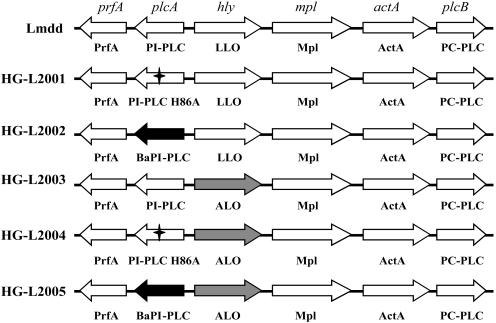
Diagram of the L. monocytogenes PrfA regulon and the mutants constructed in the present study. The star indicates a nonfunctional H86A mutation in L. monocytogenes PI-PLC (1), the black arrows indicate the replacement of L. monocytogenes PI-PLC with B. anthracis PI-PLC, and the gray arrows indicate the replacement of LLO with ALO. BaPI-PLC, B. anthracis PI-PLC.
Sodium dodecyl sulfate-polyacrylamide gel electrophoresis and Western blot.
Sodium dodecyl sulfate-polyacrylamide gel electrophoresis and the subsequent Western blotting were performed as previously described (40). The primary antibodies used in the present study were a rabbit anti-B. thuringiensis PI-PLC for detecting B. anthracis PI-PLC and a rabbit α-perfringolysin O (PFO) for detecting ALO expression. The secondary antibody was a goat anti-rabbit immunoglobulin G conjugated to horseradish peroxidase.
Expression and purification of recombinant His-tagged hemolysins.
Recombinant mature His-tagged LLO and PFO proteins were constructed, expressed, and purified as previously described (12). ALO was PCR amplified from B. anthracis chromosomal DNA with the primers 5-CGGGGGATCCGAAACACAAGCC-3 and 5-CCGGGAATTCCCTAATGACTAATAGTAGC-3 and cloned into the BamHI and EcoRI sites of the pTrcHisA expression vector (Invitrogen), resulting in an ALO fusion protein that starts at E35 for the ALO protein. Recombinant ALO was expressed and purified as previously described by Shepard et al. (32) and stored in buffer (10 mM morpholineethanesulfonic acid, 300 mM NaCl, 1 mM EDTA, 10% glycerol, and 1 mM dithiothreitol buffered to pH 6.5). Purified protein concentrations were determined by the Bradford method.
Hemolytic activity.
Hemolytic activity of bacterial broth cultural supernatants was measured by using sheep red blood cells as previously described (28). Hemolytic units were defined as the dilution of the sample at which 50% of the sheep red blood cells lysed. The specific activity of the recombinant His-tagged proteins was determined as described previously (12).
Diff-Quik staining.
Bacteria grown in J774 host cells were visualized by using a Diff-Quik staining set (Dade Diagnostics of P. R. Inc.) after bacterial infection for 6 h according to the manufacturer's instructions.
Detection of PI-PLC activity on PI.
Enzymatic activity on PI by PI-PLC was detected by using ALOA Listeria agar plates (Microbiology International, Frederick, MD). The size of an opaque halo surrounding a colony reflects the activity on PI. A quantitative method with [3H]inositol-PI was also done as previously described (13).
Detection of PI-PLC activity on glycosyl phosphatidylinositol (GPI)-anchored proteins.
Splenocytes were harvested from C57BL/6 mice. Cleavage of the GPI-anchored protein Thy1.2 on CD4+ and CD8+ T cells was measured by fluorescence-activated cell sorting (FACS). A total of 10 μl from overnight bacterial supernatant was mixed with 106 splenocytes in a well of a 96-well plate and incubated at 37°C for 1 h. Cells were then stained in phosphate-buffered saline (PBS), 1% bovine serum albumin with MAb to CD4, CD8, and Thy1.2. The cells were washed several times with PBS, 1% bovine serum albumin, fixed with 2% paraformaldehyde, and analyzed with a FACSCalibur (Becton Dickinson), and data were analyzed by using FlowJo (version 3.7; TreeStar, Inc.).
Escape from the primary phagocytic vacuole.
The escape of L. monocytogenes from the primary vacuole was determined by measuring the percentage of bacteria coated with polymerized actin filaments (stained with Alexa Fluor 568 phalloidin) in the cytosol (16). Briefly, J774 cells were plated and infected by using fluorescein isothiocyanate-labeled bacteria (38) for 90 min, and the ratio of escaped bacteria (red) over the total bacteria (green) was determined based on microscopy with appropriate filters.
Intracellular growth curve.
Stationary-phase bacterial cultures were washed and used to infect J774 macrophages on 12 mm glass coverslips at 37°C. After 1 h of infection, 50 μg of gentamicin/ml was added to the culture medium to kill the extracellular bacteria when needed. CFU per coverslip were determined by lysing host cells in sterile water and plating on brain heart infusion agar plates (34, 36).
Plaque formation.
Plaque formation assays with murine L2 fibroblasts were performed as previously described (36). Briefly, overnight murine L2 fibroblasts cultured in six-well tissue culture plates were infected with L. monocytogenes for 60 min. The monolayer was then washed with PBS and covered with 2 ml of 1% Dulbecco modified Eagle medium-containing agarose with 10 μg of gentamicin/ml. After incubation at 37°C for 3 days, a second overlay of agarose with Neutral Red was added to allow visualization of plaques. The diameters of plaques were measured and compared to that of the parental strain, Lmdd.
LDH release assay.
Lactate dehydrogenase (LDH) release from J774 host cells after bacterial infection was monitored by using the CytoTox 96 nonradioactive cytotoxicity assay kit (Promega) according to the manufacturer's instructions. Briefly, J774 cells were infected with L. monocytogenes strains at a multiplicity of infection, resulting in at least one bacterium per host cell in a 96-well plate for 5 h, and the supernatant from each well was taken for the LDH activity assay. This assay was performed in the absence of gentamicin to favor host cell lysis over rapid bacterial death (7).
FACS analysis of propidium iodide uptake.
After bacterial infection, the cytotoxicity level was detected by staining cellular DNA with the fluorescent dye propidium iodide (Molecular Probes) (11, 12). Briefly, monolayers of bone marrow-derived macrophages from C57BL/6 mice, obtained as previously described (36), were infected with L. monocytogenes strains at a multiplicity of infection of one for 30 min and then washed with PBS and incubated until 2 h postinfection at 37°C and 5% CO2. No gentamicin was added in this experiment to avoid the adverse effect of gentamicin on the most cytotoxic strains (13). The bone marrow-derived macrophages were then removed from the petri dish, washed with precooled PBS-10% FBS, stained with propidium iodide, and analyzed by FACS.
RESULTS
Construction, expression, and characterization of ALO and B. anthracis PI-PLC in L. monocytogenes.
Five strains of L. monocytogenes were constructed in which wild-type LLO and PI-PLC were replaced individually or in combination as illustrated in Fig. 1. For safety reasons, we primarily used a L. monocytogenes Δdal Δdat strain (Lmdd), which requires d-alanine for growth, as the parental strain (37). The expression of ALO and B. anthracis PI-PLC were confirmed from the bacterial culture supernatants by Western blotting with PFO and B. thuringiensis PI-PLC antibodies, respectively (data not shown).
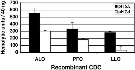
We first analyzed the hemolytic activity of supernatants from the ALO-expressing L. monocytogenes strain. Supernatants from the strain expressing ALO showed 5 to 10-fold higher hemolytic activities at pH 7.4 than those from Lmdd (data not shown). We then compared hemolytic activity using purified proteins. Recombinant ALO showed greater hemolytic specific activity than recombinant LLO and PFO (Fig. 2). We compared the activities of recombinant ALO, PFO, and LLO at pH 5.5 and 7.4. As observed previously, the ratio of activities (pH 5.5/pH 7.4) for LLO is high, 9.0, whereas that of PFO is much lower, 1.8. The ratio of ALO is similar to that of PFO, 1.8, indicating that, unlike LLO, ALO does not have an activity optimum at an acidic pH (2).
FIG. 2.
Specific hemolytic activities of recombinant cholesterol-dependent cytolysins (CDC) on sheep red blood cells at the indicated pH. Values represent the mean ± the SD of three independent experiments.
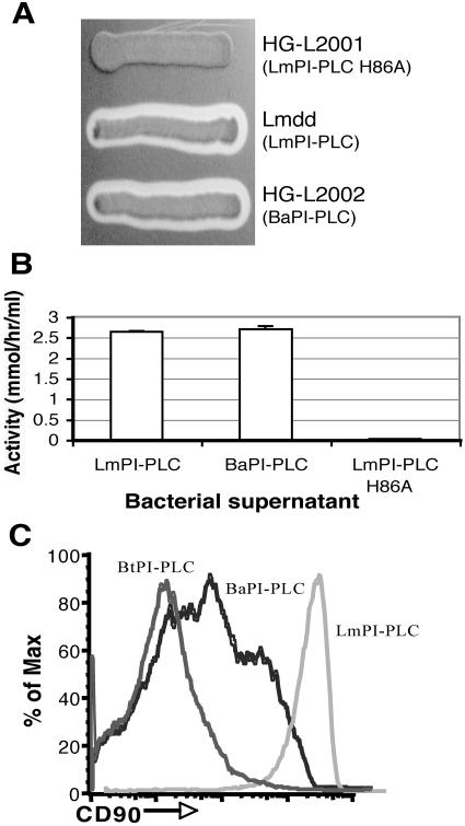
To visualize any difference in PI-PLC activities, we streaked L. monocytogenes strains on an ALOA Listeria agar plate with d-alanine and incubated them at 37°C overnight. A nonfunctional L. monocytogenes PI-PLC mutant with one amino acid change at its active site (H86A) was used as the negative control (1). The activities of B. anthracis PI-PLC and L. monocytogenes PI-PLC appeared to be similar (Fig. 3A). A quantitative assay with [3H]inositol-PI showed the same result that B. anthracis PI-PLC has almost the same activity on PI as L. monocytogenes PI-PLC (Fig. 3B). To compare their activities on GPI-anchored proteins, we measured the cleavage of Thy1 (CD90) on murine T cells. B. anthracis PI-PLC in supernatants of strain HG-L2002 showed strong activity, whereas L. monocytogenes PI-PLC showed no activity compared to a negative control (Fig. 3C). Supernatants from HG-L2005, expressing both ALO and B. anthracis PI-PLC, showed similar strong activity on Thy1 (data not shown).
FIG. 3.
Detection of PI-PLC activities. (A) Activity of indicated strains on PI using ALOA Listeria agar plates; (B) supernatant activity on PI using a quantitative [3H]inositol-PI method (13). For L. monocytogenes PI-PLC (LmPI-PLC) and B. anthracis PI-PLC (BaPI-PLC), the P value was >0.1 as determined by unpaired t test of three experiments. There is no activity detected when an inactive mutant form of L. monocytogenes PI-PLC, H86A, was used. (C) Activity on the GPI-anchored protein CD90 as determined by FACS. Left, pure B. thuringiensis PI-PLC (BtPI-PLC; 6 μg/ml); middle, supernatant of strain HG-L2002 (B. anthracis PI-PLC [BaPI-PLC]); right, supernatant of strain Lmdd (L. monocytogenes PI-PLC [LmPI-PLC]). In several separate experiments the negative control of untreated cells overlapped with the Lmdd curve (not shown).
Intracellular growth and cell-to-cell spread of L. monocytogenes strains.
The second major issue addressed in our studies was whether the heterologous expression of ALO and B. anthracis PI-PLC affected the pathogenesis of L. monocytogenes. To examine the effects of heterologous expression of B. anthracis proteins on intracellular growth, we measured the ability of these strains to escape from a primary vacuole of the J774 macrophage-like cell, their intracellular growth, and their ability to spread from cell to cell.
To determine the efficiencies of escape by different L. monocytogenes strains, we measured the fraction of bacteria stained with fluorescent phalloidin, which detects polymerized actin surrounding bacteria in the cytosol (16). At 90 min after infection, all mutant strains were found to be defective in escape compared to the Lmdd strain (Table 2). Consistent with previous observations (1), the strain expressing a full-length inactive L. monocytogenes PI-PLC performed like a plcA in-frame deletion strain (3). Escape of the B. anthracis PI-PLC-expressing strain was significantly less than either the Lmdd strain or the inactive mutant PI-PLC strain, indicating that B. anthracis PI-PLC could not functionally replace L. monocytogenes PI-PLC, although they have almost the same activities on PI (Fig. 3A and B). Escape of all of the ALO-expressing strains were significantly lower than the Lmdd strain, as has been observed with a PFO-expressing strain (16).
TABLE 2.
Measurements of L. monocytogenes phagosomal escape and cell-to-cell spread
| Strain | % Phagosomal escapea ± SD | Plaque sizeb (%) ± SD |
|---|---|---|
| Lmdd | 65 ± 9 | 100 |
| HG-L2001 | 47 ± 9* | 91 ± 3.4† |
| HG-L2002 | 37 ± 10†‡ | 61 ± 4.6† |
| HG-L2003 | 39 ± 9† | 0 |
| HG-L2004 | 28 ± 10† | 0 |
| HG-L2005 | 36 ± 13† | 0 |
That is, the percentage of total intracellular bacteria staining with Alexa Fluor 568 phalloidin at 90 min after infection of J774 cells. Two-tailed P values of unpaired t tests are indicated as follows: *, P < 0.05; †, P < 0.001 compared to values obtained with the Lmdd strain; ‡, P < 0.05 compared to values obtained with-HG-L2001. At least 200 bacterium-associated macrophages were counted for each of three experiments.
That is, the plaque size relative to that of strain Lmdd. A minimum of 10 plaques per strain per experiment were measured. Values are means of three independent experiments. Two-tailed P values of unpaired t tests are classified as follows: †, P < 0.001 compared to values obtained with the Lmdd strain.
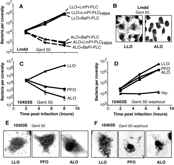
Subsequent to escape from a primary vacuole, L. monocytogenes replicates rapidly in the cytosol (5, 24). We examined the ability of mutant strains to grow in the macrophage-like cell line J774. In this assay, gentamicin (50 μg/ml) was added to the culture medium 1 h after bacterial infection in order to kill extracellular bacteria. The results demonstrated that strains with the heterologous expression of ALO showed a 1,000-fold decrease in CFU at 8 h postinfection, suggesting that ALO-expressing mutant strains were capable of permeabilizing the host cell membrane and allowing gentamicin to gain entry to intracellular bacteria (Fig. 4A). J774 cells were then infected with Lmdd expressing ALO in media containing gentamicin (50 μg/ml) and examined by light microscopy. A large number of bacteria were seen in cells infected with the parental strain for 6 h, whereas only few bacteria could be found in cells infected with the ALO-expressing strain (Fig. 4B). The decrease in CFU and the lack of bacterial cell division suggest that ALO causes membrane damage in the host cell, permitting the entry of extracellular gentamicin, which kills intracellular bacteria. The other ALO-expressing strains, HG-L2004 and HG-L2005, showed similar results (data not shown). We also inserted the gene for ALO into the wild-type strain 10403S. When growth curves were generated in the presence of gentamicin, results were similar to those obtained with strain HG-L2003 and L. monocytogenes expressing PFO (Fig. 4C and E). An additional experiment was done in which gentamicin, 50 μg/ml, was added for only a short pulse of half an hour during the early bacterial infection. In this case, the growth of the strains expressing ALO or PFO was similar to that of the wild-type 10403S (Fig. 4D). However, when observed under the microscope at 6 h postinfection, it appeared that the infected host cells had died and the bacteria were mostly extracellular (Fig. 4F). In contrast, strains expressing inactive L. monocytogenes PI-PLC or B. anthracis PI-PLC along with LLO showed almost the same rate of intracellular growth as wild-type Lmdd (Fig. 4A). Thus, heterologous expression of B. anthracis PI-PLC does not alter the early growth of L. monocytogenes in J774 cells.
FIG. 4.
Intracellular growth in J774 murine macrophage-like cells. The data shown are representative of at least three experiments. (A) Growth of Lmdd strains. (B) Light micrograph of J774 cells 6 h postinfection with Lmdd or Lmdd expressing ALO. For panels A and B, gentamicin at 50 μg/ml was added at 1 h postinfection for the duration of the experiment. (C and D) Growth of wild-type L. monocytogenes expressing LLO (10403S), PFO (DP-L4055), or ALO (DP-L4450). Gentamicin at 50 μg/ml was added at 1 h postinfection for the duration of the experiment (C) or for a short pulse from 1 to 1.5 h postinfection (D). L. monocytogenes with the LLO gene deleted (Δhly, DP-L2161) controls for killing of extracellular bacteria during the gentamicin pulse. (E and F) Light micrographs of J774 cells 6 h postinfection with WT strain 10403S or derivatives expressing either PFO or ALO. Gentamicin at 50 μg/ml was added at 1 h postinfection for the duration of the experiment (E) or for a short pulse from 1 to 1.5 h postinfection (F). LmPI-PLC, L. monocytogenes PI-PLC; BaPI-PLC, B. anthracis PI-PLC.
To further study the effects on L. monocytogenes pathogenesis of heterologous expression of B. anthracis virulence factors, we performed a plaque assay using L2 fibroblast monolayers. The plaque size reflects a bacterial strain's ability to spread from cell to cell and escape from the secondary double-membrane vacuole formed in adjacent cells. Consistent with the results presented in Fig. 4, all ALO-expressing strains showed no detectable plaques on L2 monolayers after 3 days in the presence of gentamicin (Table 2). This is consistent with the rapid killing by gentamicin of ALO-expressing strains in the primary cell after infection (Fig. 4). The strain with heterologous expression of B. anthracis PI-PLC formed plaques that were considerably smaller than either the Lmdd strain or the inactive PI-PLC strain (Table 2). Since expression of B. anthracis PI-PLC did not affect the early intracellular growth of L. monocytogenes, the reduction of plaque size indicates a defect in cell-to-cell spread.
Toxicity of L. monocytogenes strains expressing ALO.
The third major issue to be addressed by the present study was whether PI-PLC affects the permeabilization of the host cell plasma membrane by ALO. Given the ability of ALO to permeabilize the host cell membrane and allow entry of gentamicin, which kills the infecting bacteria, we compared the effects on cytotoxicity of expression of B. anthracis PI-PLC and L. monocytogenes PI-PLC in strains expressing ALO.
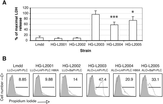
To measure differences in cytotoxicity, we first monitored the release of LDH from the cytosol of J774 cells into the tissue culture medium. Infection with the Lmdd bacteria resulted in ca. 10% LDH release during a 5-h incubation. The same was true for the other two mutant strains expressing LLO (Fig. 5A). Conversely, mutant strains expressing ALO produced much higher release of LDH. ALO in combination with L. monocytogenes PI-PLC exhibited the highest release, which was 96% of the maximal release of LDH. ALO with B. anthracis PI-PLC resulted in 74% of the maximal LDH release, and ALO in combination with inactive L. monocytogenes PI-PLC showed the lowest release, 58% (Fig. 5A).
FIG. 5.
Evaluation of the integrity of the host cell plasma membrane after L. monocytogenes infections. (A) LDH release by infected J774 cells. The release of LDH after infection of J774 cells by each strain was compared to total LDH determined after lysis with detergent. Values represent the mean ± the SD of three independent experiments. ✽, P < 0.05; ✽✽✽, P < 0.001 (as determined by unpaired t test compared to strain HG-L2003). (B) Cellular DNA staining by propidium iodide using mouse bone marrow-derived macrophages. The gray histogram represents the uninfected cells as negative control. The percentage of cells that were stained with propidium iodide is indicated. These data are representative of three experiments.
We further measured cytotoxicity by examining membrane integrity using the membrane-impermeant fluorescent dye propidium iodide (11, 12). This dye binds cellular DNA when the membrane is damaged and increases the fluorescence of host cells, which in these experiments were bone marrow-derived macrophages. The data obtained by FACS agreed with the LDH release results, showing that ALO with L. monocytogenes PI-PLC displayed the highest level of fluorescence, ALO with B. anthracis PI-PLC displayed the medium level of fluorescence, and ALO with inactive L. monocytogenes PI-PLC displayed the lowest level of fluorescence (Fig. 5B).
In summary, with both assays the highest cytotoxicity was observed upon infection with the strain expressing ALO and L. monocytogenes PI-PLC. Less cytotoxicity was observed upon infection with the strain expressing ALO and B. anthracis PI-PLC. The least cytotoxicity was seen when ALO was expressed with an inactive form of L. monocytogenes PI-PLC.
DISCUSSION
LLO and PI-PLC are two critical virulence factors that are largely responsible for mediating the escape of L. monocytogenes from host cell vacuoles (3, 6, 21, 28, 34). What roles their orthologs may play in the pathogenesis of B. anthracis is unknown. Most studies on the pathogenesis of B. anthracis have focused on the terminal stages of the disease in which anthrax toxin plays the dominant role. The recent completion of the B. anthracis genome sequence has revealed a number of proteins that are orthologous to known virulence factors of L. monocytogenes, including ALO and B. anthracis PI-PLC (29). In the present study, we characterized the properties of ALO and B. anthracis PI-PLC expressed in L. monocytogenes and investigated their effects on intracellular growth and cell-to-cell spread by using the well-established L. monocytogenes pathogenesis system. The results of our studies indicate that ALO can functionally replace LLO in mediating escape of L. monocytogenes from the primary vacuole; however, it exerts a toxic effect on the host cell (Table 2 and Fig. 4). Our results also indicate that expression of B. anthracis PI-PLC, which has strong activity on GPI-anchored proteins (Fig. 3C), hampers L. monocytogenes escape from a vacuole and reduces its cell-to-cell spread (Table 2).
LLO has uniquely evolved to decrease its toxicity in the host through having much lower activity at pH 7.4 than at the acidic pH of the phagosome (11, 12) and by having a PEST-like N-terminal sequence, which results in very low LLO levels when L. monocytogenes is growing in the host cell cytosol (7). ALO, which has 87% similarity to PFO, but only 64% similarity to LLO (18), does not have a PEST-like sequence, and its activity at pH 7.4 is almost as high as at pH 5.5 (Fig. 2). Expression of ALO by L. monocytogenes resulted in strong toxicity to both J774 cells and murine bone marrow-derived macrophages, as evidenced by permeabilization of infected cells to gentamicin (Fig. 4) and propidium iodide (Fig. 5B), and by the release of LDH from infected cells (Fig. 5A). When PFO was expressed by L. monocytogenes, similar toxicity was observed (Fig. 4E and F) (16). Mutations in PFO that altered its pH optimum to resemble that of LLO resulted in much less toxicity to the host cell (17). Mutants of LLO that increase its activity at neutral pH are more toxic (12). Therefore, the data suggest that ALO is functionally closer to PFO than LLO.
B. anthracis PI-PLC is almost identical to the well-characterized PI-PLCs from B. cereus and B. thuringiensis (14) and, like them, it is active on both PI and GPI-anchored proteins (Fig. 3 and unpublished data). L. monocytogenes expressing B. anthracis PI-PLC was able to form plaques in L2 monolayers, but the plaque size was significantly reduced compared to both strain Lmdd and a strain expressing inactive L. monocytogenes PI-PLC (Table 2). A comparison of their crystal structures has shown that L. monocytogenes PI-PLC and B. cereus PI-PLC have similar molecular structures; however, B. cereus PI-PLC has an extra β-strand (Vb) which is thought to be needed for recognition of GPI anchors (9, 23). B. anthracis PI-PLC has 97% similarity to B. cereus PI-PLC, and both have exactly the same Vb β-strand amino acid sequence. As expected, supernatants from L. monocytogenes expressing either B. cereus PI-PLC or B. anthracis PI-PLC have similar activities on the GPI-anchored protein Thy1 (unpublished data). In another study, we showed that expression of B. cereus PI-PLC inhibits escape of L. monocytogenes from a primary vacuole, blocks cell-to-cell spread, and reduces virulence in mice. We hypothesized that L. monocytogenes PI-PLC has evolved for intracellular growth and virulence by its greatly reduced activity on GPI-anchored proteins through the absence of the Vb β-strand. We speculate that cleavage of GPI-anchored proteins on the cell surface or more likely in the vacuole hampers escape and cell-to-cell spread of L. monocytogenes. The cleavage of GPI-anchored proteins by B. anthracis PI-PLC could influence the normal function of LLO directly or through host cell signals. At this stage, we also do not know which GPI-anchored proteins are cleaved by B. anthracis PI-PLC. Future studies will help to shed light on these questions.
The study of L. monocytogenes PI-PLC has been focused on its role in escape of the bacterium from the primary vacuole and its synergistic effects with LLO and PC-PLC during cell-to-cell spread (1, 3, 34). The abilities of L. monocytogenes PI-PLC to cleave PI, produce diacylglycerol, and activate protein kinase C isoforms in host cells appear to be important in its early interactions with the macrophages (38, 39). In contrast, B. anthracis PI-PLC does not complement L. monocytogenes PI-PLC in escape from the primary phagocytic vacuole; indeed, it appears to be inhibitory (Table 2).
In the present study, we used the combination of ALO with different PI-PLCs to investigate potential contributions of PI-PLC to host cell membrane damage. This method proved to be useful for evaluating the role of PI-PLC in its interplay with ALO. We demonstrated that, in combination with ALO, L. monocytogenes PI-PLC resulted in greater host membrane damage than B. anthracis PI-PLC. An early finding in the study of anthrax was an alkaline phosphatasemia produced during experimental infections of animals with B. anthracis. This was later determined to result from the cleavage of GPI-linked alkaline phosphatase by a bacterial activity (19, 35), which we have now characterized. Thus, cleavage of GPI anchors is manifested during B. anthracis infection. Its role, if any, in the pathogenesis of B. anthracis, is yet to be determined.
B. anthracis is hemolytic to human red blood cells, especially under anaerobic conditions (18). Therefore, it is possible that the expression of cytolytic genes of B. anthracis is induced under certain environmental conditions. The genes for ALO and PI-PLC, along with that for PC-PLC appear to be upregulated early during macrophage infection by germinated B. anthracis spores (18). Recent studies have suggested that the regulation of virulence genes in B. anthracis, either by a truncated pleiotropic transcriptional regulator PlcR or by the transactivator AtxA, is complex (22, 26, 33). Early B. anthracis escape from macrophage by lysis of the cell is regulated by AtxA but does not require the toxin genes expressed from pXO1 (8). Taken together, the results we have presented here suggest a role for ALO and B. anthracis PI-PLC in B. anthracis intracellular infection.
ALO has recently been shown to be an agonist for Toll-like receptor 4 (TLR4) (25). It is intriguing to note that CD14, a GPI-anchored protein, serves as a coreceptor of TLR4 (20). This suggests a site at which ALO and B. anthracis PI-PLC may function together during B. anthracis infections. To further examine the precise functions of putative B. anthracis virulence factors, it will be necessary to generate defined mutant strains and test their roles in pathogenesis in both tissue culture and animal models of infection.
Acknowledgments
We thank Richard F. Rest for providing the Sterne strain of B. anthracis, Fred Frankel for providing the L. monocytogenes Δdal Δdat strain, Rod Tweten for recombinant ALO, and Mary F. Roberts for a sample of recombinant PI-PLC from B. thuringiensis. We thank Daniel Portnoy for careful reading of the manuscript.
This study was supported by U.S. Public Health Service grants AI-056275 (H.G.), AI-27655 (to Daniel A. Portnoy) and the University of Pennsylvania Research Foundation. P.S. was also supported by a PGSB scholarship from the National Science and Engineering Research Council of Canada.
Editor: V. J. DiRita
REFERENCES
- 1.Bannam, T., and H. Goldfine. 1999. Mutagenesis of active-site histidines of Listeria monocytogenes phosphatidylinositol-specific phospholipase C: effects on enzyme activity and biological function. Infect. Immun. 67:182-186. [DOI] [PMC free article] [PubMed] [Google Scholar]
- 2.Bishop, D. K., and D. J. Hinrichs. 1987. Adoptive transfer of immunity to Listeria monocytogenes: the influence of in vitro stimulation on lymphocyte subset requirements. J. Immunol. 139:2005-2009. [PubMed] [Google Scholar]
- 3.Camilli, A., L. G. Tilney, and D. A. Portnoy. 1993. Dual roles of plcA in Listeria monocytogenes pathogenesis. Mol. Microbiol. 8:143-157. [DOI] [PMC free article] [PubMed] [Google Scholar]
- 4.Cataldi, A., E. Labruyere, and M. Mock. 1990. Construction and characterization of a protective antigen-deficient Bacillus anthracis strain. Mol. Microbiol. 4:1111-1117. [DOI] [PubMed] [Google Scholar]
- 5.Chico-Calero, I., M. Suárez, B. González-Zorn, M. Scortti, J. Slaghuis, W. Goebel, J. A. Vázquez-Boland, et al. 2002. Hpt, a bacterial homolog of the microsomal glucose-6-phosphate translocase, mediates rapid intracellular proliferation in Listeria. Proc. Natl. Acad. Sci. USA 99:431-436. [DOI] [PMC free article] [PubMed] [Google Scholar]
- 6.Cossart, P., M. F. Vicente, J. Mengaud, F. Baquero, J. C. Perez-Diaz, and P. Berche. 1989. Listeriolysin O is essential for virulence of Listeria monocytogenes: direct evidence obtained by gene complementation. Infect. Immun. 57:3629-3636. [DOI] [PMC free article] [PubMed] [Google Scholar]
- 7.Decatur, A. L., and D. A. Portnoy. 2000. A PEST-like sequence in listeriolysin O essential for Listeria monocytogenes pathogenicity. Science 290:992-995. [DOI] [PubMed] [Google Scholar]
- 8.Dixon, T. C., A. A. Fadi, T. M. Koehler, J. A. Swanson, and P. C. Hanna. 2000. Early Bacillus anthracis-macrophage interactions: intracellular survival and escape. Cell Microbiol. 2:453-463. [DOI] [PubMed] [Google Scholar]
- 9.Gandhi, A. J., B. Perussia, and H. Goldfine. 1993. Listeria monocytogenes phosphatidylinositol (PI)-specific phospholipase C has low activity on glycosyl-PI anchored proteins. J. Bacteriol. 175:8014-8017. [DOI] [PMC free article] [PubMed] [Google Scholar]
- 10.Gedde, M. M., D. E. Higgins, L. G. Tilney, and D. A. Portnoy. 2000. Role of listeriolysin O in cell-to-cell spread of Listeria monocytogenes. Infect. Immun. 68:999-1003. [DOI] [PMC free article] [PubMed] [Google Scholar]
- 11.Glomski, I. J., A. L. Decatur, and D. A. Portnoy. 2003. Listeria monocytogenes mutants that fail to compartmentalize listeriolysin O activity are cytotoxic, avirulent, and unable to evade host extracellular defenses. Infect. Immun. 71:6754-6765. [DOI] [PMC free article] [PubMed] [Google Scholar]
- 12.Glomski, I. J., M. M. Gedde, A. W. Tsang, J. A. Swanson, and D. A. Portnoy. 2002. The Listeria monocytogenes hemolysin has an acidic pH optimum to compartmentalize activity and prevent damage to infected host cells. J. Cell Biol. 156:1029-1038. [DOI] [PMC free article] [PubMed] [Google Scholar]
- 13.Goldfine, H., and C. Knob. 1992. Purification and characterization of Listeria monocytogenes phosphatidylinositol-specific phospholipase C. Infect. Immun. 60:4059-4067. [DOI] [PMC free article] [PubMed] [Google Scholar]
- 14.Griffith, O. H., J. J. Wolwerk, and A. Kuppe. 1991. Phosphatidylinositol-specific phospholipases C from Bacillus cereus and Bacillus thuringiensis. Methods Enzymol. 197:493-502. [DOI] [PubMed] [Google Scholar]
- 15.Horton, R. M., Z. Cai, S. N. Ho, and L. R. Pease. 1990. Gene splicing by overlap extension: tailor-made genes using the polymerase chain reaction. BioTechniques 8:528-535. [PubMed] [Google Scholar]
- 16.Jones, S., and D. A. Portnoy. 1994. Characterization of Listeria monocytogenes pathogenesis in a strain expressing perfringolysin O in place of listeriolysin O. Infect. Immun. 62:5608-5613. [DOI] [PMC free article] [PubMed] [Google Scholar]
- 17.Jones, S., K. Preiter, and D. A. Portnoy. 1996. Conversion of an extracellular cytolysin into a phagosome-specific lysin which supports the growth of an intracellular pathogen. Mol. Microbiol. 21:1219-1225. [DOI] [PubMed] [Google Scholar]
- 18.Klichko, V. I., J. Miller, A. Wu, S. G. Popov, and K. Alibek. 2003. Anaerobic induction of Bacillus anthracis hemolytic activity. Biochem. Biophys. Res. Commun. 303:855-862. [DOI] [PubMed] [Google Scholar]
- 19.Low, M. G. 1989. Glycosyl-phosphatidylinositol: a versatile anchor for cell surface proteins. FASEB J. 3:1600-1608. [DOI] [PubMed] [Google Scholar]
- 20.Medzhitov, R. 2001. Toll-like receptors and innate immunity. Nat. Rev. Immunol. 1:135-145. [DOI] [PubMed] [Google Scholar]
- 21.Mengaud, J., C. Braun-Breton, and P. Cossart. 1991. Identification of phosphatidylinositol-specific phospholipase C activity in Listeria monocytogenes: a novel type of virulence factor. Mol. Microbiol. 5:367-372. [DOI] [PubMed] [Google Scholar]
- 22.Mignot, T., M. Mock, D. Robichon, A. Landier, D. Lereclus, and A. Fouet. 2002. The incompatibility between the PlcR- and AtxA-controlled regulons may have selected a nonsense mutation in Bacillus anthracis. Mol. Microbiol. 42:1189-1198. [DOI] [PubMed] [Google Scholar]
- 23.Moser, J., B. Gerstel, J. E. W. Meyer, T. Chakraborty, J. Wehland, and D. W. Heinz. 1997. Crystal structure of the phosphatidylinositol-specific phospholipase C from the human pathogen Listeria monocytogenes. J. Mol. Biol. 273:269-282. [DOI] [PubMed] [Google Scholar]
- 24.O'Riordan, M., M. A. Moors, and D. A. Portnoy. 2003. Listeria intracellular growth and virulence require host-derived lipoic acid. Science 302:462-464. [DOI] [PubMed] [Google Scholar]
- 25.Park, J. M., V. H. Ng, S. Maeda, R. F. Rest, and M. Karin. 2004. Anthrolysin O and other gram-positive cytolysins are Toll-like receptor 4 agonists. J. Exp. Med. 200:1647-1655. [DOI] [PMC free article] [PubMed] [Google Scholar]
- 26.Pomerantsev, A. P., O. M. Pomerantseva, and S. H. Leppla. 2004. A spontaneous translational fusion of Bacillus cereus PlcR and PapR activates transcription of PlcR-dependent genes in Bacillus anthracis via binding with a specific palindromic sequence. Infect. Immun. 72:5814-5823. [DOI] [PMC free article] [PubMed] [Google Scholar]
- 27.Portnoy, D. A., T. Chakraborty, W. Goebel, and P. Cossart. 1992. Molecular determinants of Listeria monocytogenes pathogenesis. Infect. Immun. 60:1263-1267. [DOI] [PMC free article] [PubMed] [Google Scholar]
- 28.Portnoy, D. A., P. S. Jacks, and D. J. Hinrichs. 1988. Role of hemolysin for the intracellular growth of Listeria monocytogenes. J. Exp. Med. 167:1459-1471. [DOI] [PMC free article] [PubMed] [Google Scholar]
- 29.Read, T. D., S. N. Peterson, N. Tourasse, L. W. Baillie, I. T. Paulsen, K. E. Nelson, H. Tettelin, D. E. Fouts, J. A. Eisen, S. R. Gill, E. K. Holtzapple, O. A. Okstad, E. Helgason, J. Rilstone, M. Wu, J. F. Kolonay, M. J. Beanan, R. J. Dodson, L. M. Brinkac, M. Gwinn, R. T. Deboy, R. Madpu, S. C. Daugherty, A. S. Durkin, D. H. Haft, W. C. Nelson, J. D. Peterson, M. Pop, H. M. Khouri, D. Radune, J. L. Benton, Y. Mahamoud, L. X. Jiang, I. R. Hance, J. F. Weidman, K. J. Berry, R. D. Plaut, A. M. Wolf, K. L. Watkins, W. C. Nierman, A. Hazen, R. Cline, C. Redmond, J. E. Thwaite, O. White, S. L. Salzberg, B. Thomason, A. M. Friedlander, T. M. Koehler, P. C. Hanna, A. B. Kolsto, and C. M. Fraser. 2003. The genome sequence of Bacillus anthracis Ames and comparison to closely related bacteria. Nature 423:81-86. [DOI] [PubMed] [Google Scholar]
- 30.Ruthel, G., W. J. Ribot, S. Bavari, and T. A. Hoover. 2004. Time-lapse confocal imaging of development of Bacillus anthracis in macrophages. J. Infect. Dis. 189:1313-1316. [DOI] [PubMed] [Google Scholar]
- 31.Shannon, J. G., C. L. Ross, T. M. Koehler, and R. F. Rest. 2003. Characterization of anthrolysin O, the Bacillus anthracis cholesterol-dependent cytolysin. Infect. Immun. 71:3183-3189. [DOI] [PMC free article] [PubMed] [Google Scholar]
- 32.Shepard, L. A., A. P. Heuck, B. D. Hamman, J. Rossjohn, M. W. Parker, K. R. Ryan, A. E. Johnson, and R. K. Tweten. 1998. Identification of a membrane-spanning domain of the thiol-activated pore-forming toxin Clostridium perfringens perfringolysin O: an alpha-helical to beta-sheet transition identified by fluorescence spectroscopy. Biochem. 37:14563-14574. [DOI] [PubMed] [Google Scholar]
- 33.Slamti, L., S. Perchat, M. Gominet, G. Vilas-Boas, A. Fouet, M. Mock, V. Sanchis, J. Chaufaux, M. Gohar, and D. Lereclus. 2004. Distinct mutations in PlcR explain why some strains of the Bacillus cereus group are nonhemolytic. J. Bacteriol. 186:3531-3538. [DOI] [PMC free article] [PubMed] [Google Scholar]
- 34.Smith, G. A., H. Marquis, S. Jones, N. C. Johnston, D. A. Portnoy, and H. Goldfine. 1995. The two distinct phospholipases C of Listeria monocytogenes have overlapping roles in escape from a vacuole and cell-to-cell spread. Infect. Immun. 63:4231-4237. [DOI] [PMC free article] [PubMed] [Google Scholar]
- 35.Smith, H., J. Keppie, J. L. Stanley, and P. W. Harris-Smith. 1955. The chemical basis of the virulence of Bacillus anthracis. IV. Secondary shock as the major factor in death of guinea-pigs from anthrax. Br. J. Exp. Pathol. 36:323-335. [PMC free article] [PubMed] [Google Scholar]
- 36.Sun, A. N., A. Camilli, and D. A. Portnoy. 1990. Isolation of Listeria monocytogenes small-plaque mutants defective for intracellular growth and cell-to-cell spread. Infect. Immun. 58:3770-3778. [DOI] [PMC free article] [PubMed] [Google Scholar]
- 37.Thompson, R. J., H. G. A. Bouwer, D. A. Portnoy, and F. R. Frankel. 1998. Pathogenicity and immunogenicity of a Listeria monocytogenes strain that requires d-alanine for growth. Infect. Immun. 66:3552-3561. [DOI] [PMC free article] [PubMed] [Google Scholar]
- 38.Wadsworth, S. J., and H. Goldfine. 1999. Listeria monocytogenes phospholipase C-dependent calcium signaling modulates bacterial entry into J774 macrophage-like cells. Infect. Immun. 67:1770-1778. [DOI] [PMC free article] [PubMed] [Google Scholar]
- 39.Wadsworth, S. J., and H. Goldfine. 2002. Mobilization of protein kinase C in macrophages induced by Listeria monocytogenes affects its internalization and escape from the phagosome. Infect. Immun. 70:4650-4660. [DOI] [PMC free article] [PubMed] [Google Scholar]
- 40.Zückert, W. R., H. Marquis, and H. Goldfine. 1998. Modulation of enzymatic activity and biological function of Listeria monocytogenes broad-range phospholipase C by amino acid substitutions and by replacement with the Bacillus cereus ortholog. Infect. Immun. 66:4823-4831. [DOI] [PMC free article] [PubMed] [Google Scholar]


