Abstract
We have previously shown that infection by Trypanosoma cruzi, a parasitic protozoan, is reduced by injection of CD40 ligand (CD40L)-transfected 3T3 fibroblasts (D. Chaussabel, F. Jacobs, J. de Jonge, M. de Veerman, Y. Carlier, K. Thielemans, M. Goldman, and B. Vray, Infect. Immun. 67:1929-1934, 1999). This prompted us to transfect T. cruzi with the murine CD40L gene and to study the consequences of this transfection on the course of infection. For this, epimastigotes (Y strain) were electroporated with the pTEX vector alone or the pTEX-CD40L construct, and transfected cells were selected for their resistance to Geneticin G418. Then strain Y-, pTEX-, and pTEX-CD40L-transfected epimastigotes were transformed by metacyclogenesis into mammalian infective forms called Y, YpTEX, and YpTEX-CD40L trypomastigotes. Transfection of the CD40L gene and expression of the CD40L protein were assessed by reverse transcription-PCR and Western blot analysis. The three strains of parasites were infective in vitro for mouse peritoneal macrophages. When organisms were inoculated into mice, a very low level of parasitemia and no mortality were seen with the YpTEX-CD40L strain compared to the Y and YpTEX strains. Furthermore, the proliferative capacity and the secretion of gamma interferon were both preserved in spleen cells (SCs) from YpTEX-CD40L-infected mice but not with SCs from Y- and YpTEX-infected mice. These results suggest that the CD40L produced by transfected T. cruzi is involved in the modulation of an antiparasite immune response. Moreover, mice surviving YpTEX-CD40L infection resisted a challenge infection with the wild-type strain. Taken together, our data demonstrate the feasibility of generating a T. cruzi strain expressing a bioactive host costimulatory molecule that counteracts the immunodeficiency induced by the parasite during infection and enhances protective immunity against a challenge infection.
CD40 is a cell surface receptor belonging to the tumor necrosis factor (TNF) receptor family. It is expressed by various endothelial and epithelial cells and by immunocompetent cells, such as B lymphocytes, activated CD4+ and CD8+ T lymphocytes, dendritic cells (DCs), follicular DCs, monocytes, and macrophages. On the other hand, its ligand, the CD40 ligand (CD40L) (or CD154), is a costimulatory protein that belongs to the TNF family. It is expressed by activated CD4+ T lymphocytes, B lymphocytes, DCs, NK cells, monocytes, and macrophages, among others (2, 51, 57).
The CD40/CD40L interaction has a potent immunomodulatory capacity that has been widely documented (13, 18, 20, 22, 40, 46, 58) and triggers a pleiotropic pathway involved in both humoral and cellular immunity. By exerting potent biological activity for CD4+ T cells and antigen-presenting cells, such as DCs and macrophages, this pathway plays a major role in anti-infective host defense. Indeed, CD40 ligation results in the secretion of multiple cytokines, such as gamma interferon (IFN-γ), by immunocompetent cells. Furthermore, CD40 expression on CD8+ T cells seems to be involved in CD8+ T-cell memory generation (6, 10-11, 23).
Trypanosoma cruzi, the etiological agent of Chagas' disease, is a hemoflagellate parasitic protozoan that infects humans as well as domestic and wild mammals (43). Transmission of this organism occurs after the bite of an infected blood-sucking bug (family Reduvidae) which harbors epimastigotes in its gut and which releases metacyclic (infective) trypomastigotes in its feces or urine. Infective trypomastigotes enter mammalian hosts through skin abrasions or locally disrupted mucosal epithelia. In the vertebrate host, trypomastigotes are the circulating blood forms that infect several types of cells (macrophages, fibroblasts, nerve cells, muscle cells), while amastigotes are intracellular multiplication forms (49). Experimental infection of BALB/c mice mimics the human disease. This model has an acute phase with parasitemia and mortality, followed by a chronic phase during which parasites become undetectable in peripheral blood while persisting in tissues and inducing pathological manifestations (8, 30).
We have shown previously that injection of CD40L-transfected 3T3 fibroblasts at the time of T. cruzi inoculation dramatically reduces both the parasitemia and mortality rate of T. cruzi-infected mice (13). Because of the critical role of the CD40/CD40L pathway in the induction and effector phases of immune responses, these data prompted us to investigate the feasibility of transfecting T. cruzi with the gene encoding CD40L and to analyze the consequences of infecting mice inoculated with the transfected parasite by monitoring parasitic and immunologic parameters.
MATERIALS AND METHODS
Epimastigote and trypomastigote forms of T. cruzi.
T. cruzi epimastigotes (Y strain), the vector form, was a kind gift from D. Le Ray, Tropical Medicine Institute, Antwerp, Belgium. They were grown in liver infusion-tryptose (LIT) medium at 28°C (9). T. cruzi trypomastigotes (Y strain) were maintained by weekly intraperitoneal inoculation into male BALB/c mice (6 to 8 weeks old; Bantin & Kingman Universal, Hull, Humberside, United Kingdom) with 100 blood form trypomastigotes in 0.2 ml of phosphate-buffered saline (PBS) on day 0. For challenge infections, the Tehuantepec strain of T. cruzi was maintained in mice like the Y strain and used as previously described (13, 33, 52, 53, 56).
Parasitemia of infected mice was monitored by counting the trypomastigotes in blood samples collected by means of tail incisions every week. Survival rates were determined daily. To monitor splenomegaly, a major feature of acute infection, spleens were harvested from mice (two or three mice per group per week) and weighed (14).
To obtain large quantities of parasites, trypomastigotes (2.5 × 10⁵ parasites/rat) were inoculated into irradiated (7 Gy) F344 Fischer rats (Iffa Credo, Brussels, Belgium). Trypomastigotes were obtained from the blood (containing 10 U heparin/ml) of infected rats by ion-exchange chromatography on DEAE cellulose (Whatman DE 52) equilibrated with PBS-glucose at pH 7.4 (33). Trypomastigotes were centrifuged (15 min, 1,800 × g, 4°C), resuspended in endotoxin-free PBS-glucose, and lysed by six freeze-thaw cycles to obtain T. cruzi lysate that served as an antigen. The protein concentration of the lysate was adjusted to 20 μg/ml.
The procedures used for maintenance and care of mice and rats complied with the guidelines of the Free University of Brussels Ethics Committee for the humane use of laboratory animals.
Construction of pTEX-CD40L vector and transfection of T. cruzi epimastigotes.
The murine CD40L cDNA (2) was amplified by reverse transcription (RT)-PCR using mRNA derived from activated EL4 cells as previously described (13, 28). For cloning in the pTEX vector (2), the murine CD40L insert was purified as an XbaI-SalI fragment from pBluescript-CD40L (a kind gift from K. Thielemans, Laboratorium Fysiologie, Faculteit Geneeskunde, Vrije Universiteit Brussel, Brussels, Belgium) and ligated into an SpeI-SalI-digested vector. The resulting pTEX-CD40L plasmid was used as a shuttle vector for transfection of CD40L in T. cruzi. Transfection experiments were performed as follows. Epimastigotes were grown to the log phase in LIT medium and then washed once in LIT medium, resuspended at a density of 108 cells/ml, and incubated on ice for 10 min. A cell suspension (350 μl) was mixed with 50 μl (1 mg/ml) of plasmid DNA, and electroporation was performed at 350 V and 1,500 μF with two successive pulses. Samples were then cooled on ice for 5 min, transferred to a flask containing 5 ml of LIT medium containing 10% fetal bovine serum, and incubated at 28°C for 24 h, and then Geneticin (G418 sulfate; Alexis Corporation, San Diego, Calif.) was added to a final concentration of 50 μg/ml. The concentration of G418 was increased gradually to 200 μg/ml during 3 to 4 weeks to allow identification of resistant epimastigotes.
Metacyclogenesis.
Differentiation of epimastigotes to mammalian infective metacyclic trypomastigotes was carried out in vitro by using a chemically defined medium as previously described (5, 15). During the first step of the differentiation process, the epimastigotes adhered to the culture flask, and 1 to 4 days later metacyclic trypomastigotes were observed in the culture medium. The transfected trypomastigotes were recovered from the supernatant and maintained in culture using VN5 cells (Vero cells transfected with the neo gene coding for neomycin phosphotransferase, which inhibits Geneticin).
Detection of CD40L gene and CD40L protein in transfected T. cruzi.
Total RNA was extracted from T. cruzi by using an RNeasy kit (QIAGEN Benelux, Venlo, The Netherlands) according to the manufacturer's instructions. For cDNA synthesis, 5 μg of RNA was reverse transcribed using an oligo(dT) primer. Amplification of CD40L cDNA was performed with the following primers: 5′ATAGAAACATACAGCCAACCTTC 3′ (reverse) and 5′ AGTTTGAGTAAGCCAAAAGATGAG 3′ (forward).
For Western blot analysis, total-parasite extracts were solubilized in Laemmli buffer, and 10 μl (corresponding to 2 × 10⁶ parasites) was separated by sodium dodecyl sulfate-polyacrylamide gel electrophoresis and then transferred to nitrocellulose. Specific anti-mCD40L goat polyclonal antibody (Santa Cruz Biotechnology, Inc., Heidelberg, Germany) was used at a final concentration of 1 μg/ml. Horseradish peroxidase-labeled rabbit anti-goat antibody (Sigma Chemical Co., St. Louis, Mo.) was used as the secondary antibody, and proteins were visualized by enhanced chemiluminescence (Pierce, Rockford, Ill.).
To detect the release of CD40L molecules by YpTEX-CD40L-transfected parasites, trypomastigotes were isolated (150 × 106 cells/ml in Dulbecco modified Eagle medium) and incubated for 20 h at 37°C. The viability of trypomastigotes was 100%, as shown by the 3-(4,5-dimethylthiazol-2-yl)-2,5-diphenyltetrazolium bromide (MTT) (Sigma) test (38). After centrifugation (10 min, 2,000 × g), the supernatants were harvested and frozen at −20°C until they were used. Fifteen microliters was analyzed by Western blotting using the reagents described above.
Intracellular multiplication of CD40L-transfected trypomastigotes of T. cruzi.
The capacity of CD40L-transfected trypomastigotes to multiply intracellularly was evaluated by using mouse peritoneal macrophages (MPM). BALB/c mice were killed by cervical dislocation, and MPM were harvested by washing the peritoneal cavities twice with ice-cold Hanks' balanced salt solution without Ca2+ and Mg2+. MPM were collected by centrifugation at 400 × g for 10 min at 4°C. Sterile distilled water (1 ml) was added to the pellet for 30 s to lyse the red cells. MPM were immediately suspended in Hanks' balanced salt solution without Ca2+ and Mg2+ and centrifuged as described above. The resulting pellet was suspended in culture medium (RPMI 1640 medium) supplemented with 25 mM HEPES, 2 mM glutamine, 10% fetal calf serum, penicillin (100 IU/ml), and streptomycin (100 μg/ml). All these reagents were obtained from GIBCO, Grand Island, N.Y. MPM were then allowed to adhere (2 × 10⁵ MPM/well) in 96-well microplates (Nunc, Roskilde, Denmark) for 2 h at 37°C in a 5% CO2 water-saturated atmosphere in culture medium. Nonadherent cells were removed by washing with culture medium at 37°C before addition of appropriate solutions diluted in culture medium. Trypomastigotes (Y-, pTEX-, or pTEX-CD40L-transfected trypomastigotes) were added to MPM at a parasite-to-cell ratio of 10:1. After 16 h, the cultures were washed to remove all free parasites. After 48 h, the cells were fixed with methanol and stained with Giemsa stain. At least 200 MPM were microscopically counted to determine the percentage of infected cells and the mean number of amastigotes per infected MPM (54).
Measurement of cell proliferation by [3H]thymidine incorporation.
Mice were killed by cervical dislocation. Spleens were harvested and weighed. Suspensions of spleen cells (SCs) were obtained by spleen dilaceration. After centrifugation on a Ficoll gradient (Lymphoprep Axis-Shield), SCs were resuspended in RPMI 1640 medium (GIBCO) and counted. Some of these SCs were cultured in microplates (2.5 × 10⁵ SCs/well), and supernatants were harvested to measure IFN-γ production (see below). Proliferation of SCs from noninfected or infected mice was measured using the remaining SCs. These cells were cultured in 96-well plates (Nunc) at a final concentration of 2.5 × 10⁵ SCs in 100 μl per well. Stimulation was performed by adding 100 μl of T. cruzi lysate (20 μg/ml) to SCs, and the microplates were incubated at 37°C in an humidified atmosphere containing 5% CO2 for 72 h. Then cells were pulsed with 1 μl of [methyl-3H]thymidine (1 mCi/ml; MP Biomedicals Irvine, Calif.). After 16 h, SCs were harvested with a cell harvester on nitrocellulose filters (Packard Instrument Company Inc., Downers Grove, Ill.), and radioactivity was measured with an automated scintillation counter (Packard microplate scintillation counter).
Determination of IFN-γ by ELISA.
IFN-γ was measured in SC culture supernatants (100 μl) by an enzyme-linked immunosorbent assay (ELISA) performed according to the manufacturer's instructions (CytoSets-Biosource International, Camarillo, Calif.). Experiments were repeated three times, and assays were performed in triplicate. Optical densities at 450 nm were measured with a microplate reader (Packard Spectra Count). Cytokine levels were calculated by reference to standard curves prepared with culture medium.
Levels of anti-T. cruzi antibodies.
Blood samples from noninfected and infected mice were harvested by cardiac puncture, and, after clotting at room temperature and centrifugation, serum was frozen at −20°C until it was used. Levels of anti-T. cruzi antibodies were measured by an ELISA using 96-well microplates (Maxisorp; Nunc) precoated overnight at 4°C with T. cruzi lysate (50 μg/ml). After washing, serum samples (dilution, 1/1,000) were added, and the microplates were incubated for 2 h at room temperature. After washing, biotin-conjugated anti-mouse immunoglobulin antibody (dilution, 1/1,000; Dako A/S, Glostrup, Denmark) was added, and the microplates were incubated at 37°C for 1 h; this was followed by addition of streptavidin-horseradish peroxidase (dilution, 1/1,000; Dako) and additional incubation at 37°C for 1 h. After addition of hydrogen peroxide (10 μl; 30%; Merck) and 2,2-azino-bis(3-ethylbenz-thiazoline-6-sulfonic acid) (ABTS) (100 mg/ml; Sigma), the optical density at 405 nm was measured with an ELISA reader (Packard Spectra Count).
Statistical analysis.
Statistical comparisons of the data were carried out by using the Mann-Whitney U test. The survival analyses were carried out by means of Kaplan-Meier curves and Gehan's generalized Wilcoxon test. The statistical analyses were carried out using Statistica (Statsoft, Tulsa, Okla.).
RESULTS
Construction of pTEX-CD40L expression vector and generation of CD40L-transfected epimastigotes.
The murine CD40L cDNA was amplified by RT-PCR from mRNA derived from murine thymoma line EL4 (2, 28). The full-length CD40L was then cloned as an XbaI-SalI fragment at SpeI-SalI sites in the multiple-cloning site of the pTEX vector that was flanked by the untranslated 5′ and 3′ regions of the T. cruzi glyceraldehyde-3-phosphate dehydrogenase gene and the neomycin phosphotransferase gene (neo) as a selectable marker (24). This shuttle vector, which replicates in Escherichia coli and T. cruzi, was used as a vehicle for expression of CD40L in T. cruzi.
Epimastigotes were electroporated with the pTEX-CD40L construct or the pTEX vector alone, and transfected cells were selected for their resistance to Geneticin G418. Wild-type parasites cultured in the presence of G418 (50 μg/ml) died 10 to 15 days later, whereas parasites transfected with either pTEX or pTEX-CD40L survived, indicating that resistance to G418 was conferred by the neo gene product. To generate stable transfectants, parasites were grown in the presence of gradually increasing concentrations of G418 ranging from 0.1 to 1 mg/ml. Under this antibiotic pressure, no detectable differences in the growth rates of parasites were observed between epimastigotes transfected with pTEX and epimastigotes transfected with the pTEX-CD40L construct (data not shown).
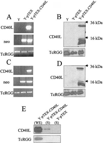
The CD40L transcript and protein were then analyzed in stable transfected epimastigotes using RT-PCR and Western blotting. Total RNA was extracted from wild-type epimastigotes (strain Y) or epimastigotes transfected with pTEX or with pTEX-CD40L, and reverse transcription was performed using the oligo(dT) primer. The cDNA obtained was used as a template for PCR amplification with a different set of specific primers. As shown in Fig. 1A, a specific fragment of the expected size (780 bp) corresponding to CD40L cDNA was detected only in CD40L-transfected epimastigotes. As a control, the neo cDNA (820 bp) was detected in epimastigotes transfected with either pTEX or pTEX-CD40L, whereas cDNA corresponding to the chromosome-encoded TcRGG gene (1,030 bp) was detected in all epimastigotes tested (39).
FIG. 1.
RT-PCR and Western blot analysis. RNA and whole-cell extracts from wild-type strain Y-transfected, pTEX-transfected (YpTEX), or YpTEX-CD40L-transfected (YpTEX-CD40L) epimastigotes were used for RT-PCR (A) and Western blot analysis (B). Trypomastigotes derived from these epimastigotes were used for RT-PCR (C) and Western blot analysis (D). Specific primers were used to amplify the cDNA indicated (neo, neomycin phosphotransferase). Whole-cell extracts (WE) or supernatants (S) from YpTEX- and YpTEX-CD40L-transfected trypomastigotes were analyzed by Western blotting (E). For protein analysis, CD40L was detected using a specific antibody, and an anti-TcRGG polyclonal antibody was used as a control.
For CD40L protein analysis, a whole-cell extract from epimastigotes grown to the log phase was analyzed by Western blotting using specific anti-CD40L antibodies (Fig. 1B). Two bands at approximately 30 kDa and 14 kDa corresponding to CD40L were detected in the protein extracts from the pTEX-CD40L-transfected cell line but not in protein extracts from the wild-type- or pTEX-transfected cell line. As a control, anti-TcRGG antibody revealed a specific band in all protein extracts tested.
Metacyclogenesis and characterization of CD40L-transfected trypomastigotes.
Transformation by metacyclogenesis of wild-type-, pTEX-, and pTEX-CD40L-transfected epimastigotes to mammalian infective metacyclic trypomastigotes was carried out in vitro using a defined medium (5, 15). We maintained transfected metacyclic forms in culture using VN5 cells expressing neomycin phosphotransferase. This process allowed us to achieve intracellular multiplication of amastigotes under selective drug pressure and consequently to ensure that CD40L was expressed in the derived trypomastigotes. To verify this, transfected trypomastigotes were analyzed by RT-PCR and Western blotting using different probes (Fig. 1C and D). CD40L cDNA was specifically amplified by RT-PCR from pTEX-CD40L-transfected trypomastigotes (designated YpTEX-CD40L) but not from wild-type-transfected trypomastigotes (designated Y) or pTEX-transfected trypomastigotes (designated YpTEX), whereas neo cDNA was detected in both transfected parasites (Fig. 1C). Western blot analysis performed with whole-cell lysates revealed two specific bands corresponding to 30 kDa and 14 kDa in only the pTEX-CD40L-transfected trypomastigotes (Fig. 1D). Anti-TcRGG antibody used as a control revealed a specific band in all protein extracts tested. To determine whether CD40L is expressed even in the absence of drug selection, first CD40L-transfected trypomastigotes were cultured with MPM without G418. One week later, trypomastigotes in the culture supernatant were harvested and then tested by Western blotting. The data confirmed that there was expression of CD40L compared to YpTEX-transfected trypomastigotes (referred to hereafter as YpTEX trypomastigotes) maintained under the same conditions. Second, similar results were obtained for YpTEX-CD40L-transfected trypomastigotes harvested after three or four successive passages in mice.
Immunolocalization of CD40L in transfected trypomastigotes.
To localize the CD40L molecule expressed by YpTEX-CD40L-transfected trypomastigotes (referred to hereafter as YpTEX CD40L trypomastigotes) immunofluorescence and a flow cytometry analysis were performed using the anti-CD40L antibody used in the Western blot test and then fluorescein isothiocyanate-conjugated rabbit anti-goat antibody. No significant staining was observed at the surface of the transfected parasites (data not shown). However, when YpTEX-CD40L trypomastigotes were permeabilized by treatment with Triton X-100 (1%) to allow penetration of the antibody probes into the cytoplasm, not surprisingly, a significant signal was detected by flow cytometry analysis compared to the signal for the YpTEX trypomastigotes (data not shown). The lack of detection of CD40L at the surface of the transfected parasites could be attributed to the rapid turnover of the molecule at the cell surface, leading to its release into the medium. To test this possibility, purified transfected parasites were incubated for 20 h in Dulbecco modified Eagle medium. At the end of this incubation, 100% of the parasites were still alive, as shown by the MTT test (38). Supernatants harvested from YpTEX and YpTEX-CD40L trypomastigotes were then tested by Western blotting for CD40L reactivity (Fig. 1E). Surprisingly, CD40L (30 kDa) was found in the supernatant of the YpTEX-CD40L trypomastigotes. As a control, no reactivity was detected in the supernatant of YpTEX trypomastigotes, while a positive reaction was observed in whole-cell extract of YpTEX-CD40L trypomastigotes, as shown in Fig. 1D. Another control using antibody directed against TcRGG, a cytoplasmic molecule with a potential nuclear sequence (39), revealed an expected positive signal in the whole-cell extract but not in the supernatant. These data indicate that CD40L can be secreted by transfected parasites.
CD40L-transfected T. cruzi trypomastigotes: infectious capacity in vitro, kinetics of parasitemia, and cumulative mortality in vivo.
We first tested the infectious capacity of transfected parasites in vitro using MPM. The three strains of trypomastigotes (Y, YpTEX, and YpTEX-CD40L) were added to MPM at a parasite-to-cell ratio of 10:1 and incubated for 48 h at 37°C. The percentages of infected MPM were 87.0 ± 2.1, 75.5 ± 3.8, and 78.3 ± 2.1% for the Y, YpTEX, and YpTEX-CD40L strains, respectively. Compared to the Y strain, no significant difference was seen with YpTEX (P > 0.05) or with YpTEX-CD40L (P > 0.05). However, the mean number of amastigotes per infected MPM was significantly higher for Y (6.9 ± 0.4) than for YpTEX (3.8 ± 0.1) and YpTEX-CD40L (3.2 ± 0.1) (P = 0.0001). These data are means ± standard errors of the means of two independent experiments (n = 11). It is important that no significant difference was observed with parasites transfected with either the pTEX or pTEX-CD40L vector (P > 0.05). These data confirmed the infectious capacity of transfected parasites previously shown with VN5 cells.
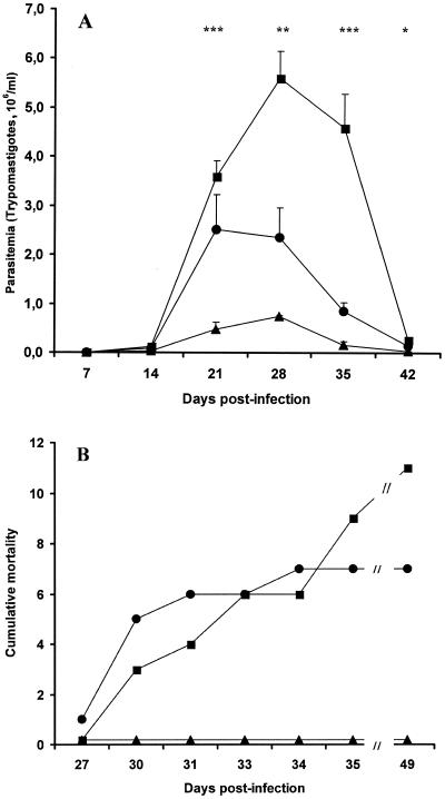
We then investigated parasitic parameters of the three strains during the course of experimental infections in mice. Three groups of 38 mice were inoculated with Y, YpTEX, and YpTEX-CD40L (1 × 10³ trypomastigotes per mouse). Parasitemia was monitored every week. Mice that were infected with the YpTEX-CD40L strain exhibited very low levels of parasitemia, while mice that were infected with the Y strain had the usual pattern of parasitemia. Interestingly, an intermediate level of parasitemia was observed for the group of YpTEX-infected mice (Fig. 2A). The surviving mice were counted every day. In each group of infected mice, 18 mice were sacrificed to harvest the spleens. The sacrificed mice were excluded from calculation of the survival rate. These experiments showed that all 20 mice infected with the YpTEX-CD40L strain survived. In contrast, 11 and 7 mice died in the groups infected with the Y and YpTEX strains, respectively (Fig. 2B). Multiple-group analysis of the survival curves confirmed that there were significant differences, as follows: for YpTEX-CD40L versus Y, P = 0.0002; for YpTEX-CD40L versus YpTEX, P = 0.004; and for YpTEX versus Y, P = 0.62 (not significant).
FIG. 2.
Parasitemia and cumulative mortality of mice infected with the Y, YpTEX, and YpTEX-CD40L strains. (A) Three groups (38 mice per group) were infected on day 0 with the Y (▪), YpTEX (•), or YpTEX-CD40L (▴) strain. Parasitemia was recorded every week. The data are means ± standard errors of the means of two independent experiments in which 15 and 23 mice per group were used. Because some mice died in the course of infection or were sacrificed to harvest the spleens, 36, 30, 28, 26, 20, and 10 mice were in the group infected with strain Y on days 7, 14, 21, 28, 35, and 42 postinfection, respectively; 36, 30, 28, 26, 18, and 15 mice were in the group infected with YpTEX on days 7, 14, 21, 28, 35, and 42 postinfection, respectively; and 36, 30, 28, 26, 24, and 22 mice were in the group infected with YpTEX-CD40L on days 7, 14, 21, 28, 35, and 42 postinfection, respectively. For comparisons of YpTEX-CD40L and YpTEX, one asterisk indicates that the P value is <0.05, two asterisks indicate that the P value is <0.01, and three asterisks indicate that the P value is <0.001 (as determined by the Mann-Whitney U test). (B) Three groups (38 mice per group) were infected on day 0 with the Y (▪), YpTEX (•), or YpTEX-CD40L (▴) strain. The number of surviving mice was recorded every day. Sacrificed mice were excluded from calculation of the survival rate. The data are from two independent experiments. The results are expressed as the number of mice that died (cumulative mortality) during the course of infection. For YpTEX-CD40L versus Y, P = 0.0002; for YpTEX-CD40L versus YpTEX, P = 0.004; for YpTEX versus Y, P = 0.62 (not significant) (as determined by Gehan's Wilcoxon test).
The kinetics of mouse weight may be considered representative of the state of health. In two independent experiments, mice (15 mice per group) were infected with the Y, YpTEX, or YpTEX-CD40L strain. The levels of parasitemia and mortality were similar to those observed in previous experiments (data not shown). In addition, mice were weighed every week. As expected, a sharp decrease in weight was observed for Y-infected mice. A slower decrease in weight was observed for the YpTEX-infected mice. For the YpTEX-CD40L-infected mice, the weight increased until day 21 postinfection, as it did in the control group of noninfected mice, in which the weight increased regularly. Then it decreased slightly until it was 26.0 ± 0.7 g, while the weights were 19.9 ± 1.2 g and 31.2 ± 0.3 g for the YpTEX-infected mice and the noninfected mice at day 35 postinfection. In summary, the weight kinetics of the infected mice corroborated the infection pattern.
Taken together, these data indicate that mice infected with the YpTEX-CD40L strain were able to control the parasitemia and to survive the acute phase of the T. cruzi infection with a minimal lost of weight.
The spleens harvested from the sacrificed mice were weighed, and the peak weight was reached at day 21 postinfection. There were no statistically significant differences between the three groups of mice; the weights were 654.8 ± 102.8 mg, 680.4 ± 106.4 mg, and 627.4 ± 101.7 mg for spleens from mice infected with Y, YpTEX, and YpTEX-CD40L, respectively. These data are means ± standard deviations from two independent experiments (n = 5). They corresponded to the usual splenomegaly observed in the course of infection (14).
Proliferation of SCs activated with T. cruzi lysate.
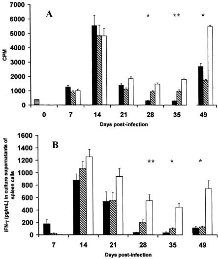
To further investigate the immunological parameters, spleens were harvested, and SCs were isolated and incubated with T. cruzi lysates (Fig. 3A). At day 14 postinfection, a high level of proliferation was observed with SCs from mice infected with the three strains. Then the indexes decreased sharply at day 21 postinfection. These data reflect the well-known immunosuppressive effect of infection upon SC proliferation (50). The proliferation was close to that of the control (day 0) for SCs from Y-infected mice at days 28 and 35 postinfection. For YpTEX-infected mice, the SC proliferation was stable at days 21, 28, 35, and 49 postinfection and remained as low as it was at day 7 postinfection. In contrast, the proliferation was partially inhibited in the case of the YpTEX-CD40L-infected mice, particularly at days 28, 35, and 49 postinfection. Taken together, these data suggest that SC proliferation, which is a marker of immunosuppression usually observed during the acute phase of the infection, was less inhibited in the course of the YpTEX-CD40L infection.
FIG. 3.
Proliferation SCs activated with T. cruzi lysates and production of IFN-γ by SCs from mice infected with the Y, YpTEX, and YpTEX-CD40L strains. Three groups of mice (66 mice per group) were infected on day 0 with the Y (solid bars), YpTEX (diagonally cross-hatched bars), and YpTEX-CD40L (open bars) strains. One group of five mice (horizontally cross-hatched bar) was not infected and served as a control (day 0 postinfection). Every week, five mice were sacrificed. (A) SCs were harvested and incubated with T. cruzi lysate. (B) IFN-γ was measured by ELISA in the culture supernatants of SCs stimulated with T. cruzi lysates. The data are means and standard errors of the means of three independent experiments. For comarisons of YpTEX-CD40L and YpTEX, one asterisk indicates that the P value is <0.05 and two asterisks indicate that the P value is <0.01 (as determined by the Mann-Whitney U test).
IFN-γ production by SCs.
IFN-γ production was measured in the supernatant of SCs stimulated with T. cruzi lysate. As shown in Fig. 3B, the production of IFN-γ increased first at day 14 postinfection for SCs from mice infected with the three strains. Then it decreased at day 21 postinfection for SCs from Y- and YpTEX- infected mice, and it remained at a very low level until day 49 postinfection. In contrast, for SCs from YpTEX-CD40L-infected mice, the production of IFN-γ was enhanced, particularly at days 28, 35, and 49 postinfection. This significant IFN-γ production fits well with the elevated cell proliferation observed at the same time with the YpTEX-CD40L strain (Fig. 3 A).
Kinetics of anti-T. cruzi antibody levels.
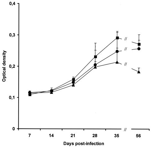
Samples of serum were harvested by cardiac puncture from Y-, YpTEX-, or YpTEX-CD40L infected mice and tested at a 1/1,000 dilution by an ELISA against T. cruzi antigen extract. The optical densities corresponding to the specific anti-T. cruzi antibody levels are shown in Fig. 4. As expected, the anti-T. cruzi antibody levels increased progressively during the course of infection. However, despite the very low level of parasitemia observed previously (Fig. 2A), the levels of anti-T. cruzi antibodies were similar in the sera of YpTEX- and YpTEX-CD40L-infected mice.
FIG. 4.
Kinetics of anti-T. cruzi antibody levels. Samples of serum were harvested by cardiac puncture from mice used in three independent experiments and infected with the Y (▪), YpTEX (•), and YpTEX-CD40L (▴) strains. Using an ELISA, the optical densities corresponding to the anti-T. cruzi antibody levels were measured. Seven samples of serum from uninfected mice served as a control (optical density, 0.107). The data are means and standard errors of the means (n = 5). There was no statistically significant difference between YpTEX-CD40L and YpTEX (as determined by a Mann-Whitney U test).
Protective capacity of YpTEX-CD40L-transfected trypomastigotes.
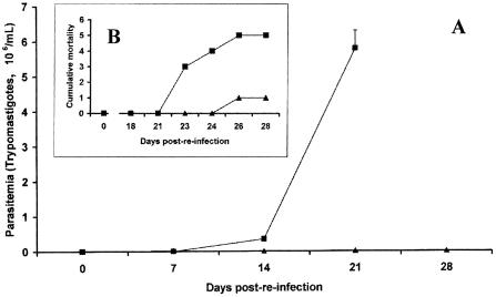
To determine the potential of the vaccine approach based on YpTEX-CD40L-transfected parasites, mice were infected with YpTEX-CD40L-transfected T. cruzi (day 0). When parasitemia was undetectable in circulating blood (starting on day 39 postinfection), the mice were intraperitoneally challenged at day 55 postinfection (day 0 of the challenge infection) with a different strain of T. cruzi trypomastigotes (Tehuantepec strain; 100 trypomastigotes). As shown in Fig. 5A, no parasites were detected in three mice, and a very low-level, transient parasitemia (not exceeding 0.051 × 10⁶ trypomastigotes/ml) was seen in three other mice in the 4 weeks following this challenge infection. This indicates that mice were effectively immunized. Control mice infected at the same time with the same inoculum exhibited the usual high level of parasitemia, confirming that the inoculum was effectively infectious. Only one mouse died in the group of YpTEX-CD40L-infected mice, while all the control mice died before day 26 postinfection (Fig. 5B).
FIG. 5.
Protective capacity of YpTEX-CD40L-transfected trypomastigotes. A group of six mice (▴) that survived after YpTEX-CD40L infection were challenged intraperitoneally at day 55 postinfection (day 0 of reinfection) with a different strain of T. cruzi trypomastigotes (Tehuantepec strain; 100 trypomastigotes). Five mice (control group) (▪) were infected at the same time with the same inoculum. (A) Parasitemia. (B) Cumulative mortality. The data are representative of one of two experiments.
DISCUSSION
In the present study, we generated a clone of T. cruzi (YpTEX-CD40L) that synthesizes murine CD40L molecules by using the pTEX shuttle vector that is widely used to express heterologous DNA sequences in the trypanosome genetic environment (24). Because CD40L participates in the triggering of the immune response via its interaction with its cognate CD40 receptor expressed on various immunocompetent cells, we also investigated the immunomodulation induced by this CD40L-transfected parasite during the course of infection in mice.
To our knowledge, this is the first report about a transfected protozoan parasite producing a biologically active host-costimulatory molecule (CD40L). The production of CD40L by transfected parasites was assessed by RT-PCR and Western blotting both in epimastigotes and in the derived trypomastigotes obtained by in vitro metacyclogenesis. Moreover, the stability of CD40L production in transfected parasites was confirmed after several passages in mice, indicating that plasmid DNA was stably established even in the absence of drug selection. The stability of such a transfection is in line with the results of other studies in which shuttle vector pTEX was used to transfect different molecules in T. cruzi (8, 35, 37).
Western blot analysis showed that, besides the 30-kDa fragment corresponding to the expected molecular size of CD40L, a lower band at 14 kDa was also present for both epimastigotes and trypomastigotes. This band could have been due to a degradation product or to an internal ATG start (Met 147), resulting in the carboxy-terminal part of CD40L. This was suggested by the presence of a Shine-Dalgarno-like sequence close to Met-147, 5′-AAA GGA TAT TAT ACC ATG. A fragment that had a similar molecular size was reported in another study of recombinant soluble CD40L produced in E. coli (32). This soluble extracellular carboxy-terminal region of CD40L encompassing TNF homology sequences and containing receptor-binding domains can be expressed as a soluble molecule with biological activity (17, 36).
Our data showed that mice inoculated with the YpTEX-CD40L strain of T. cruzi exhibited reduced parasitemia and no mortality, reflecting the biological activity of CD40L produced by transfected parasites. This feature suggests that the molecule was properly processed and presented to trigger immunocompetent cells.
Attempts have been made to localize the CD40L molecule in different parasite extracts by Western blotting, and we have detected the molecule in the nonsoluble membrane-enriched fraction (data not shown). This suggests that the parasite can effectively recognize a higher eukaryotic peptide signal that allows export of the host molecule to the membrane. No significant signal has been observed at the surface of the transfected parasite using the same specific anti-CD40L antibody in immunofluorescence and flow cytometry analyses. However, CD40L was detected by Western blotting in culture supernatant, supporting the possibility that the transfected CD40L molecule undergoes rapid turnover at the cell surface that leads to secretion of the molecule. Other studies have shown that host cytokines, such as IFN-γ, can be properly processed and secreted into media across the parasite membrane in Leishmania major (48) and Plasmodium knowlesi (41).
Although CD40L exists in nature predominantly as a membrane-anchored molecule, the soluble form of CD40L may be biologically active (2, 17, 32, 36). It is likely that CD40L can be released from the parasites in the course of infection, activates immunocompetent cells, and promotes an effective and protective immune response against T. cruzi infection. It is also possible that CD40L can be released following lysis of part of the parasite population during infection and could contribute to such enhanced immunity. The infective capacity of the YpTEX and YpTEX-CD40L trypomastigotes was tested by incubating them with MPM, which are usual host cells for T. cruzi (55). Like the Y strain, these two strains were able to infect more than 70% of MPM. The mean number of YpTEX amastigotes per infected cell was similar to the number obtained with YpTEX-CD40L parasites, suggesting that CD40L does not interfere with the multiplication rate of the transfected parasite. However, the mean numbers of amastigotes for the transfected parasites remained lower than the number of amastigotes for wild-type strain Y. This suggests that the presence of the plasmid DNA in the parasite might slow the multiplication of the amastigotes.
Despite the similar size of the inoculum (1 × 10³ trypomastigotes), reduced parasitemia and no mortality were observed in YpTEX-CD40L-infected mice. This is in line with previous results showing that CD40L-transfected fibroblasts have a protective effect when they are injected together with T. cruzi trypomastigotes (13). Our results suggest that synthesis of CD40L molecules occurred effectively in the transfected YpTEX-CD40L strain in vivo. It is likely that immunocompetent cells were overstimulated by the parasite itself expressing and releasing CD40L as early as the inoculation occurred and then during its tremendous expansion in blood and various organs and tissues (8, 59), so that many of the escape mechanisms usually induced by the nontransfected parasites were bypassed.
The YpTEX strain induced a level of parasitemia lower than that induced by the Y strain. This was probably due to the genetic transfection that attenuated its virulence, as observed for in vitro infection of MPM. This suggests that the presence of foreign plasmid DNA in the parasite might interfere with the multiplication of the amastigotes in vivo. Because the YpTEX-CD40L strain induced a level of parasitemia lower than that induced by the YpTEX strain, expression of CD40L is likely to be involved in the reduction of the infectivity of the YpTEX-CD40L strain.
As is the case for many other pathogens, the evolutionary adaptation of T. cruzi allows the parasite to evade the immune system and to induce dramatic immunodeficiency by using various molecular strategies (1, 12, 29, 47, 52, 53, 56). CD40L-transfected parasites exhibited less pronounced suppression of SC proliferation than the wild-type Y and YpTEX strains. Indeed, the sharply reduced SC proliferation observed with the Y, YpTEX, and YpTEX-CD40L strains at day 21 postinfection was very similar to the results described previously (50). However, the proliferation of SCs from YpTEX-CD40L-infected mice was greater than that observed with the Y and YpTEX strains at days 28, 35, and 49 postinfection. It is likely that the expression of CD40L molecules by the parasite itself accounts for sustained T-cell activation and therefore counteracts the immunodeficiency induced by parasite-derived molecules, such as glycoinositolphospholipids (7, 19, 25, 34, 52, 53). In relation to the higher SC proliferation, the production of IFN-γ, a cardinal cytokine in the anti-infective immune response, was clearly enhanced in supernatants of SCs from YpTEX-CD40L-infected mice from day 21 to day 49 postinfection. IFN-γ production was shown to be related to CD40/CD40L ligation (10). This suggests that parasites expressing CD40L are able to stimulate a more efficient type 1 immune response.
Various immunocompetent cells expressing CD40 are probably activated by the YpTEX-CD40L trypomastigotes. Although the expression of CD40 molecules seems to be reduced in infected DCs and macrophages (7, 42, 52), it should be considered that their activation could be overstimulated by YpTEX-CD40L-transfected parasites and therefore their immunological function might be partially restored.
CD8+ lymphocytes seem to be of prime importance in the control of infection by destroying major histocompatibility complex class I-positive infected cells (16) and by producing a large amount of IFN-γ (31). In addition, CD8+ T-cell memory can be generated with the “help” of CD4 via the CD40 molecule (6). Therefore, the CD40/CD40L pathway plays a central role in the immune response mediated by both CD4+ and CD8+ T cells (21). Further investigations are required to determine the immunocompetent cells involved in the reduced infection induced by the YpTEX-CD40L strain. Preliminary experiments indicated that the percentage of CD8+ SCs increased during the course of infection, particularly in mice inoculated with the YpTEX-CD40L strain. Moreover, the flow cytometry analysis showed that the high level of production of IFN-γ was mainly due to CD8+ cells (data not shown). Ongoing investigations should clarify the role of these cells in the control of CD40L-transfected parasite infection.
In the course of T. cruzi murine infection, protective antibodies specific for T. cruzi are produced (26, 27). This production is correlated with the level of parasitemia reached during the acute phase of infection (30). Our results show that, despite a very low level of parasitemia, a significant level of T. cruzi-specific antibodies was induced by the YpTEX-CD40L strain. CD40L is indeed involved in the stimulation of the humoral response through T-cell-dependent B-cell activation, cytokine-mediated B-cell activation, and immunoglobulin isotype switching (3, 4). Future investigations will be concerned with analysis of such a humoral response generated against YpTEX-CD40L-transfected parasites and the potential involvement of these antibodies in protective immunity.
A successful experimental vaccine based on live modified parasites should lead to 100% survival in the preinfection stage and to stimulation of a protective immune response. Preinfection of mice with the YpTEX-CD40L-transfected parasites clearly resulted in a very low level of parasitemia and an absence of mortality. Furthermore, the mice developed sustained immunity against the parasite.
Further investigations related to functional activities of delivered host costimulatory CD40L have shown that surviving mice infected with YpTEX-CD40L-transfected parasites resist a challenge infection. This indicates that the YpTEX-CD40L-transfected parasites strongly stimulate a protective immune response. Such data open new perspectives for a novel vaccine approach based on a CD40L-transfected pathogen. Future work will focus on characterization of the immunological mechanisms involved in such protection and on comparing these mechanisms to those described with other known attenuated parasites (44, 45).
In conclusion, we generated T. cruzi expressing a host costimulatory molecule and demonstrated that CD40L-transfected parasites can counteract the immunodeficiency induced by the parasite itself. These data open new perspectives for immunomodulation regarding the control of parasite infection.
Acknowledgments
This work was supported by a grant from the Centre de Recherche Interuniversitaire en Vaccinologie.
We are very grateful to Y. de Launoit (Laboratoire de Virologie Moléculaire, Faculté de Médecine, ULB, Brussels, Belgium) for kindly providing the VN5 cells, to D. Le Ray (Tropical Medicine Institute, Antwerp, Belgium) for providing the T. cruzi Y strain, and to K. Thielemans (Laboratorium Fysiologie, Faculteit Geneeskunde, Vrije Universiteit Brussel, Brussels, Belgium) for providing CD40L cDNA. We acknowledge the help of Christine Decaestecker (Laboratoire de Toxicologie, Institut de Pharmacie, ULB, Brussels, Belgium) with the statistical analysis and the help of Iolanda Mazza in preparation of the manuscript.
Editor: J. F. Urban, Jr.
REFERENCES
- 1.Abrahamsohn, I. A., and R. L. Coffman. 1995. Cytokine and nitric oxide regulation of the immunosuppression in Trypanosoma cruzi infection. J. Immunol. 155:3955-3963. [PubMed] [Google Scholar]
- 2.Armitage, R. J., W. C. Fanslow, L. Strockbine, T. A. Sato, K. N. Clifford, B. M. Macduff, D. M. Anderson, S. D. Gimpel, T. Davis-Smith, and C. R. Maliszewski. 1992. Molecular and biological characterization of a murine ligand for CD40. Nature 357:80-82. [DOI] [PubMed] [Google Scholar]
- 3.Armitage, R. J., and M. R. Alderson. 1995. B-cell stimulation. Curr. Opin. Immunol. 7:2-247. [DOI] [PubMed] [Google Scholar]
- 4.Bishop, G. A., and B. S. Hostager. 2001. Signaling by CD40 and its mimics in B cell activation. Immunol. Res. 24:97-109. [DOI] [PubMed] [Google Scholar]
- 5.Bonaldo, M. C., T. Souto-Padron, W. de Souza, and S. Goldenberg. 1988. Cell-substrate adhesion during Trypanosoma cruzi differentiation. J. Cell Biol. 106:1349-1358. [DOI] [PMC free article] [PubMed] [Google Scholar]
- 6.Bourgeois, C., B. Rocha, and C. Tanchot. 2002. A role for CD40 expression on CD8+ T cells in the generation of CD8+ T cell memory. Science 297:2060-2063. [DOI] [PubMed] [Google Scholar]
- 7.Brodskyn, C., J. Patricio, R. Oliveira, L. Lobo, A. Arnholdt, L. Mendonca-Previato, A. Barral, and M. Barral-Netto. 2002. Glycoinositolphospholipids from Trypanosoma cruzi interfere with macrophages and dendritic cell responses. Infect. Immun. 70:3736-3737. [DOI] [PMC free article] [PubMed] [Google Scholar]
- 8.Buckner, F. S., A. J. Wilson, and W. C. Van Voorhis. 1999. Detection of live Trypanosoma cruzi in tissues of infected mice by using histochemical stain for beta-galactosidase. Infect. Immun. 67:403-409. [DOI] [PMC free article] [PubMed] [Google Scholar]
- 9.Camargo, E. P. 1964. Growth and differentiation in Trypanosoma cruzi. I. Origin of metacyclic trypanosomes in liquid media. Rev. Inst. Med. Trop. Sao Paulo 12:93-100. [PubMed] [Google Scholar]
- 10.Cella, M., D. Scheidegger, K. Palmer-Lehmann, P. Lane, A. Lanzavecchia, and G. Alber. 1996. Ligation of CD40 on dendritic cells triggers production of high levels of interleukin-12 and enhances T cell stimulatory capacity: T-T help via APC activation. J. Exp. Med. 184:747-752. [DOI] [PMC free article] [PubMed] [Google Scholar]
- 11.Cella, M., F. Facchetti, A. Lanzavecchia, and M. Colonna. 2000. Plasmacytoid dendritic cells activated by influenza virus and CD40L drive a potent TH1 polarization. Nat. Immunol. 1:305-310. [DOI] [PubMed] [Google Scholar]
- 12.Cerrone, M. C., D. M. Ritter, and R. E. Kuhn. 1992. Effect of antigen-specific T helper cells or interleukin-2 on suppressive ability of macrophage subsets detected in spleens of Trypanosoma cruzi-infected mice as determined by limiting dilution-partition analysis. Infect. Immun. 60:1489-1498. [DOI] [PMC free article] [PubMed] [Google Scholar]
- 13.Chaussabel, D., F. Jacobs, J. de Jonge, M. de Veerman, Y. Carlier, K. Thielemans, M. Goldman, and B. Vray. 1999. CD40 ligation prevents Trypanosoma cruzi infection through interleukin-12 upregulation. Infect. Immun. 67:1929-1934. [DOI] [PMC free article] [PubMed] [Google Scholar]
- 14.Chaussabel, D., B. Pajak, V. Vercruysse, C. Bisseye, V. Garze, M. Habib, M. Goldman, M. Moser, and B. Vray. 2003. Alteration of migration and maturation of dendritic cells and T-cell depletion in the course of experimental Trypanosoma cruzi infection. Lab. Investig. 83:1373-1382. [DOI] [PubMed] [Google Scholar]
- 15.Contreras, V. T., J. M. Salles, N. Thomas, C. M. Morel, and S. Goldenberg. 1985. In vitro differentiation of Trypanosoma cruzi under chemically defined conditions. Mol. Biochem. Parasitol. 16:315-327. [DOI] [PubMed] [Google Scholar]
- 16.DosReis, G. A. 1997. Cell-mediated immunity in experimental Trypanosoma cruzi infection. Parasitol. Today 13:335-342. [DOI] [PubMed] [Google Scholar]
- 17.Fanslow, W. C., S. Srinivasan, R. Paxton, M. G. Gibson, M. K. Spriggs, and R. J. Armitage. 1994. Structural characteristics of CD40 ligand that determine biological function. Semin. Immunol. 6:267-278. [DOI] [PubMed] [Google Scholar]
- 18.Ferlin, W. G., W. T. von der, F. Cottrez, D. A. Ferrick, R. L. Coffman, and M. C. Howard. 1998. The induction of a protective response in Leishmania major-infected BALB/c mice with anti-CD40 mAb. Eur. J. Immunol. 28:525-531. [DOI] [PubMed] [Google Scholar]
- 19.Gomes, N. A., J. O. Previato, B. Zingales, L. Mendonca-Previato, and G. A. DosReis. 1996. Down-regulation of T lymphocyte activation in vitro and in vivo induced by glycoinositolphospholipids from Trypanosoma cruzi. Assignment of the T cell-suppressive determinant to the ceramide domain. J. Immunol. 156:628-635. [PubMed] [Google Scholar]
- 20.Grewal, I. S., P. Borrow, E. G. Pamer, M. B. Oldstone, and R. A. Flavell. 1997. The CD40-CD154 system in anti-infective host defense. Curr. Opin. Immunol. 9:491-497. [DOI] [PubMed] [Google Scholar]
- 21.Grewal, I. S., and R. A. Flavell. 1998. CD40 and CD154 in cell-mediated immunity. Annu. Rev. Immunol. 16:111-135. [DOI] [PubMed] [Google Scholar]
- 22.Gurunathan, S., K. R. Irvine, C. Y. Wu, J. I. Cohen, E. Thomas, C. Prussin, N. P. Restifo, and R. A. Seder. 1998. CD40 ligand/trimer DNA enhances both humoral and cellular immune responses and induces protective immunity to infectious and tumor challenge. J. Immunol. 161:4563-4571. [PMC free article] [PubMed] [Google Scholar]
- 23.Heinzel, F. P., R. M. Rerko, and A. M. Hujer. 1998. Underproduction of interleukin-12 in susceptible mice during progressive leishmaniasis is due to decreased CD40 activity. Cell. Immunol. 184:129-142. [DOI] [PubMed] [Google Scholar]
- 24.Kelly, J. M., H. M. Ward, M. A. Miles, and G. Kendall. 1992. A shuttle vector which facilitates the expression of transfected genes in Trypanosoma cruzi and Leishmania. Nucleic Acids Res. 20:3963-3969. [DOI] [PMC free article] [PubMed] [Google Scholar]
- 25.Kierszenbaum, F., and M. B. Sztein. 2001. Down-regulation of human B lymphocyte activities by a Trypanosoma cruzi membrane glycoprotein. Int. J. Parasitol. 31:265-271. [DOI] [PubMed] [Google Scholar]
- 26.Krautz, G. M., J. C. Kissinger, and A. U. Krettli. 2000. The targets of the lytic antibody response against Trypanosoma cruzi. Parasitol. Today 16:31-34. [DOI] [PubMed] [Google Scholar]
- 27.Kumar, S., and R. L. Tarleton. 1998. The relative contribution of antibody production and CD8+ T cell function to immune control of Trypanosoma cruzi. Parasite Immunol. 20:207-216. [DOI] [PubMed] [Google Scholar]
- 28.Lane, P., T. Brocker, S. Hubele, E. Padovan, A. Lanzavecchia, and F. McConnell. 1993. Soluble CD40 ligand can replace the normal T cell-derived CD40 ligand signal to B cells in T cell-dependent activation. J. Exp. Med. 177:1209-1213. [DOI] [PMC free article] [PubMed] [Google Scholar]
- 29.Lopes, M. F., and G. A. DosReis. 1996. Trypanosoma cruzi-induced immunosuppression: selective triggering of CD4+ T-cell death by the T-cell receptor-CD3 pathway and not by the CD69 or Ly-6 activation pathway. Infect. Immun. 64:1559-1564. [DOI] [PMC free article] [PubMed] [Google Scholar]
- 30.Marinho, C. R., M. R. D'Imperio Lima, M. G. Grisotto, and J. M. Alvarez. 1999. Influence of acute-phase parasite load on pathology, parasitism, and activation of the immune system at the late chronic phase of Chagas' disease. Infect. Immun. 67:308-318. [DOI] [PMC free article] [PubMed] [Google Scholar]
- 31.Martin, D., and R. Tarleton. 2004. Generation, specificity, and function of CD8 T cells in Trypanosoma cruzi infection. Immunol. Rev. 201:304-317. [DOI] [PubMed] [Google Scholar]
- 32.Mazzei, G. J., M. D. Edgerton, C. Losberger, S. Lecoanet-Henchoz, P. Graber, A. Durandy, J. F. Gauchat, A. Bernard, B. Allet, and J. Y. Bonnefoy. 1995. Recombinant soluble trimeric CD40 ligand is biologically active. J. Biol. Chem. 270:7025-7028. [DOI] [PubMed] [Google Scholar]
- 33.Metz, G., Y. Carlier, and B. Vray. 1993. Trypanosoma cruzi upregulates nitric oxide release by IFN-gamma-preactivated macrophages, limiting cell infection independently of the respiratory burst. Parasite Immunol. 15:693-699. [DOI] [PubMed] [Google Scholar]
- 34.Millar, A. E., M. Wleklinski-Lee, and S. J. Kahn. 1999. The surface protein superfamily of Trypanosoma cruzi stimulates a polarized Th1 response that becomes anergic. J. Immunol. 162:6092-6099. [PubMed] [Google Scholar]
- 35.Miyahira, Y., S. Kobayashi, T. Takeuchi, T. Kamiyama, T. Nara, J. Nakajima-Shimada, and T. Aoki. 1999. Induction of CD8+ T cell-mediated protective immunity against Trypanosoma cruzi. Int. Immunol. 11:133-141. [DOI] [PubMed] [Google Scholar]
- 36.Morris, A. E., R. L. Remmele, Jr., R. Klinke, B. M. Macduff, W. C. Fanslow, and R. J. Armitage. 1999. Incorporation of an isoleucine zipper motif enhances the biological activity of soluble CD40L (CD154). J. Biol. Chem. 274:418-423. [DOI] [PubMed] [Google Scholar]
- 37.Norris, K. A. 1998. Stable transfection of Trypanosoma cruzi epimastigotes with the trypomastigote-specific complement regulatory protein cDNA confers complement resistance. Infect. Immun. 66:2460-2465. [DOI] [PMC free article] [PubMed] [Google Scholar]
- 38.Olivares Fontt, E., P. De Baetselier, C. Heirman, K. Thielemans, R. Lucas, and B. Vray. 1998. Effects of granulocyte-macrophage colony-stimulating factor and tumor necrosis factor alpha on Trypanosoma cruzi trypomastigotes. Infect. Immun. 66:2722-2727. [DOI] [PMC free article] [PubMed] [Google Scholar]
- 39.Ouaissi, A., B. Vergnes, M. Borges, and E. Guilvard. 2000. Identification and molecular characterization of two novel Trypanosoma cruzi genes encoding polypeptides sharing sequence motifs found in proteins involved in RNA editing reactions. Gene 253:271-280. [DOI] [PubMed] [Google Scholar]
- 40.Oz, H. S., W. T. Hughes, E. K. Thomas, and C. J. McClain. 2002. Effects of immunomodulators on acute Trypanosoma cruzi infection in mice. Med. Sci. Monit. 8:BR208-BR211.12070425 [Google Scholar]
- 41.Ozwara, H., A. van der Wel, C. H. Kocken, and A. W. Thomas. 2003. Heterologous promoter activity in stable and transient Plasmodium knowlesi transgenes. Mol. Biochem. Parasitol. 130:61-64. [DOI] [PubMed] [Google Scholar]
- 42.Planelles, L., M. C. Thomas, C. Maranon, M. Morell, and M. C. Lopez. 2003. Differential CD86 and CD40 co-stimulatory molecules and cytokine expression pattern induced by Trypanosoma cruzi in APCs from resistant or susceptible mice. Clin. Exp. Immunol. 131:41-47. [DOI] [PMC free article] [PubMed] [Google Scholar]
- 43.Prata, A. 2001. Clinical and epidemiological aspects of Chagas disease. Lancet Infect. Dis. 1:92-100. [DOI] [PubMed] [Google Scholar]
- 44.Revelli, S., L. Gomez, J. Wietzerbin, O. Bottasso, and M. A. Basombrio. 1999. Levels of tumor necrosis factor alpha, gamma interferon, and interleukins 4,6, and 10 as determined in mice infected with virulent or attenuated strains of Trypanosoma cruzi. Parasitol. Res. 85:147-150. [DOI] [PubMed] [Google Scholar]
- 45.Soares, M. B., R. Goncalves, A. S. Pyrrho, D. A. Costa, C. N. Paiva, and C. R. Gattass. 2003. Balanced cytokine-producing pattern in mice immunized with an avirulent Trypanosoma cruzi. An. Acad. Bras. Cienc. 75:167-172. [DOI] [PubMed] [Google Scholar]
- 46.Soong, L., J. C. Xu, I. S. Grewal, P. Kima, J. Sun, B. J. Longley, N. H. Ruddle, D. McMahon-Pratt, and R. A. Flavell. 1996. Disruption of CD40-CD40 ligand interactions results in an enhanced susceptibility to Leishmania amazonensis infection. Immunity 4:263-273. [DOI] [PubMed] [Google Scholar]
- 47.Tarleton, R. L. 1988. Trypanosoma cruzi-induced suppression of IL-2 production. II. Evidence for a role for suppressor cells. J. Immunol. 140:2769-2773. [PubMed] [Google Scholar]
- 48.Tobin, J. F., S. L. Reiner, F. Hatam, S. Zheng, C. L. Leptak, D. F. Wirth, and R. M. Locksley. 1993. Transfected Leishmania expressing biologically active IFN-gamma. J. Immunol. 150:5059-5069. [PubMed] [Google Scholar]
- 49.Tyler, K. M., and D. M. Engman. 2001. The life cycle of Trypanosoma cruzi revisited. Int. J. Parasitol. 31:472-480. [DOI] [PubMed] [Google Scholar]
- 50.Vandekerckhove, F., A. Darji, M. T. Rivera, Y. Carlier, B. Vray, A. Billiau, and P. De Baetselier. 1994. Modulation of T-cell responsiveness during Trypanosoma cruzi infection: analysis in different lymphoid compartments. Parasite Immunol. 16:77-85. [DOI] [PubMed] [Google Scholar]
- 51.van Kooten, C., and J. Banchereau. 2000. CD40-CD40 ligand. J. Leukoc. Biol. 67:2-17. [DOI] [PubMed] [Google Scholar]
- 52.Van Overtvelt, L., N. Vanderheyde, V. Verhasselt, J. Ismaili, L. De Vos, M. Goldman, F. Willems, and B. Vray. 1999. Trypanosoma cruzi infects human dendritic cells and prevents their maturation: inhibition of cytokines, HLA-DR, and costimulatory molecules. Infect. Immun. 67:4033-4040. [DOI] [PMC free article] [PubMed] [Google Scholar]
- 53.Van Overtvelt, L., M. Andrieu, V. Verhasselt, F. Connan, J. Choppin, V. Vercruysse, M. Goldman, A. Hosmalin, and B. Vray. 2002. Trypanosoma cruzi down-regulates lipopolysaccharide-induced MHC class I on human dendritic cells and impairs antigen presentation to specific CD8(+) T lymphocytes. Int. Immunol. 14:1135-1144. [DOI] [PubMed] [Google Scholar]
- 54.Vray, B., P. De Baetselier, A. Ouaissi, and Y. Carlier. 1991. Trypanosoma cruzi but not Trypanosoma brucei fails to induce a chemiluminescent signal in a macrophage hybridoma cell line. Infect. Immun. 59:3303-3308. [DOI] [PMC free article] [PubMed] [Google Scholar]
- 55.Vray, B. 2002. Macrophages in parasitic infection. p. 253-304. In B. Burke and C. E. Lewis (ed.), The macrophage. Oxford University Press, Oxford, UK.
- 56.Vray, B., I. Camby, V. Vercruysse, T. Mijatovic, N. V. Bovin, P. Ricciardi-Castagnoli, H. Kaltner, I. Salmon, H. J. Gabius, and R. Kiss. 2004. Upregulation of galectin-3 and its ligands by Trypanosoma cruzi infection with modulation of adhesion and migration of murine dendritic cells. Glycobiology 14:647-657. [DOI] [PubMed] [Google Scholar]
- 57.Wykes, M. 2003. Why do B cells produce CD40 ligand? Immunol. Cell Biol. 81:328-331. [DOI] [PubMed] [Google Scholar]
- 58.Xiang, R., F. J. Primus, J. M. Ruehlmann, A. G. Niethammer, S. Silletti, H. N. Lode, C. S. Dolman, S. D. Gillies, and R. A. Reisfeld. 2001. A dual-function DNA vaccine encoding carcinoembryonic antigen and CD40 ligand trimer induces T cell-mediated protective immunity against colon cancer in carcinoembryonic antigen-transgenic mice. J. Immunol. 167:4560-4565. [DOI] [PubMed] [Google Scholar]
- 59.Younes-Chennoufi, A., M. Hontebeyrie-Joskowicz, V. Tricottet, H. Eisen, M. Reynes, and G. Said. 1988. Persistence of Trypanosoma cruzi antigens in the inflammatory lesions of chronically infected mice. Trans. R. Soc. Trop. Med. Hyg. 82:77-83. [DOI] [PubMed] [Google Scholar]


