

Abstract
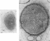
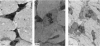
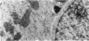
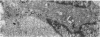
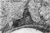
Optic nerves from perfusion-fixed human embryos of 28,50,75,105,120 and 165 mm crown-rump length were examined in the electron microscope. The number of glial cells per section was found to increase steadily from 10 weeks post-conception to 18 weeks and a close correlation (r = 0.92) was found between the percentage vascularity and the glial population. Mitotic figures were present in all optic nerves examined. From 14 weeks onwards all glial cells, except pericytes, were found to be fibrous astrocytes. The human fibrous astrocyte appears to pass through the following stages of development: (1) Astrocytic precursors (dark glioblasts) have a dense cytoplasmic matrix with few organelles, although a single cilium is frequently present.(2) Concomitant with the increase in vascularization of the optic nerve found between 12 and 14 weeks glycogen granules increase in the cytoplasm of astrocytic precursors, followed by microfibrils, which appear first in the processes and later extend into the perikaryon. (3) With the appearance of glycogen granules the cytoplasmic organelles, particularly mitochondria, increase in amount and the cytoplasmic matrix gradually becomes less dense. (4) With increasing age fewer organelles are found in astrocytic processes, which become thinner and densely packed with microfibrils.
Full text
PDF
Images in this article


Selected References
These references are in PubMed. This may not be the complete list of references from this article.
- BARNES B. G. Ciliated secretory cells in the pars distalis of the mouse hypophysis. J Ultrastruct Res. 1961 Oct;5:453–467. doi: 10.1016/s0022-5320(61)80019-1. [DOI] [PubMed] [Google Scholar]
- Blunt M. J., Baldwin F., Wendell-Smith C. P. Gliogenesis and myelination in kitten optic nerve. Z Zellforsch Mikrosk Anat. 1972;124(3):293–310. doi: 10.1007/BF00355032. [DOI] [PubMed] [Google Scholar]
- Caley D. W., Maxwell D. S. An electron microscopic study of the neuroglia during postnatal development of the rat cerebrum. J Comp Neurol. 1968 May;133(1):45–70. doi: 10.1002/cne.901330104. [DOI] [PubMed] [Google Scholar]
- Davison P. F., Huneeus F. C. Fibrillar proteins from squid axons. II. Microtubule protein. J Mol Biol. 1970 Sep 28;52(3):429–439. doi: 10.1016/0022-2836(70)90411-0. [DOI] [PubMed] [Google Scholar]
- Dobbing J., Sands J. Timing of neuroblast multiplication in developing human brain. Nature. 1970 May 16;226(5246):639–640. doi: 10.1038/226639a0. [DOI] [PubMed] [Google Scholar]
- Keene M. F. Some Observations on Myelination in the Human Central Nervous System. J Anat. 1931 Oct;66(Pt 1):1–13. [PMC free article] [PubMed] [Google Scholar]
- Mori S., Leblond C. P. Electron microscopic features and proliferation of astrocytes in the corpus callosum of the rat. J Comp Neurol. 1969 Oct;137(2):197–225. doi: 10.1002/cne.901370206. [DOI] [PubMed] [Google Scholar]
- Peters A., Vaughn J. E. Microtubules and filaments in the axons and astrocytes of early postnatal rat optic nerves. J Cell Biol. 1967 Jan;32(1):113–119. doi: 10.1083/jcb.32.1.113. [DOI] [PMC free article] [PubMed] [Google Scholar]
- Phillips D. E. An electron microscopic study of macroglia and microglia in the lateral funiculus of the developing spinal cord in the fetal monkey. Z Zellforsch Mikrosk Anat. 1973 Jun 28;140(2):145–167. doi: 10.1007/BF00306691. [DOI] [PubMed] [Google Scholar]
- Sorokin S. P. Reconstructions of centriole formation and ciliogenesis in mammalian lungs. J Cell Sci. 1968 Jun;3(2):207–230. doi: 10.1242/jcs.3.2.207. [DOI] [PubMed] [Google Scholar]
- Sturrock R. R. Histogenesis of the anterior limb of the anterior commissure of the mouse brain. 3. An electron microscopic study of gliogenesis. J Anat. 1974 Feb;117(Pt 1):37–53. [PMC free article] [PubMed] [Google Scholar]
- Vaughn J. E. An electron microscopic analysis of gliogenesis in rat optic nerves. Z Zellforsch Mikrosk Anat. 1969;94(3):293–324. doi: 10.1007/BF00319179. [DOI] [PubMed] [Google Scholar]
- Vaughn J. E., Peters A. Electron microscopy of the early postnatal development of fibrous astrocytes. Am J Anat. 1967 Jul;121(1):131–152. doi: 10.1002/aja.1001210109. [DOI] [PubMed] [Google Scholar]
- Wuerker R. B., Kirkpatrick J. B. Neuronal microtubules, neurofilaments, and microfilaments. Int Rev Cytol. 1972;33:45–75. doi: 10.1016/s0074-7696(08)61448-5. [DOI] [PubMed] [Google Scholar]