Abstract
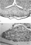
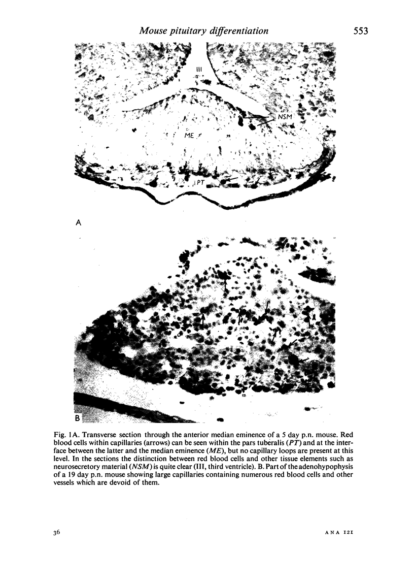
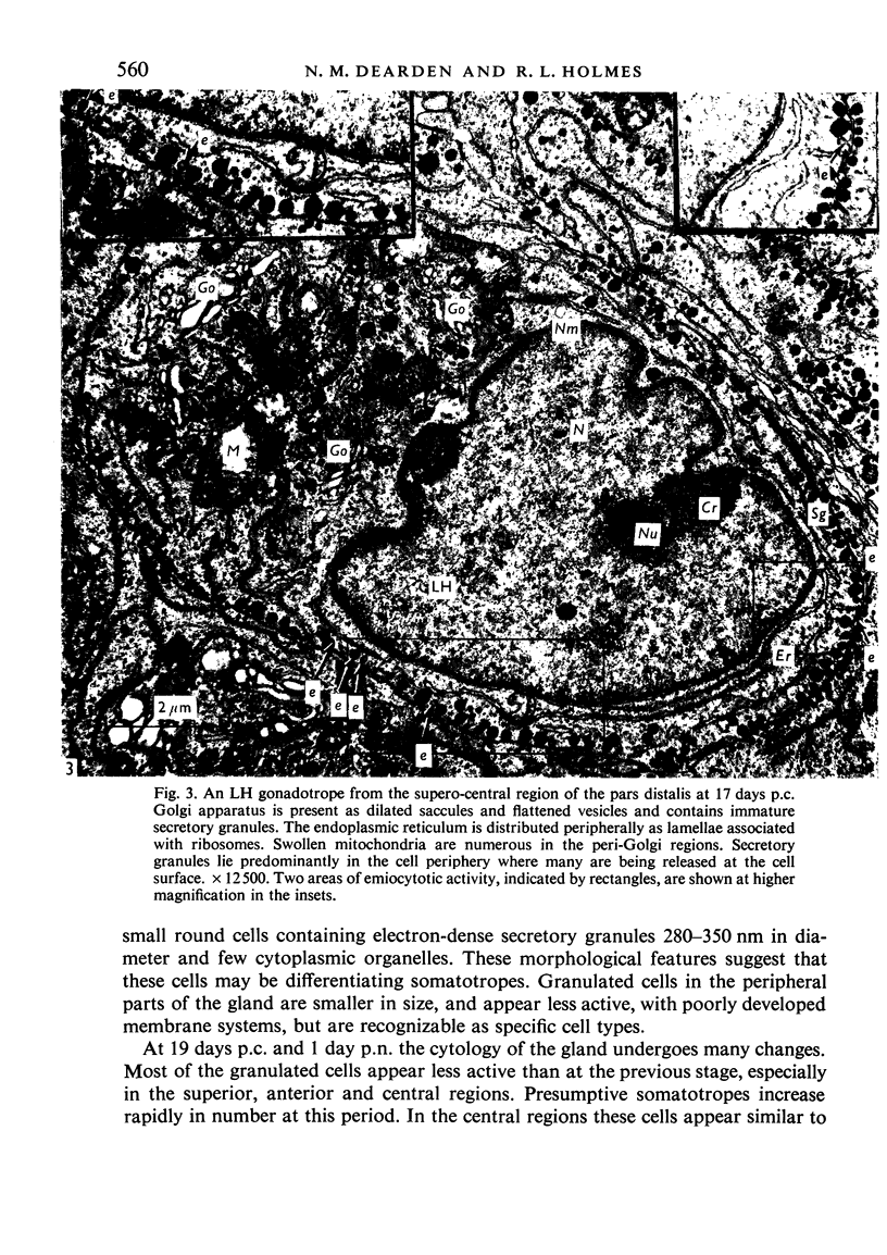
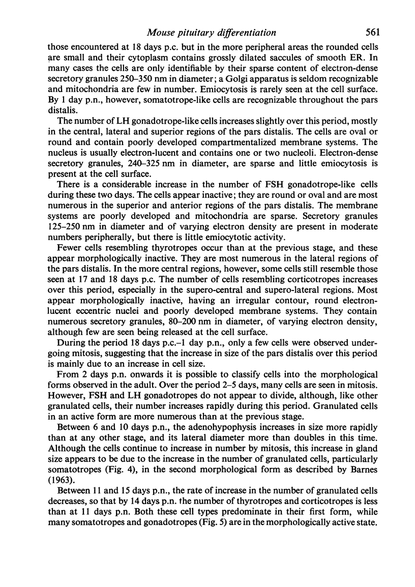
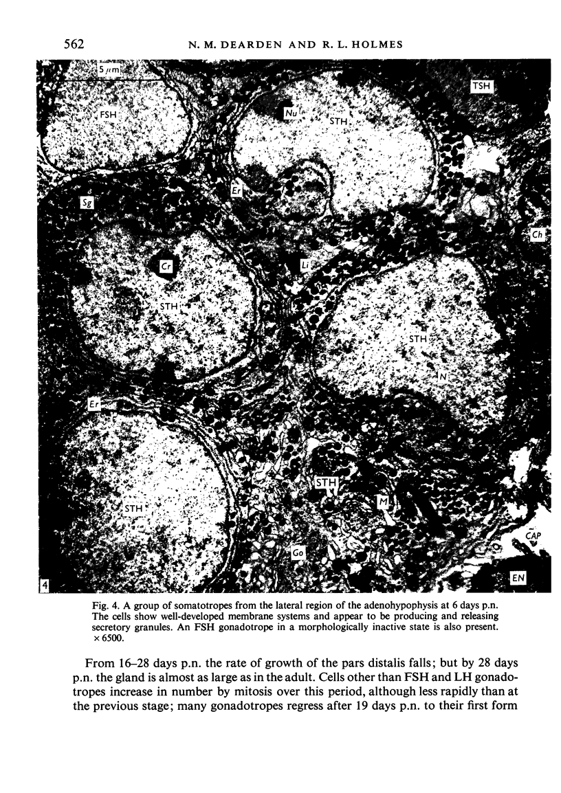
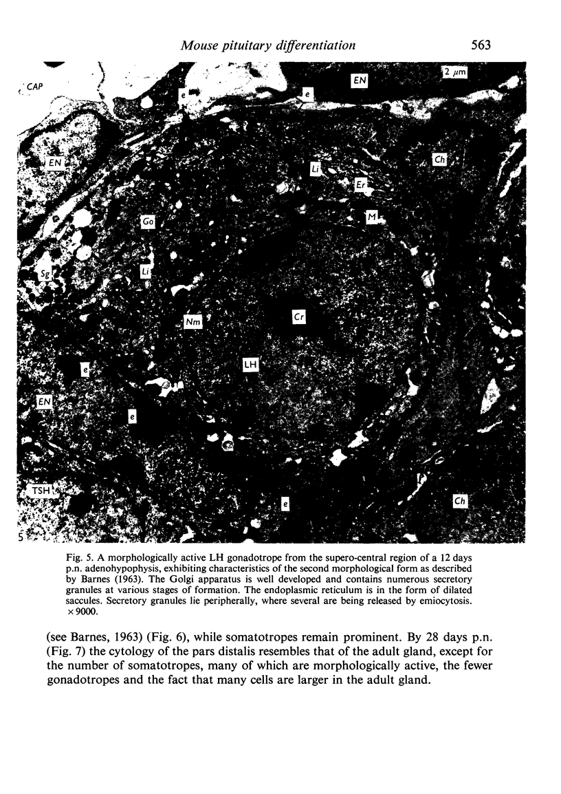
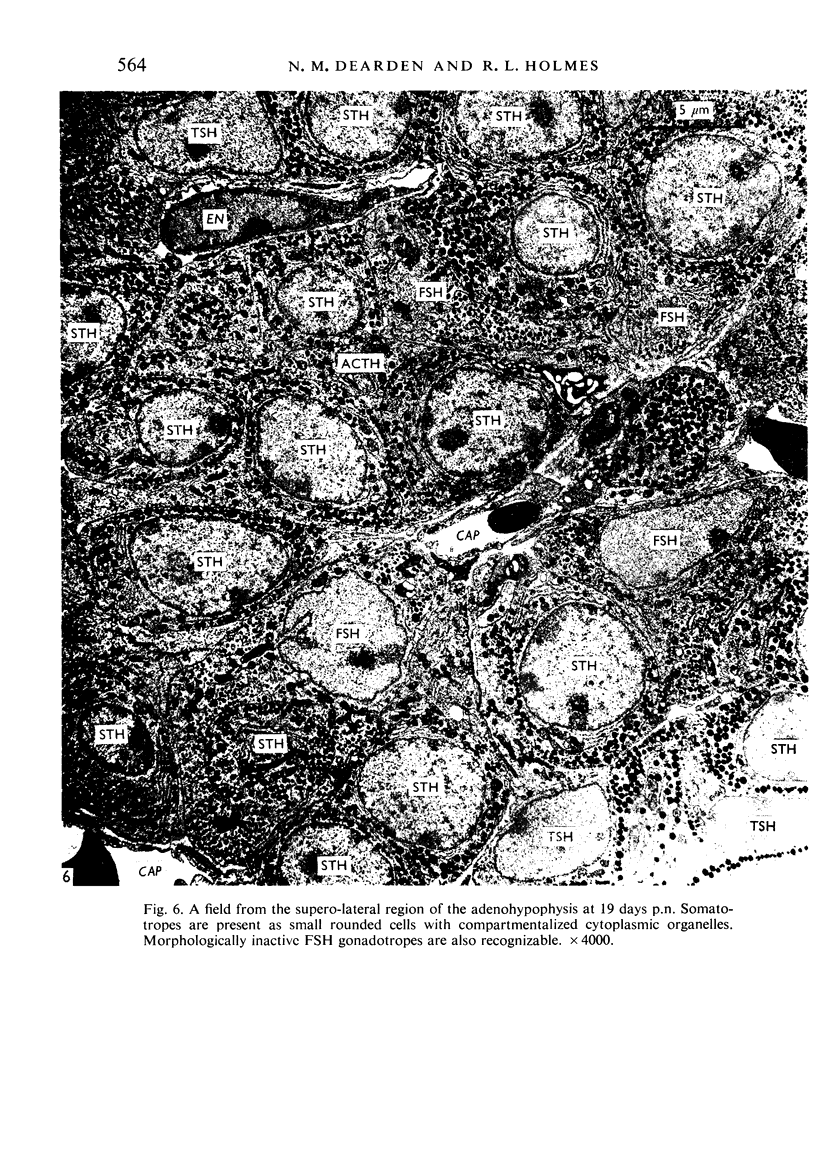
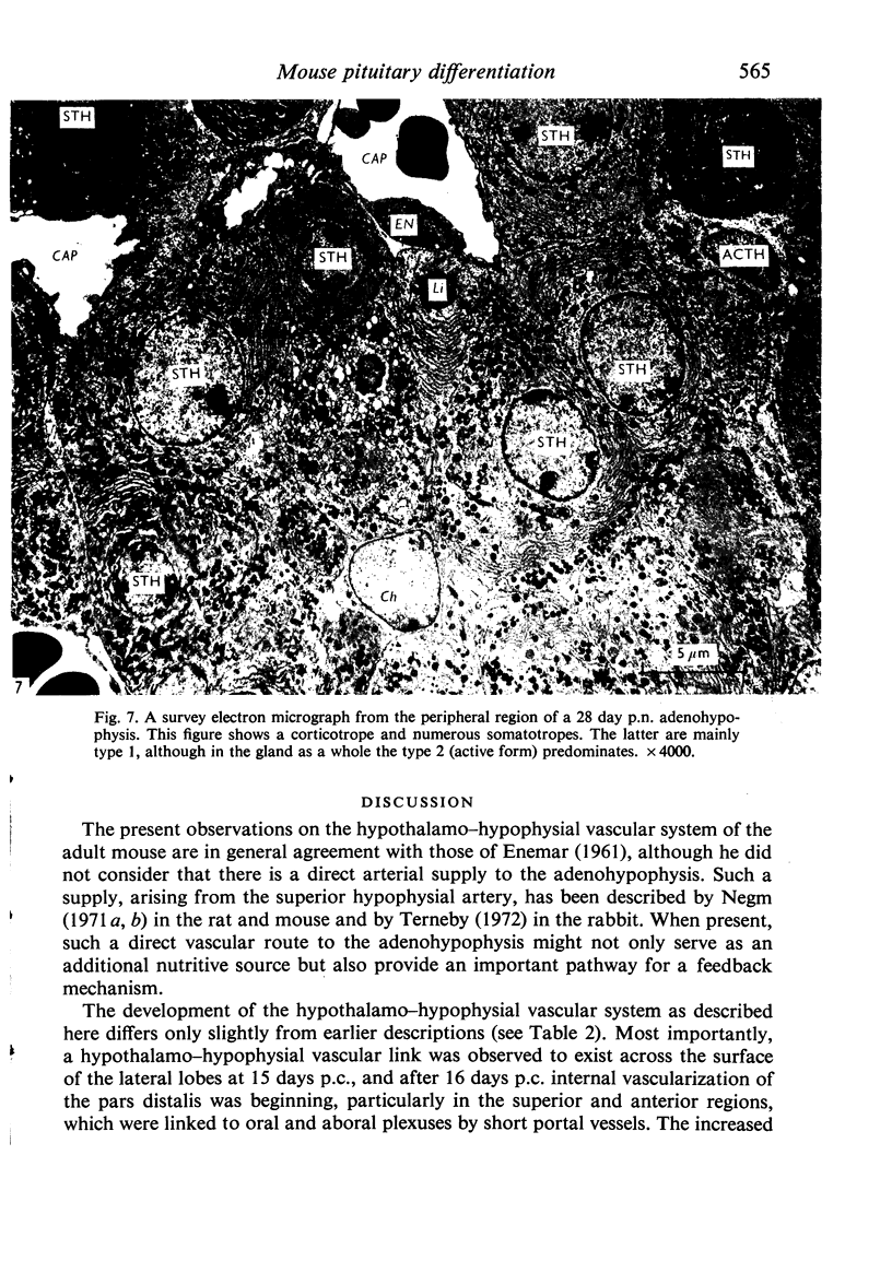
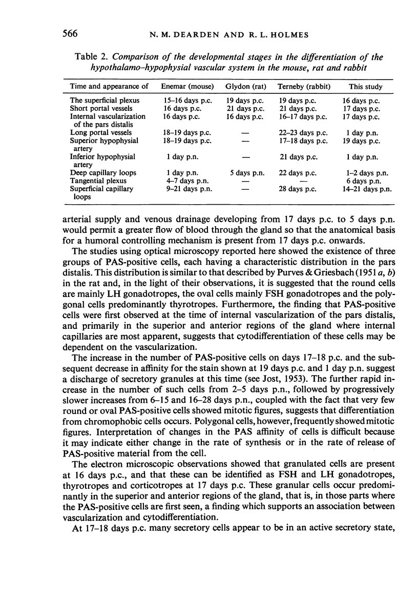
Pituitaries of fetal and postnatal (15 days p.c.-28 days p.n.) and adult (male) mice were studied by light and electron microscopy to correlate the developmental pattern of the hypothalamo-hypophysial vascular system with the time of onset of function of the adenohypophysis. The superior and anterior regions of the adenohypophysis become vascularized at 17 days p.c., when portal vessels extend from oral primary plexus to the pars distalis for the first time. Adenohypophysial vascularity and the number of portal vessels steadily increase to reach the adult pattern at 5 days p.n. At 1 day p.n. deep capillary loops appear in the caudal regions of the oral primary plexus; a capillary (tangential) plexus underlies the ependymal lining of the third ventricle by 6 days p.n. Superficial capillary loops were not observed until the third postnatal week. Granulation of secretory cells commences at 16 days p.c., predominantly in the upper and anterior adenohypophysis; at 17 days thyrotropes, gonadotropes and corticotropes are recognizable and by morphological criteria appear actively secretory on days 17-18 p.c., although few appear active at 19 days p.c. and 1 day p.n. Somatotropes are first seen at 18 days p.c., predominantly in the central and lateral regions of the pars distalis. Active secretory cells increase in number over the period 2-10 days p.n., but after 11 days p.n. thyrotropes and corticotropes seem to become progressively less active; fewer gonadotropes are seen after 15 days p.n., and these apparently become progressively less active from day 19. Most somatotropes appear active until 28 days p.n. The observations suggest that hypothalamic control of adenohypophysial function may exist in the mouse from 17 days p.c.
Full text
PDF
Images in this article
Selected References
These references are in PubMed. This may not be the complete list of references from this article.
- Eurenius L., Jarskär R. Electron microscope studies on the development of the external zone of the mouse median eminence. Z Zellforsch Mikrosk Anat. 1971;122(4):488–502. doi: 10.1007/BF00936083. [DOI] [PubMed] [Google Scholar]
- GLYDON R. S. The development of the blood supply of the pituitary in the albino rat, with special reference to the portal vessels. J Anat. 1957 Apr;91(2):237–244. [PMC free article] [PubMed] [Google Scholar]
- HALASZ B., PUPP L., UHLARIK S. Hypophysiotrophic area in the hypothalamus. J Endocrinol. 1962 Oct;25:147–154. doi: 10.1677/joe.0.0250147. [DOI] [PubMed] [Google Scholar]
- HYMER W. C., McSHAN W. H. Isolation of rat pituitary granules and the study of their biochemical properties and hormonal activities. J Cell Biol. 1963 Apr;17:67–86. doi: 10.1083/jcb.17.1.67. [DOI] [PMC free article] [PubMed] [Google Scholar]
- Holmes R. L. The vascular pattern of the median eminence of the hypophysis in the macaque. Folia Primatol (Basel) 1967;7(3):216–230. doi: 10.1159/000155121. [DOI] [PubMed] [Google Scholar]
- Negm I. M. The vascular blood supply of the pituitary and its development. Acta Anat (Basel) 1971;80(4):604–619. doi: 10.1159/000143716. [DOI] [PubMed] [Google Scholar]
- PURVES H. D., GRIESBACH W. E. The site of thyrotrophin and gonadotrophin production in the rat pituitary studied by McManus-Hotchkiss staining for glycoprotein. Endocrinology. 1951 Aug;49(2):244–264. doi: 10.1210/endo-49-2-244. [DOI] [PubMed] [Google Scholar]
- REYNOLDS E. S. The use of lead citrate at high pH as an electron-opaque stain in electron microscopy. J Cell Biol. 1963 Apr;17:208–212. doi: 10.1083/jcb.17.1.208. [DOI] [PMC free article] [PubMed] [Google Scholar]
- Schechter J. The cytodifferentiation of the rabbit pars distalis: an electron microscopic study. Gen Comp Endocrinol. 1971 Feb;16(1):1–20. doi: 10.1016/0016-6480(71)90202-4. [DOI] [PubMed] [Google Scholar]
- Terneby U. K. The development of the hypophysial vascular system in the rabbit, with particular regard to the primary plexus and portal vessels. J Neurovisc Relat. 1972;32(14):311–346. doi: 10.1007/BF02327928. [DOI] [PubMed] [Google Scholar]
- YAMADA K., SANO M., ITO T. A postnatal histogenetic study of the anterior pituitary of the mouse. Okajimas Folia Anat Jpn. 1957 Aug;30(2-3):177–195. doi: 10.2535/ofaj1936.30.2-3_177. [DOI] [PubMed] [Google Scholar]
- Yamada K., Yamashita K. An electron microscopic study on the possible site of production of ACTH in the anterior pituitary of mice. Z Zellforsch Mikrosk Anat. 1967;80(1):29–43. doi: 10.1007/BF00331475. [DOI] [PubMed] [Google Scholar]


