

Abstract
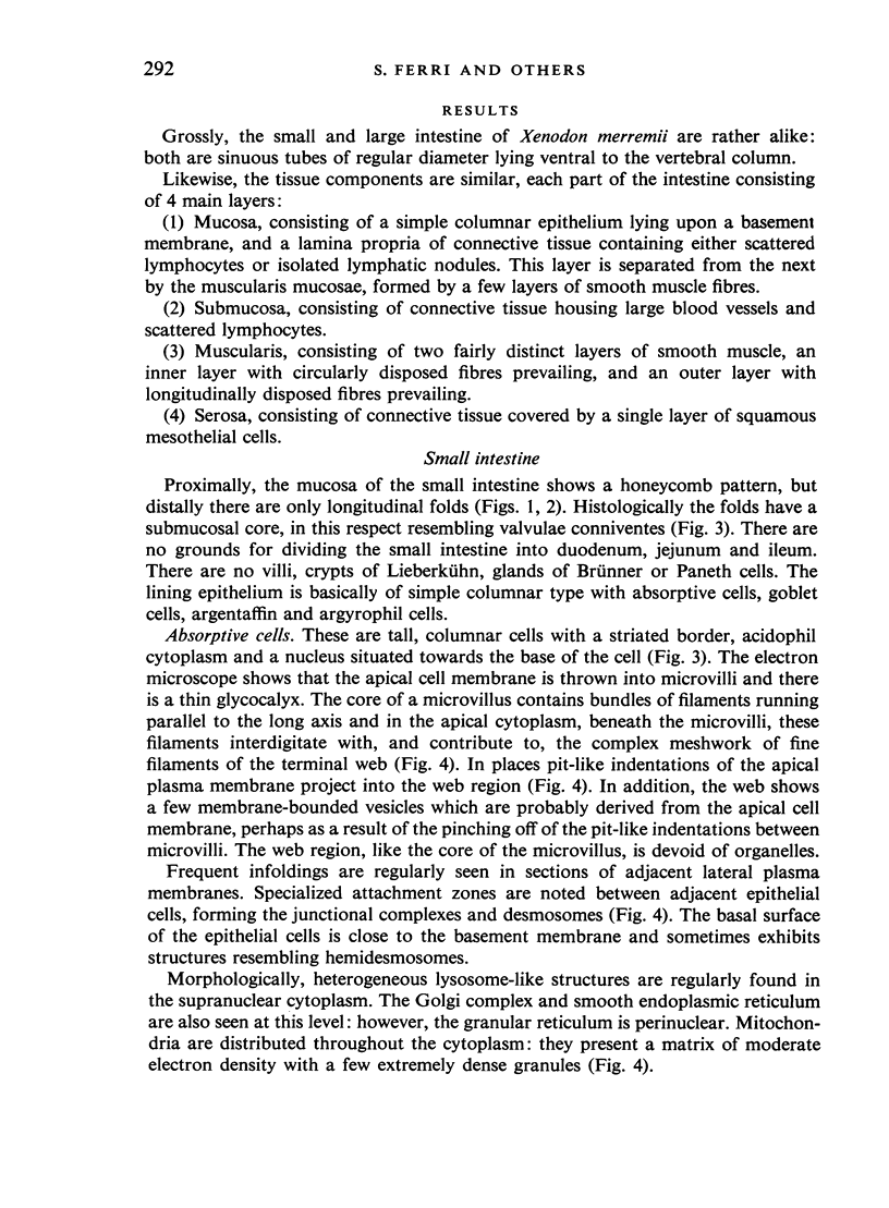
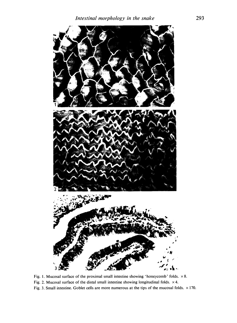
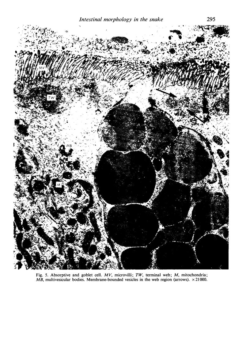
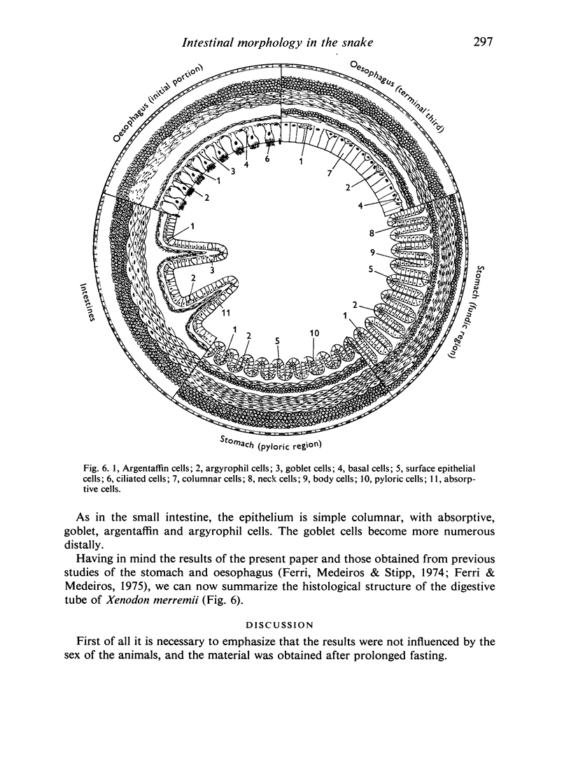
The small and large intestines of Xenodon merremii have a similar structure. They are separated by a sphincter of thickened circular muscle. The mucosa of the proximal part of the small intestine is raised into a honeycomb pattern, but distally there are only longitudinal folds. The lining epithelium throughout is of a simple columnar type, with absorptive, goblet, argentaffin and argyrophil cells, but no Paneth cells, villi or crypts of Lieberkühn are present.
Full text
PDF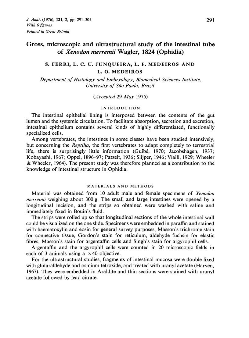
Images in this article

Selected References
These references are in PubMed. This may not be the complete list of references from this article.
- BEHNKE O. A PRELIMINARY REPORT ON "MICROTUBULES" IN UNDIFFERENTIATED AND DIFFERENTIATED VERTEBRATE CELLS. J Ultrastruct Res. 1964 Aug;11:139–146. doi: 10.1016/s0022-5320(64)80098-8. [DOI] [PubMed] [Google Scholar]
- Bennett G. Migration of glycoprotein from golgi apparatus to cell coat in the columnar cells of the duodenal epithelium. J Cell Biol. 1970 Jun;45(3):668–673. doi: 10.1083/jcb.45.3.668. [DOI] [PMC free article] [PubMed] [Google Scholar]
- Bonneville M. A., Weinstock M. Brush border development in the intestinal absorptive cells of Xenopus during metamorphosis. J Cell Biol. 1970 Jan;44(1):151–171. doi: 10.1083/jcb.44.1.151. [DOI] [PMC free article] [PubMed] [Google Scholar]
- FARQUHAR M. G., PALADE G. E. Junctional complexes in various epithelia. J Cell Biol. 1963 May;17:375–412. doi: 10.1083/jcb.17.2.375. [DOI] [PMC free article] [PubMed] [Google Scholar]
- FREEMAN J. A. Fine structure of the goblet cell mucous secretory process. Anat Rec. 1962 Dec;144:341–357. doi: 10.1002/ar.1091440406. [DOI] [PubMed] [Google Scholar]
- Ferri S., Medeiros L. O., Stipp A. C. Gastric wall histological analysis and cellular types distribution in Xenodon merremii Wagler, 1924 (Ophidia). Gegenbaurs Morphol Jahrb. 1974;120(5):627–637. [PubMed] [Google Scholar]
- Freeman J. A. Goblet cell fine structure. Anat Rec. 1966 Jan;154(1):121–147. doi: 10.1002/ar.1091540111. [DOI] [PubMed] [Google Scholar]
- Ito S. Structure and function of the glycocalyx. Fed Proc. 1969 Jan-Feb;28(1):12–25. [PubMed] [Google Scholar]
- Ito S. The enteric surface coat on cat intestinal microvilli. J Cell Biol. 1965 Dec;27(3):475–491. doi: 10.1083/jcb.27.3.475. [DOI] [PMC free article] [PubMed] [Google Scholar]
- Johnson C. F. Disaccharidase: localization in hamster intestine brush borders. Science. 1967 Mar 31;155(3770):1670–1672. doi: 10.1126/science.155.3770.1670. [DOI] [PubMed] [Google Scholar]
- Kobayashi S. An electron microscope study of the intestinal mucosa of the snake, Elaphe quadrivirgata (BOIE). Arch Histol Jpn. 1967 Nov;28(5):525–536. doi: 10.1679/aohc1950.28.525. [DOI] [PubMed] [Google Scholar]
- PADYKULA H. A., STRAUSS E. W., LADMAN A. J., GARDNER F. H. A morphologic and histochemical analysis of the human jejunal epithelium in nontropical sprue. Gastroenterology. 1961 Jun;40:735–765. [PubMed] [Google Scholar]
- PALAY S. L., KARLIN L. J. An electron microscopic study of the intestinal villus. I. The fasting animal. J Biophys Biochem Cytol. 1959 May 25;5(3):363–372. doi: 10.1083/jcb.5.3.363. [DOI] [PMC free article] [PubMed] [Google Scholar]
- SANBORN E., KOEN P. F., MACNABB J. D., MOORE G. CYTOPLASMIC MICROTUBULES IN MAMMALIAN CELLS. J Ultrastruct Res. 1964 Aug;11:123–138. doi: 10.1016/s0022-5320(64)80097-6. [DOI] [PubMed] [Google Scholar]
- STRAUSS E. W. The absorption of fat by intestine of golden hamster in vitro. J Cell Biol. 1963 Jun;17:597–607. doi: 10.1083/jcb.17.3.597. [DOI] [PMC free article] [PubMed] [Google Scholar]
- Singh I. The depletion of the granules of argyrophile cells of dog duodenum by reserpine. Z Zellforsch Mikrosk Anat. 1970;107(1):111–118. doi: 10.1007/BF00338962. [DOI] [PubMed] [Google Scholar]
- TRIER J. S. STUDIES ON SMALL INTESTINAL CRYPT EPITHELIUM. I. THE FINE STRUCTURE OF THE CRYPT EPITHELIUM OF THE PROXIMAL SMALL INTESTINE OF FASTING HUMANS. J Cell Biol. 1963 Sep;18:599–620. doi: 10.1083/jcb.18.3.599. [DOI] [PMC free article] [PubMed] [Google Scholar]
- Trier J. S., Rubin C. E. Electron microscopy of the small intestine: a review. Gastroenterology. 1965 Nov;49(5):574–603. [PubMed] [Google Scholar]