Abstract
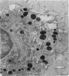
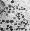
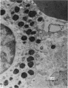
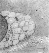
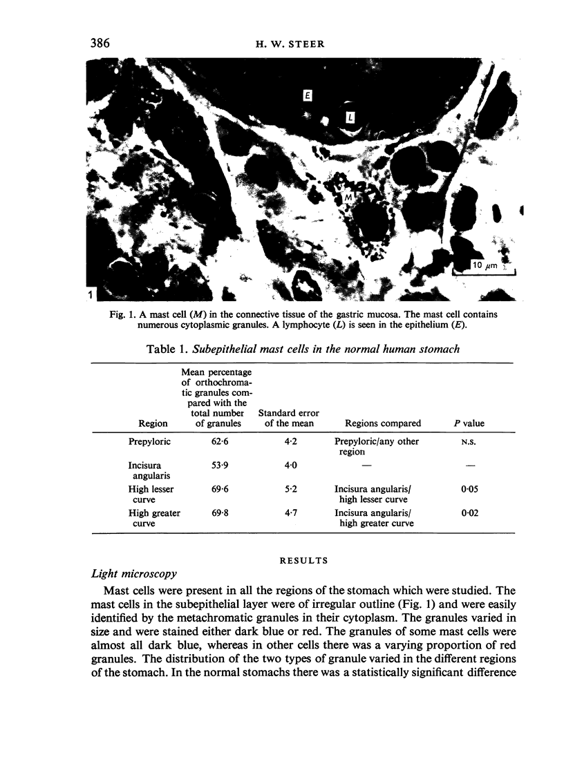
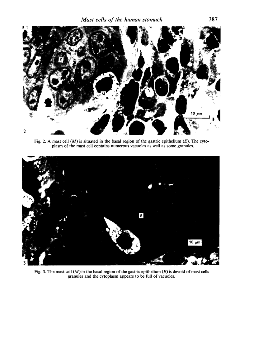
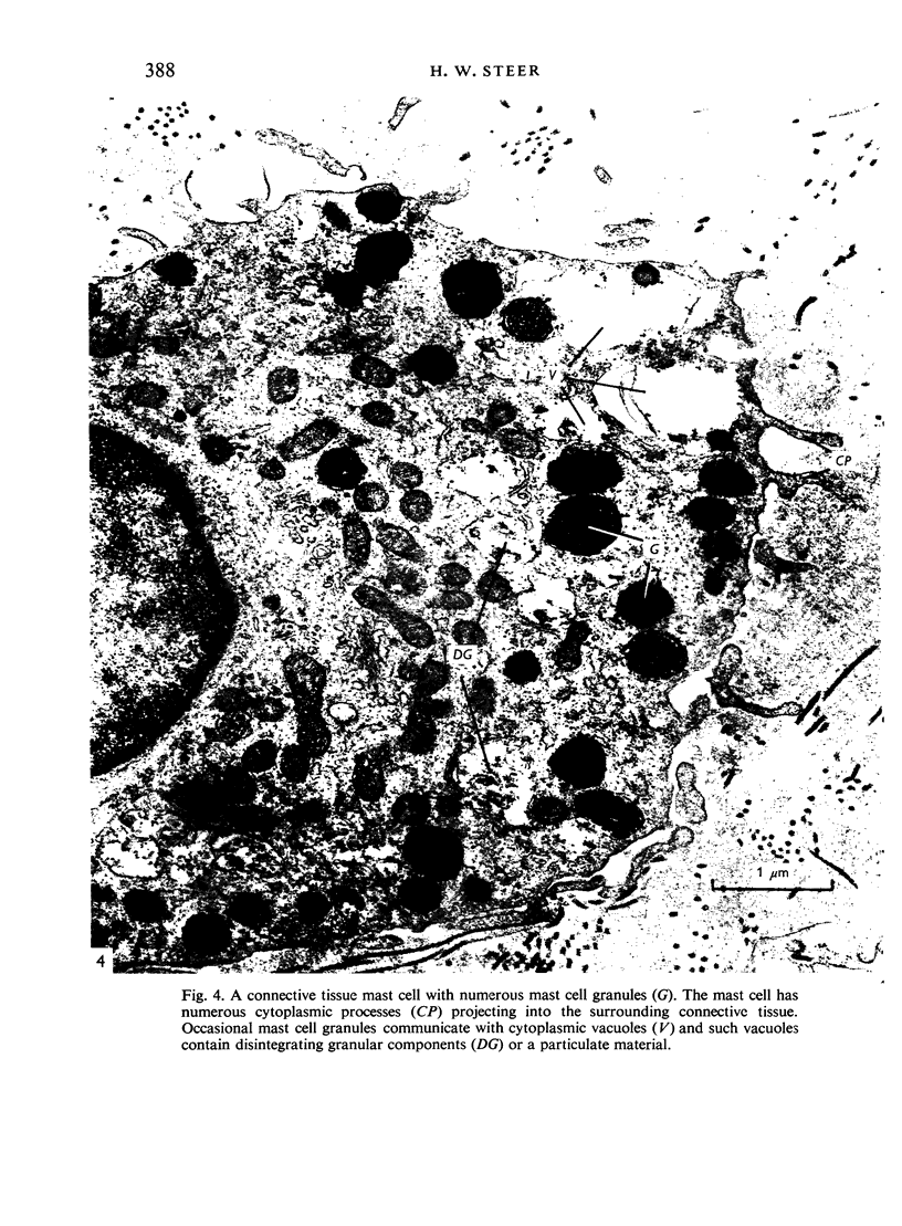
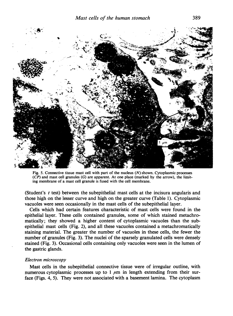
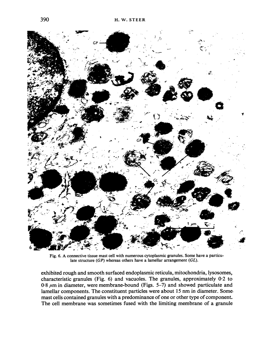
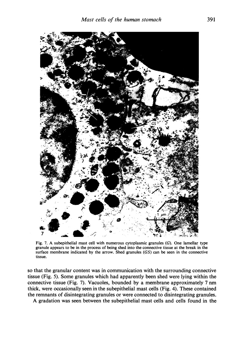
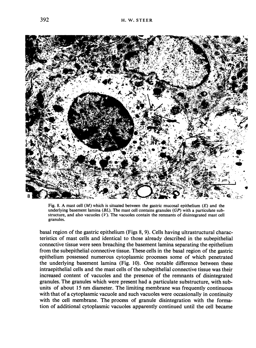
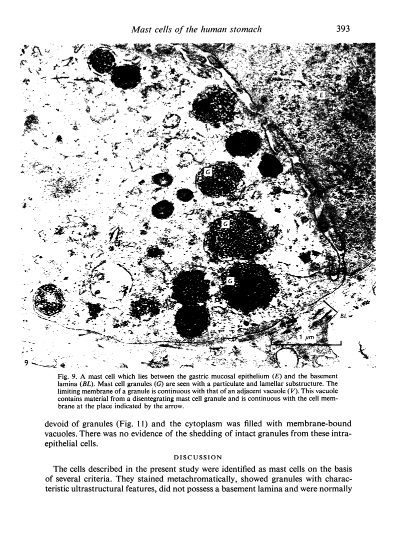
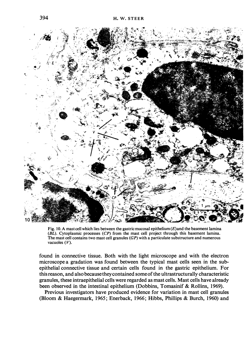
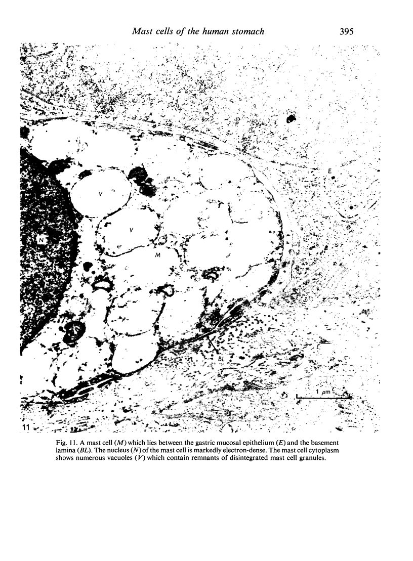
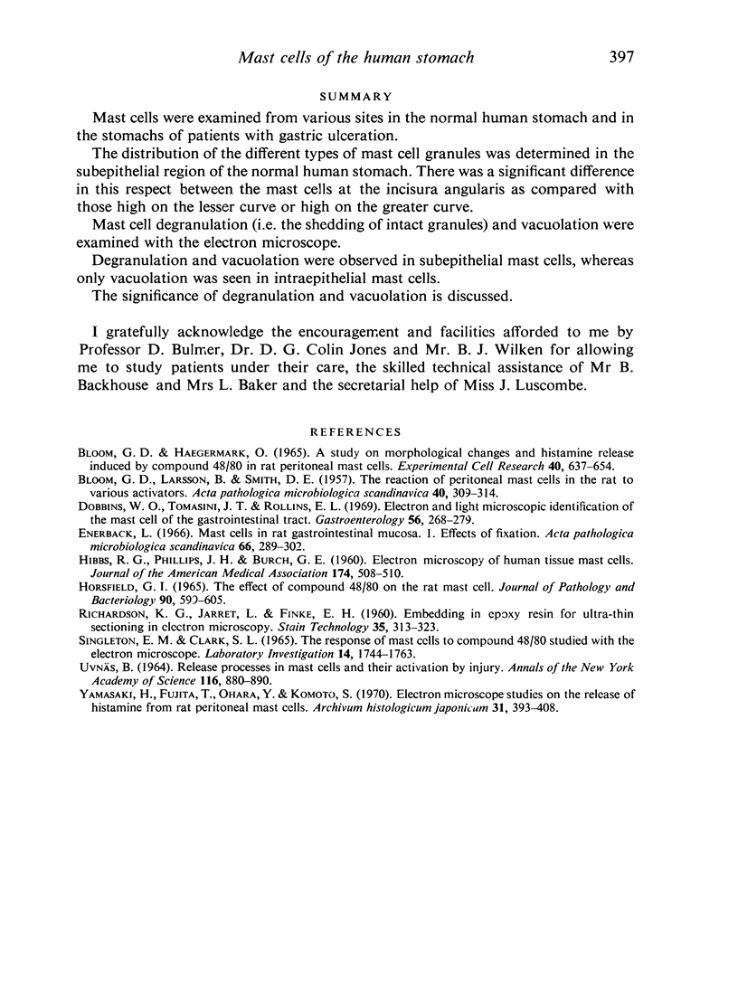
Mast cells were examined from various sites in the normal human stomach and in the stomachs of patients with gastric ulceration. The distribution of the different types of mast cell granules was determined in the subepithelial region of the normal human stomach. There was a significant difference in this respect between the mast cells at the incisura angularis as compared with those high on the lesser curve or high on the greater curve. Mast cell degranulation (i.e. the shedding of intact granules) and vacuolation were examined with the electron microscope. Degranulation and vacuolation were observed in subepithelial mast cells, whereas only vacuolation was seen in intraepithelial mast cells. The significance of degranulation and vacuolation is discussed.
Full text
PDF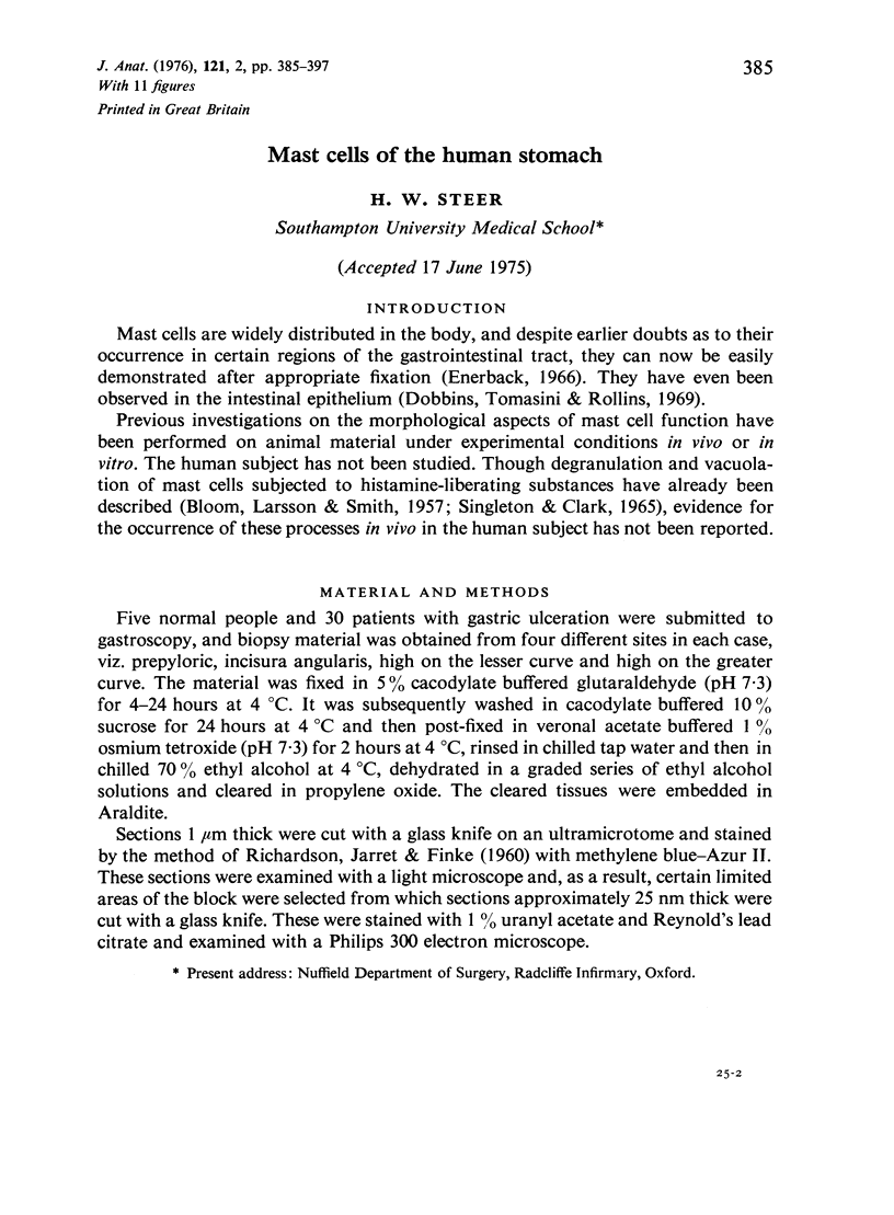
Images in this article
Selected References
These references are in PubMed. This may not be the complete list of references from this article.
- BLOOM G., LARSSON B., SMITH D. E. The reaction of peritoneal mast cells in the rat to various activators. Acta Pathol Microbiol Scand. 1957;40(4):309–314. [PubMed] [Google Scholar]
- Bloom G. D., Haegermark O. A study on morphological changes and histamine release induced by compound 48/80 in rat peritoneal mast cells. Exp Cell Res. 1965 Dec;40(3):637–654. doi: 10.1016/0014-4827(65)90241-7. [DOI] [PubMed] [Google Scholar]
- Dobbins W. O., 3rd, Tomasini J. T., Rollins E. L. Electron and light microscopic identification of the mast cell of the gastrointestinal tract. Gastroenterology. 1969 Feb;56(2):268–279. [PubMed] [Google Scholar]
- Enerbäck L. Mast cells in rat gastrointestinal mucosa. I. Effects of fixation. Acta Pathol Microbiol Scand. 1966;66(3):289–302. doi: 10.1111/apm.1966.66.3.289. [DOI] [PubMed] [Google Scholar]
- HIBBS R. G., PHILLIPS J. H., BURCH G. E. Electron microscopy of human tissue mast cells. JAMA. 1960 Oct 1;174:508–510. doi: 10.1001/jama.1960.63030050006012. [DOI] [PubMed] [Google Scholar]
- Horsfield G. I. The effect of compound 48/80 on the rat mast cell. J Pathol Bacteriol. 1965 Oct;90(2):599–605. doi: 10.1002/path.1700900228. [DOI] [PubMed] [Google Scholar]
- RICHARDSON K. C., JARETT L., FINKE E. H. Embedding in epoxy resins for ultrathin sectioning in electron microscopy. Stain Technol. 1960 Nov;35:313–323. doi: 10.3109/10520296009114754. [DOI] [PubMed] [Google Scholar]
- Singleton E. M., Clark S. L., Jr The response of mast cells to compound 48/80 studied with the electron microscope. Lab Invest. 1965 Oct;14(10):1744–1763. [PubMed] [Google Scholar]
- Yamasaki H., Fujita T., Oara Y., Komoto S. Electron microscope studies on the release of histamine from rat peritoneal mast cells. Arch Histol Jpn. 1970 Mar;31(3):393–408. doi: 10.1679/aohc1950.31.393. [DOI] [PubMed] [Google Scholar]


