Abstract
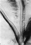
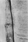
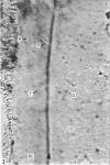
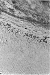
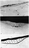
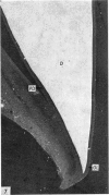
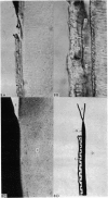
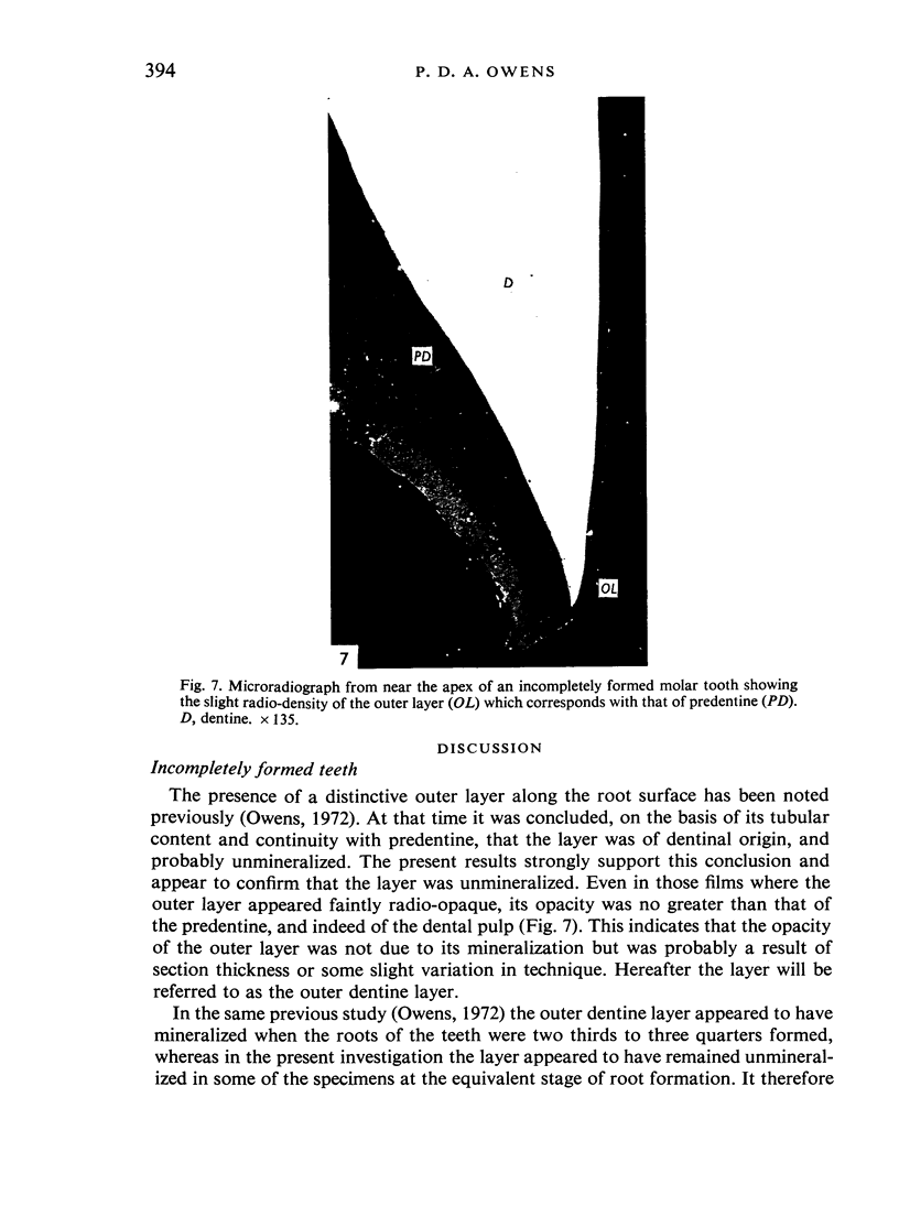
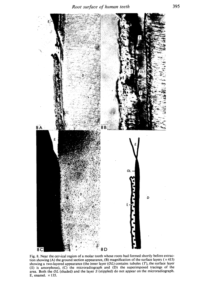
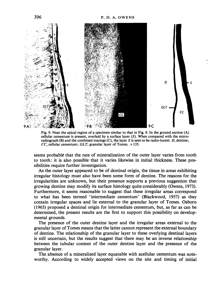
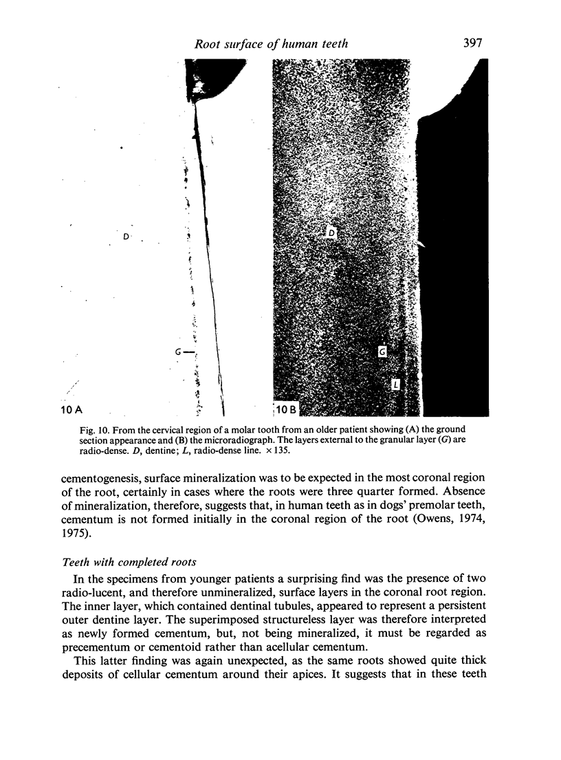
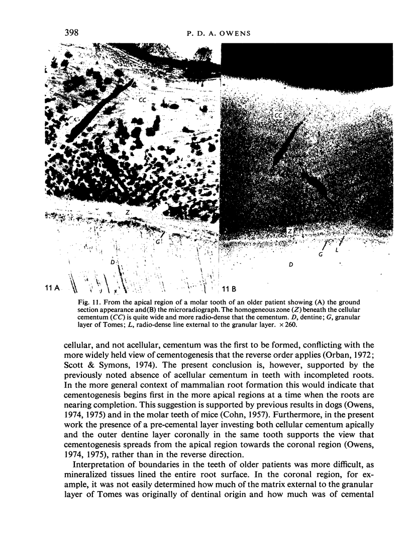
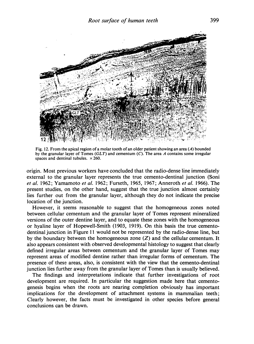
In an attempt to clarify the nature of the human cemento-dentinal junction, ground sections of incompletely formed and fully formed extracted teeth were prepared and their histology compared with their microradiographic appearances. The results showed that incompletely formed teeth possess distinctive surface layers outside the granular layer of Tomes. The evidence indicates that these layers are of dentinal origin; their presence during development supports previous explanations by the author of the hyaline layer of Hopewell-Smith and of so-called intermediate cementum. The results also indicate that the granular layer of Tomes does not represent the outer limit of root dentine. The relationship of these surface layers to the definitive cementum which is present in fully formed teeth was studied in both young and older patients. From the results it was concluded that cementum formation begins in the more apical region of the teeth at a time when root formation is well advanced, and that it spreads towards the crown rather than in the generally accepted reverse direction.
Full text
PDF
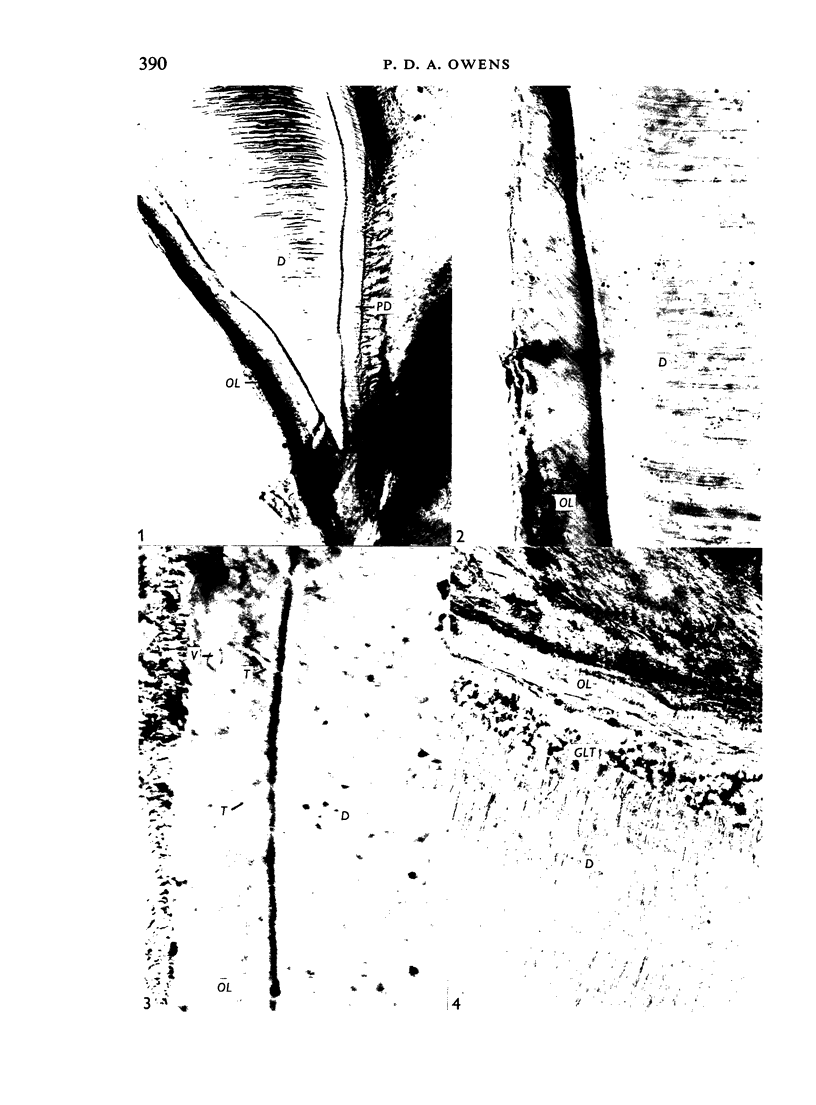
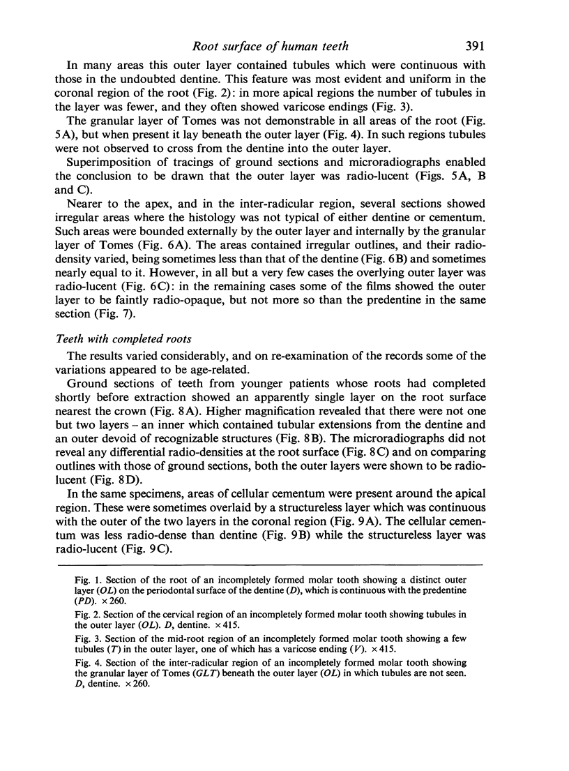
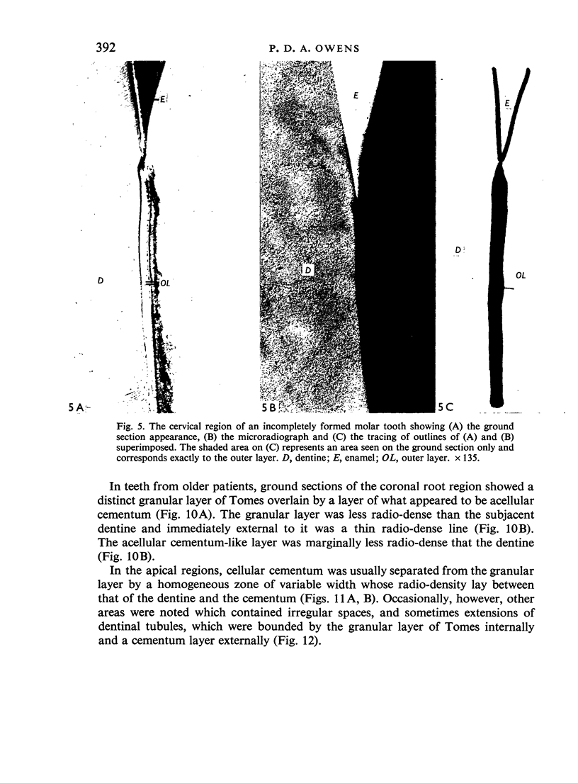
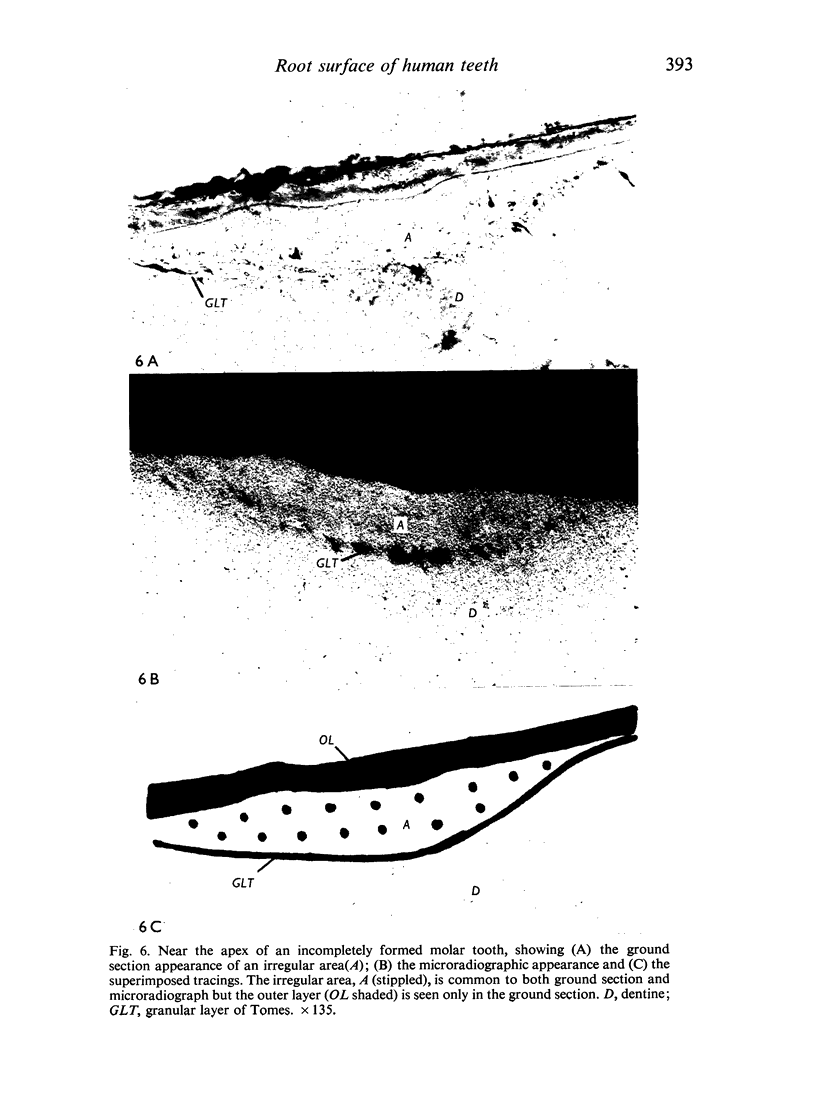

Images in this article
Selected References
These references are in PubMed. This may not be the complete list of references from this article.
- Anneroth G., Bergman G., Welander E. Microradiographic study of dental tissues using soft and ultrasoft x-rays. Odontol Revy. 1966;17(2):131–148. [PubMed] [Google Scholar]
- COHN S. A. Development of the molar teeth in the albino mouse. Am J Anat. 1957 Sep;101(2):295–319. doi: 10.1002/aja.1001010205. [DOI] [PubMed] [Google Scholar]
- Furseth R. A microradiographic and electron microscopic study of the cementum of human deciduous teeth. Acta Odontol Scand. 1967 Dec;25(6):613–645. doi: 10.3109/00016356709019780. [DOI] [PubMed] [Google Scholar]
- Kawasaki K. On the configuration of incremental lines in human dentine as revealed by tetracycline labelling. J Anat. 1975 Feb;119(Pt 1):61–66. [PMC free article] [PubMed] [Google Scholar]
- Owens P. D. A light microscopic study of the development of the roots of premolar teeth in dogs. Arch Oral Biol. 1974 Jul;19(7):525–538. doi: 10.1016/0003-9969(74)90068-5. [DOI] [PubMed] [Google Scholar]
- Owens P. D. Light microscopic observations on the formation of the layer of Hopewell-Smith in human teeth. Arch Oral Biol. 1972 Dec;17(12):1785–1788. doi: 10.1016/0003-9969(72)90243-9. [DOI] [PubMed] [Google Scholar]
- Owens P. D. Mineralization in the roots of human deciduous teeth demonstrated by tetracycline labelling. Arch Oral Biol. 1973 Jul;18(7):889–897. doi: 10.1016/0003-9969(73)90059-9. [DOI] [PubMed] [Google Scholar]
- Owens P. D. Patterns of mineralization in the roots of premolar teeth in dogs. Arch Oral Biol. 1975 Nov;20(11):709–712. doi: 10.1016/0003-9969(75)90039-4. [DOI] [PubMed] [Google Scholar]
- Ten Cate A. R. An analysis of Tomes' granular layer. Anat Rec. 1972 Feb;172(2):137–147. doi: 10.1002/ar.1091720202. [DOI] [PubMed] [Google Scholar]


