

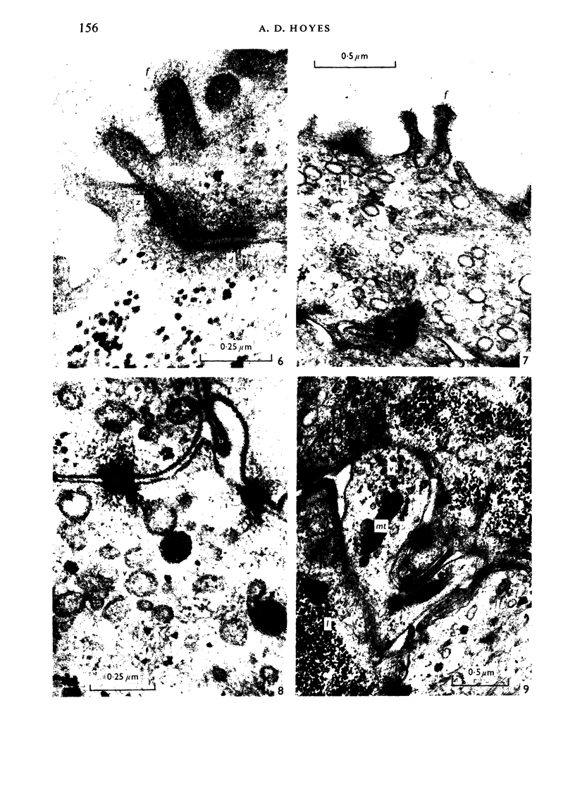
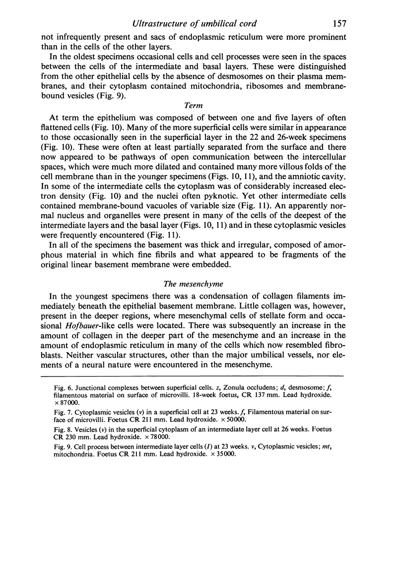
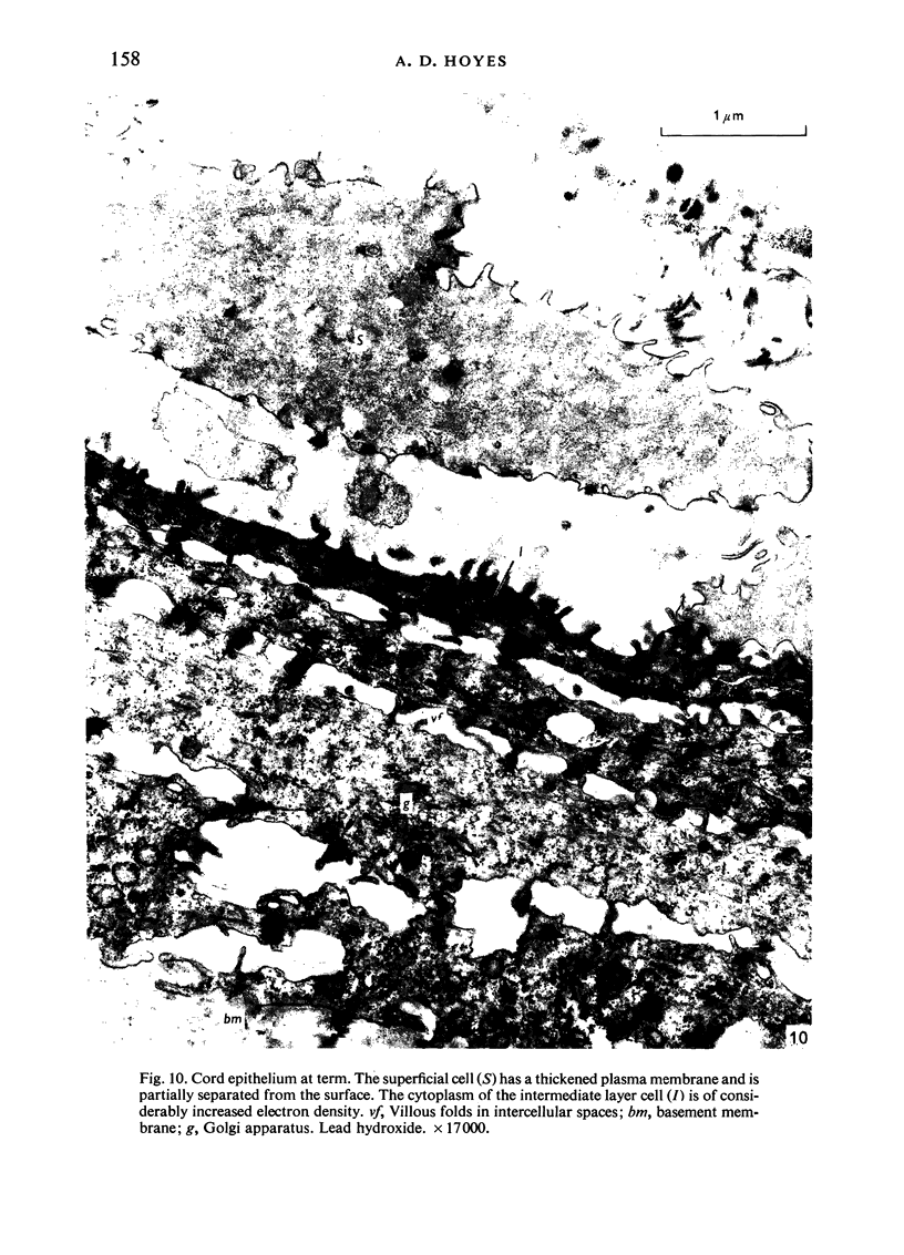
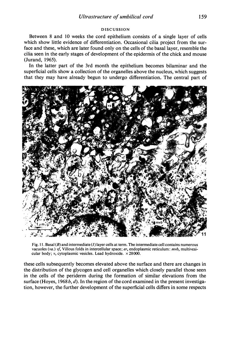
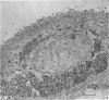
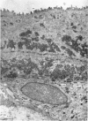
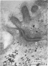
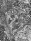
Full text
PDF

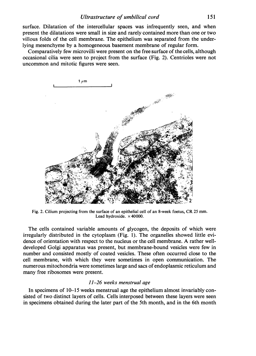

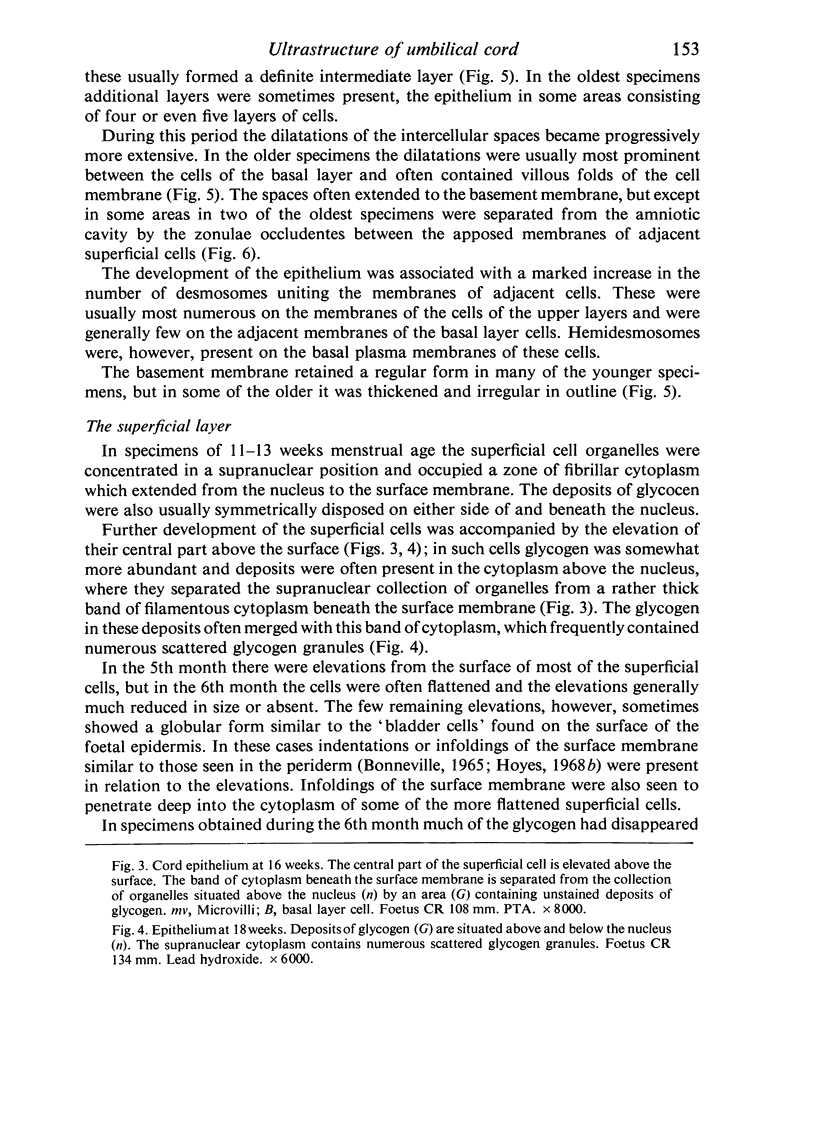


Images in this article


Selected References
These references are in PubMed. This may not be the complete list of references from this article.
- Breathnach A. S., Wyllie L. M. Fine structure of cells forming the surface layer of the epidermis in human fetuses at fourteen and twelve weeks. J Invest Dermatol. 1965 Sep;45(3):179–189. doi: 10.1038/jid.1965.116. [DOI] [PubMed] [Google Scholar]
- GORDON G. B., MILLER L. R., BENSCH K. G. FIXATION OF TISSUE CULTURE CELLS FOR ULTRASTRUCTURAL CYTOCHEMISTRY. Exp Cell Res. 1963 Aug;31:440–443. doi: 10.1016/0014-4827(63)90024-7. [DOI] [PubMed] [Google Scholar]
- HUTCHINSON D. L., GRAY M. J., PLENTL A. A., ALVAREZ H., CALDEYRO-BARCIA R., KAPLAN B., LIND J. The role of the fetus in the water exchange of the amniotic fluid of normal and hydramniotic patients. J Clin Invest. 1959 Jun;38(6):971–980. doi: 10.1172/JCI103880. [DOI] [PMC free article] [PubMed] [Google Scholar]
- Hashimoto K., Gross B. G., DiBella R. J., Lever W. F. The ultrastructure of the skin of human embryos. IV. The epidermis. J Invest Dermatol. 1966 Oct;47(4):317–335. doi: 10.1038/jid.1966.150. [DOI] [PubMed] [Google Scholar]
- Hoyes A. D. Acid mucopolysaccharide in human fetal epidermis. J Invest Dermatol. 1967 Jun;48(6):598–601. [PubMed] [Google Scholar]
- Hoyes A. D. Electron microscopy of the surface layer (periderm) of human foetal skin. J Anat. 1968 Sep;103(Pt 2):321–336. [PMC free article] [PubMed] [Google Scholar]
- Hoyes A. D. Fine structure of human amniotic epithelium in early pregnancy. J Obstet Gynaecol Br Commonw. 1968 Sep;75(9):949–962. doi: 10.1111/j.1471-0528.1968.tb01620.x. [DOI] [PubMed] [Google Scholar]
- KARNOVSKY M. J. Simple methods for "staining with lead" at high pH in electron microscopy. J Biophys Biochem Cytol. 1961 Dec;11:729–732. doi: 10.1083/jcb.11.3.729. [DOI] [PMC free article] [PubMed] [Google Scholar]
- LEESON C. R., LEESON T. S. THE FINE STRUCTURE OF THE RAT UMBILICAL CORD AT VARIOUS TIMES OF GESTATION. Anat Rec. 1965 Feb;151:183–197. doi: 10.1002/ar.1091510209. [DOI] [PubMed] [Google Scholar]
- PALADE G. E. A study of fixation for electron microscopy. J Exp Med. 1952 Mar;95(3):285–298. doi: 10.1084/jem.95.3.285. [DOI] [PMC free article] [PubMed] [Google Scholar]
- PLENTL A. A. Transfer of water across the perfused umbilical cord. Proc Soc Exp Biol Med. 1961 Jul;107:622–626. doi: 10.3181/00379727-107-26707. [DOI] [PubMed] [Google Scholar]
- SCHRAMM B. [The cutaneous sheath of the umbilical cord and its significance]. Gynecol Obstet (Paris) 1962 Sep-Oct;61:556–562. [PubMed] [Google Scholar]