

Abstract
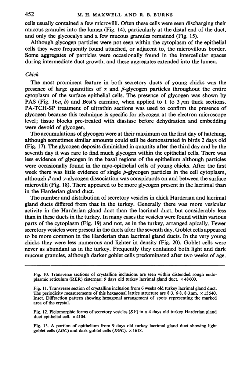
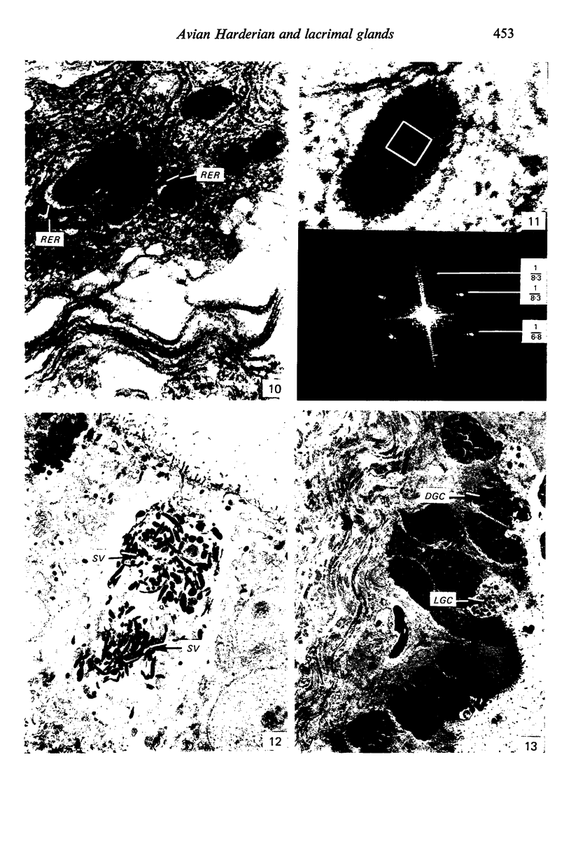
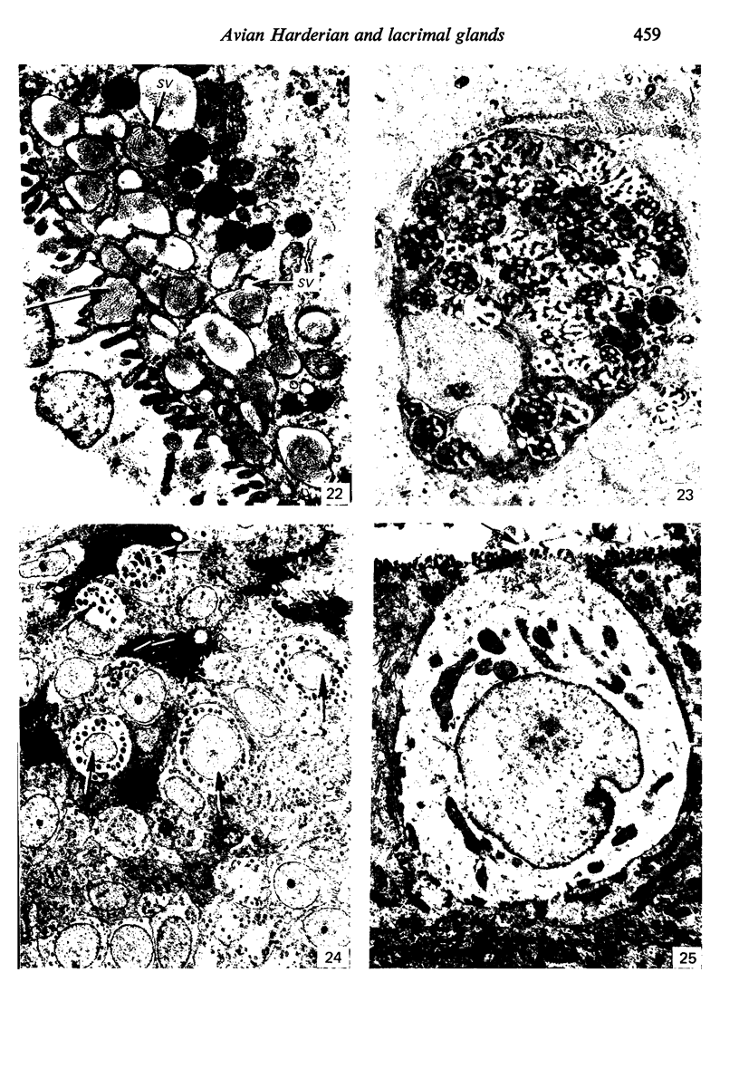
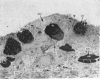
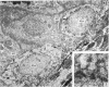
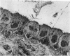
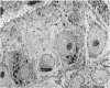
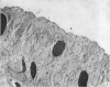
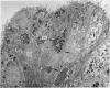
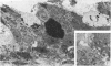
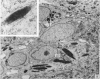
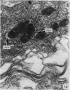
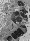
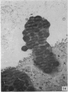
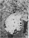
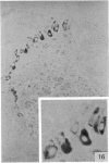
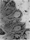
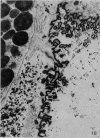
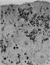
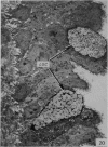
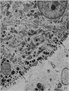
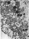
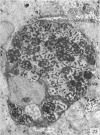
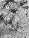
The ultrastructure of the epithelium in the Harderian and lacrimal gland ducts of the turkey, fowl and duck has been investigated. The findings establish that the ducts are fundamentally similar in morphology, although species differences occur. The duct epithelia are composed of cuboidal, columnar or pseudostratisfied columnar cells with many secretory vesicles as well as goblet cells. PAS-positive fibrillary rods and crystalline inclusions occupied the cisternae of the rough endoplasmic reticulum of the epithelial cells of turkeys. The crystals, which were thought to evolve from the rods, had a mean periodicity of 7.0 nm. In chicks between 1 and 3 days of age, but not in poults and ducklings, the duct epithelia displayed large intracytoplasmic deposits of glycogen which disappeared by the seventh day.
Full text
PDF


Images in this article


Selected References
These references are in PubMed. This may not be the complete list of references from this article.
- Ackerman G. A. Ultrastructural localization of glycogen in erythrocytes and developing erythrocytic cells in normal human bone marrow. Z Zellforsch Mikrosk Anat. 1973 Jul 16;140(4):433–444. doi: 10.1007/BF00306670. [DOI] [PubMed] [Google Scholar]
- Alexander J. H., Young J. A., van Lennep E. W. The ultrastructure of the duct system in the rat extraorbital lacrimal gland. Z Zellforsch Mikrosk Anat. 1973 Nov 15;144(4):453–466. doi: 10.1007/BF00307373. [DOI] [PubMed] [Google Scholar]
- Burns R. B., Maxwell M. H. The structure of the Harderian and lacrimal gland ducts of the turkey, fowl and duck. A light microscope study. J Anat. 1979 Mar;128(Pt 2):285–292. [PMC free article] [PubMed] [Google Scholar]
- Burns R. B. The structure of the lachrymal glands of the domestic fowl and of the duck. Res Vet Sci. 1976 Nov;21(3):292–299. [PubMed] [Google Scholar]
- FREEMAN B. M. THE IMPORTANCE OF GLYCOGEN AT THE TERMINATION OF THE EMBRYONIC EXISTENCE OF GALLUS DOMESTICUS. Comp Biochem Physiol. 1965 Feb;14:217–222. doi: 10.1016/0010-406x(65)90197-0. [DOI] [PubMed] [Google Scholar]
- Kühnel W., Beier H. M. Morphologie und Cytochemie der Harderschen Drüse von Anatiden. Z Zellforsch Mikrosk Anat. 1973 Jul 31;141(2):255–283. [PubMed] [Google Scholar]
- Maxwell M. H. Two rapid and simple methods used for the removal of resins from 1.0 micron thick epoxy sections. J Microsc. 1978 Mar;112(2):253–255. doi: 10.1111/j.1365-2818.1978.tb01174.x. [DOI] [PubMed] [Google Scholar]
- Mueller A. P., Sato K., Glick B. The chicken lacrimal gland, gland of Harder, caecal tonsil, and accessory spleens as sources of antibody-producing cells. Cell Immunol. 1971 Apr;2(2):140–152. doi: 10.1016/0008-8749(71)90033-5. [DOI] [PubMed] [Google Scholar]
- REYNOLDS E. S. The use of lead citrate at high pH as an electron-opaque stain in electron microscopy. J Cell Biol. 1963 Apr;17:208–212. doi: 10.1083/jcb.17.1.208. [DOI] [PMC free article] [PubMed] [Google Scholar]
- Rothwell B., Wight P. A., Burns R. B., Mackenzie G. M. The Harderian glands of the domestic fowl. 3. Ultrastructure. J Anat. 1972 Jul;112(Pt 2):233–250. [PMC free article] [PubMed] [Google Scholar]
- WATSON M. L. Staining of tissue sections for electron microscopy with heavy metals. J Biophys Biochem Cytol. 1958 Jul 25;4(4):475–478. doi: 10.1083/jcb.4.4.475. [DOI] [PMC free article] [PubMed] [Google Scholar]
- Wight P. A., Mackenzie G. M., Rothwell B., Burns R. B. The Harderian glands of the domestic fowl. II. Histochemistry. J Anat. 1971 Dec;110(Pt 3):323–333. [PMC free article] [PubMed] [Google Scholar]