

Abstract
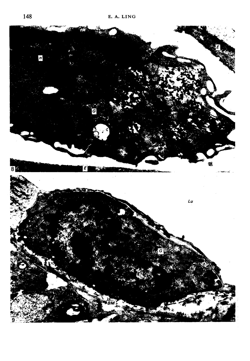
A single dose of colloidal carbon was given intravascularly to young adult rats in order to label circulating monocytes. Two days after injection dorsal rhizotomies were performed on the fifth to eighth cervical nerves on the right side. The rats were killed 1, 3, 4 and 8 days later. Electron microscopic examination of the spinal cord showed wide-spread tissue degeneration on the operated side in the dorsolateral fasciculus, the dorsal horn and the dorsal neuronal white column, the changes in the last named being the most severe. A variety of non-neuronal elements was found in the dorsolateral fasciculus and dorsal horn. These included astrocytes, oligodendrocytes, microglia-like cells, plasma cells, mast cells, polymorphonuclear leucocytes, monocytes and macrophages. Monocytes and macrophages were most common 3 and 4 days after operation. Some of these cells carried intracytoplasmic carbon particles. Carbon-labelled monocytes were observed in blood vessel lumina, perivascularly and in the neuropil. Monocytes crossing blood vessel walls were also encountered, indicating that the neuropil monocytes were derived from circulating cells. Macrophages were characterized by pleomorphic phagosomes which seemed to be composed largely of myelin remnants. The presence of carbon particles in their cytoplasm, and also their general similarity to monocytes, suggested that they originated from the latter. Local microglial cells were considered to be another source of macrophages. Indeed, there were present some microglia-like cells which were regarded as 'activated microglia' as they showed morphological resemblances to microglia on the one hand and to macrophages on the other. In particular their cytoplasm always included phagosomes. It is concluded that the macrophages which appear in the altered spinal cord following rhizotomy are derived both from circulating monocytes and from indigenous microglia.
Full text
PDF


Images in this article


Selected References
These references are in PubMed. This may not be the complete list of references from this article.
- ADRIAN E. K., Jr, WALKER B. E. Incorporation of thymidine-H3 by cells in normal and injured mouse spinal cord. J Neuropathol Exp Neurol. 1962 Oct;21:597–609. doi: 10.1097/00005072-196210000-00007. [DOI] [PubMed] [Google Scholar]
- Adrian E. K., Jr, Smothermon R. D. Leucocytic infiltration into the hypoglossal nucleus following injury to the hypoglossal nerve. Anat Rec. 1970 Jan;166(1):99–115. doi: 10.1002/ar.1091660108. [DOI] [PubMed] [Google Scholar]
- Adrian E. K., Jr, Williams M. G. Cell proliferation in injured spinal cord. An electron microscopic study. J Comp Neurol. 1973 Sep 1;151(1):1–24. doi: 10.1002/cne.901510102. [DOI] [PubMed] [Google Scholar]
- Berner A., Torvik A., Stenwig A. E. Origin of macrophages in traumatic lesions and Wallerian degeneration in peripheral nerves. Acta Neuropathol. 1973;25(3):228–236. doi: 10.1007/BF00685202. [DOI] [PubMed] [Google Scholar]
- Blakemore W. F. Microglial reactions following thermal necrosis of the rat cortex: an electron microscope study. Acta Neuropathol. 1972;21(1):11–22. doi: 10.1007/BF00687996. [DOI] [PubMed] [Google Scholar]
- Booz K. H., Felsing T. Uber ein transitorisches, perinatales subependymales Zellsystem der weissen Ratte. Z Anat Entwicklungsgesch. 1973;141(3):275–288. [PubMed] [Google Scholar]
- Cammermeyer J. The life history of the microglial cell: a light microscopic study. Neurosci Res (N Y) 1970;3:43–129. doi: 10.1016/b978-0-12-512503-1.50008-6. [DOI] [PubMed] [Google Scholar]
- Feigin I. Mesenchymal tissues of the nervous system. The indigenous origin of brain macrophages in hypoxic states and in multiple sclerosis. J Neuropathol Exp Neurol. 1969 Jan;28(1):6–24. doi: 10.1097/00005072-196901000-00002. [DOI] [PubMed] [Google Scholar]
- Huntington H. W., Terry R. D. The origin of the reactive cells in cerebral stab wounds. J Neuropathol Exp Neurol. 1966 Oct;25(4):646–653. doi: 10.1097/00005072-196610000-00010. [DOI] [PubMed] [Google Scholar]
- Imamoto K., Leblond C. P. Presence of labeled monocytes, macrophages and microglia in a stab wound of the brain following an injection of bone marrow cells labeled with 3H-uridine into rats. J Comp Neurol. 1977 Jul 15;174(2):255–279. doi: 10.1002/cne.901740205. [DOI] [PubMed] [Google Scholar]
- Imamoto K., Leblond C. P. Radioautographic investigation of gliogenesis in the corpus callosum of young rats. II. Origin of microglial cells. J Comp Neurol. 1978 Jul 1;180(1):139–163. doi: 10.1002/cne.901800109. [DOI] [PubMed] [Google Scholar]
- KONIGSMARK B. W., SIDMAN R. L. ORIGIN OF BRAIN MACROPHAGES IN THE MOUSE. J Neuropathol Exp Neurol. 1963 Oct;22:643–676. doi: 10.1097/00005072-196310000-00006. [DOI] [PubMed] [Google Scholar]
- Kitamura T., Hattori H., Fujita S. Autoradiographic studies on histogenesis of brain macrophages in the mouse. J Neuropathol Exp Neurol. 1972 Jul;31(3):502–518. doi: 10.1097/00005072-197207000-00008. [DOI] [PubMed] [Google Scholar]
- Ling E. A. Brain macrophages in rats following intravenous labelling of mononuclear leucocytes with colloidal carbon. J Anat. 1978 Jan;125(Pt 1):101–106. [PMC free article] [PubMed] [Google Scholar]
- Ling E. A. Electron microscopic studies of macrophages in Wallerian degeneration of rat optic nerve after intravenous injection of colloidal carbon. J Anat. 1978 May;126(Pt 1):111–121. [PMC free article] [PubMed] [Google Scholar]
- Ling E. A. Light and electron microscopic demonstration of some lysosomal enzymes in the amoeboid microglia in neonatal rat brain. J Anat. 1977 Jul;123(Pt 3):637–648. [PMC free article] [PubMed] [Google Scholar]
- Ling E. A., Paterson J. A., Privat A., Mori S., Leblond C. P. Investigation of glial cells in semithin sections. I. Identification of glial cells in the brain of young rats. J Comp Neurol. 1973 May 1;149(1):43–71. doi: 10.1002/cne.901490104. [DOI] [PubMed] [Google Scholar]
- Ling E. A. Some aspects of amoeboid microglia in the corpus callosum and neighbouring regions of neonatal rats. J Anat. 1976 Feb;121(Pt 1):29–45. [PMC free article] [PubMed] [Google Scholar]
- Ling E. A. Study in the changes of the proportions and numbers of the various glial cell types in the spinal cord of neonatal and young adult rats. Acta Anat (Basel) 1976;96(2):188–195. doi: 10.1159/000144672. [DOI] [PubMed] [Google Scholar]
- Ling E. A., Tan C. K. Amoeboid microglial cells in the corpus callosum of neonatal rats. Arch Histol Jpn. 1974 Mar;36(4):265–280. doi: 10.1679/aohc1950.36.265. [DOI] [PubMed] [Google Scholar]
- Nathaniel E. J., Nathaniel D. R. Oligodendroglial response to degeneration of dorsal root fibers in adult rat spinal cord. Exp Neurol. 1977 Feb;54(2):217–232. doi: 10.1016/0014-4886(77)90266-7. [DOI] [PubMed] [Google Scholar]
- Olsson Y., Sjöstrand J. Origin of macrophages in Wallerian degereration of peripheral nerves demonstrated autoradiographically. Exp Neurol. 1969 Jan;23(1):102–112. doi: 10.1016/0014-4886(69)90037-5. [DOI] [PubMed] [Google Scholar]
- Skoff R. P. The fine structure of pulse labeled (3-H-thymidine cells) in degenerating rat optic nerve. J Comp Neurol. 1975 Jun 15;161(4):595–611. doi: 10.1002/cne.901610408. [DOI] [PubMed] [Google Scholar]
- Stenwig A. E. The origin of brain macrophages in traumatic lesions, Wallerian degeneration, and retrograde degeneration. J Neuropathol Exp Neurol. 1972 Oct;31(4):696–704. doi: 10.1097/00005072-197210000-00011. [DOI] [PubMed] [Google Scholar]
- Vaughn J. E., Hinds P. L., Skoff R. P. Electron microscopic studies of Wallerian degeneration in rat optic nerves. I. The multipotential glia. J Comp Neurol. 1970 Oct;140(2):175–206. doi: 10.1002/cne.901400204. [DOI] [PubMed] [Google Scholar]