



Abstract
Background
This study aimed to assess the expression level of long non-coding RNA H19 (lncRNA H19) in peripheral blood mononuclear cells (PBMCs) of postmenopausal patients with type 2 diabetes mellitus (T2DM) with comorbid osteoporosis (T2DM-OP), and to evaluate its diagnostic value and its role in the pathogenesis of T2DM-OP.
Methods
A total of 150 postmenopausal patients with T2DM were recruited from the First People’s Hospital of Lianyungang City, China. Based on the bone mineral density (BMD) results measured by dual-energy X-ray absorptiometry (DXA), the patients were stratified into the T2DM-OP group (n = 108) and the T2DM group (n = 42). Clinical data and laboratory parameters were collected, and quantitative reverse transcription polymerase chain reaction (qRT-PCR) was used to quantitatively determine the expression of lncRNA H19 in PBMCs.
Results
When compared with the T2DM group, the expression level of lncRNA H19 in PBMCs of T2DM-OP patients was significantly lower (P < 0.001). The results of bivariate analysis revealed that lncRNA H19 was positively correlated with albumin (ALB, R = 0.311) and BMD (R = 0.696), while negatively correlated with age (R = -0.514), glycated hemoglobin A1c (HbA1c, R = -0.272), fasting blood glucose (FBG, R = -0.275), and transforming growth factor-β1 (TGF-β1, R = -0.392). All of these correlations were statistically significant (R < 0.001). Binary logistic regression analysis identified age, body mass index (BMI), HbA1c, TGF-β1, and lncRNA H19 as independent risk factors for T2DM-OP. Additionally, multiple linear regression analysis demonstrated that age, HbA1c, FBG, TGF-β1, and BMD were key determinants of the expression of lncRNA H19. Receiver operating characteristic (ROC) curve analysis indicated that lncRNA H19 had high diagnostic accuracy for postmenopausal T2DM-OP, with an area under the curve (AUC) of 0.833.
Conclusions
The expression level of lncRNA H19 in PBMCs of postmenopausal T2DM-OP patients was significantly reduced. This reduction could contribute to the pathogenesis of T2DM-OP through the regulation of modulating glucose metabolism and the lncRNA H19/TGF-β1 signaling pathway. Moreover, its expression level may serve as a highly promising non-invasive biomarker with high diagnostic accuracy for the early detection of postmenopausal T2DM-OP.
Trial registration
Retrospectively registered (ChiCTR2300074807).
Keywords: Type 2 diabetes mellitus, Osteoporosis, Postmenopausal, Peripheral blood mononuclear cells, LncRNA H19, TGF-β1
Introduction
Type 2 diabetes mellitus (T2DM) is a prevalent chronic disease associated with various complications. Its primary characteristics include hyperglycemia, relative insulin deficiency, or insulin resistance [1]. Osteoporosis (OP) is primarily characterized by reduced bone strength and an increased risk of fractures. Increased bone fragility is a typical feature of OP, whereas decreased bone mineral density (BMD) and damage to the bone microstructure are its primary pathological features [2–4]. Both environmental and genetic factors contribute to OP [5]. Type 2 diabetes mellitus with comorbid osteoporosis (T2DM-OP) is defined as a serious chronic skeletal complication in diabetic patients, resulting from chronic hyperglycemia, the accumulation of advanced glycation end products (AGEs), oxidative stress, and other pathological factors [6]. Postmenopausal women are predisposed to OP [4, 7]. In contrast, women with ovarian dysfunction and estrogen deficiency are more vulnerable to oxidative stress and metabolic disorders of AGEs following the onset of T2DM. This pathological cascade leads to an imbalanced state in which bone resorption exceeds bone formation, resulting in accelerated bone loss and reduced BMD, ultimately promoting the development of OP [8].
With the acceleration of population aging and the increasing prevalence of diabetes, the incidence of OP associated with T2DM is rising annually [9]. Approximately 50% of postmenopausal women globally are affected by OP [10]. Postmenopausal OP is a chronic, progressive disease with a high risk of pathological fractures. Osteoporotic fractures reduce the quality of life and are associated with a higher incidence, mortality, and a substantial economic burden [11]. This significantly impacts the health of postmenopausal women and poses a major public health challenge. Evidence indicates that approximately one-third to one-half of women over 50 years of age will sustain osteoporotic fractures [12, 13]. Osteoporotic vertebral fractures are difficult to detect, and their incidence is anticipated to rise annually, affecting numerous postmenopausal women [14]. Measurement of BMD via dual-energy X-ray absorptiometry (DXA) is currently the clinical gold standard for OP diagnosis [15]. Although this technique offers advantages such as high objectivity, sensitivity, reproducibility, and accurate quantification, its clinical application is restricted by complex operations and high costs. Owing to limited technological and medical resources in some Chinese hospitals, this technique cannot be widely implemented, leading to limited clinical utility. Therefore, there is an urgent requirement for novel non-invasive biomarkers capable of predicting the severity of disease progression.
Long non-coding RNAs (lncRNAs) are transcripts exceeding 200 nucleotides in length without protein-coding capacity. Previous research has demonstrated that the long non-coding RNA H19 (lncRNA H19)/transforming growth factor-β1 (TGF-β1) signaling pathway regulates bone metabolic processes [16]. LncRNA H19 can regulate osteoblast differentiation via the Wnt/β-Catenin signaling pathway, thereby promoting bone remodeling [17]. As a member of the TGF-β superfamily, TGF-β1 exhibits multifunctional characteristics that govern cell proliferation and differentiation. This cytokine is predominantly distributed in bone tissue and serves as a pivotal regulatory factor for bone turnover, which plays a critical role in the pathogenesis of OP [18].
Thus, is lncRNA H19 a crucial factor in the pathogenesis of postmenopausal T2DM-OP? Can its expression level serve as a clinical biomarker for early disease prognosis? In this study, we quantitatively determined the expression level of lncRNA H19 in peripheral blood mononuclear cells (PBMCs) of postmenopausal women with T2DM, including those with and without OP. Additionally, we assessed its correlation with clinical indicators. This study aims to clarify the diagnostic value and mechanistic role of lncRNA H19 dysregulation in postmenopausal T2DM-OP.
Materials and methods
Study population and design
In this study, 150 postmenopausal women with T2DM were enrolled from the Department of Endocrinology at the First People’s Hospital of Lianyungang City between May and October 2023. According to the diagnostic criteria of the American Diabetes Association [19], diabetes was defined as fasting blood glucose (FBG) ⩾ 7.0 mmol/L, glycated hemoglobin A1c (HbA1c) ⩾ 6.5%, or the glucose level in the oral glucose tolerance test (OGTT) 2 hours after glucose load ⩾ 11.1 mmol/L. BMD measurements of the femur (femoral neck and total hip) and lumbar spine (L1-L4) were obtained using DXA and were expressed in g/cm2. The T-value was recorded, which was defined as the standard deviation (SD) difference between the subject’s BMD and the sex-matched peak bone mass of a healthy young population. To ensure measurement consistency, all DXA scans were performed and calibrated by a certified technician using the same equipment throughout the study. Based on European guidance for the diagnosis and management of OP in postmenopausal women [20], OP was defined as the situation where the average BMD at any measurement location (lumbar spine L1-L4, femoral neck, or total hip) was at least 2.5 SD lower than the reference value of young people. Based on this criterion, 150 postmenopausal women with T2DM were stratified into the T2DM group (n = 42) and the T2DM-OP group (n = 108).
The exclusion criteria were as follows: (1) Type 1 diabetes mellitus (T1DM); (2) Acute or chronic infectious diseases; (3) Severe acute complications of diabetes (e.g., diabetic ketoacidosis); (4) Concurrent endocrine disorders that affect bone metabolism (e.g., hyperparathyroidism, hyperthyroidism, Cushing’s syndrome, or bone-related autoimmune diseases); (5) A history of OP and currently receiving pharmacotherapy; (6) Malignancies; (7) Recent (< 6 months) use of bone-modifying agents (e.g., vitamin D, calcium supplements, or corticosteroids); (8) Severe damage to the heart, kidneys, liver, or other organs; (9) Chronic nicotine and alcohol dependence for at least 5 years; (10) Prolonged bedridden status; (11) Surgical menopause (e.g., post-oophorectomy or hysterectomy).
Sample size justification
The sample size of this study was primarily determined based on clinical feasibility. According to the outpatient data of the Department of Endocrinology (2022–2023), there was an average of 30–40 eligible postmenopausal patients with T2DM per month. During the 6-month study period, 150 eligible patients were consecutively enrolled, covering over 80% of potential candidates to ensure sample representativeness.
To verify whether the sample size was sufficient to detect the observed differences, a post-hoc power analysis was performed using the Mann-Whitney test module in G*Power 3.1 software. Based on the effect size, sample size, and significance level (α = 0.05, two-tailed) of lncRNA H19 expression, the statistical power (1-β) exceeded 80%. This indicates that the current sample size was sufficient to reliably detect differential expression of lncRNA H19 between the two groups. For diagnostic performance analysis, the Obuchowski method [21] was employed to verify that a sample size of 150 patients could detect biomarkers with an area under the curve (AUC) of at least 0.8 in over 90% of cases, thereby ensuring the reliability of receiver operating characteristic (ROC) curve results.
Data collection
Clinical and anthropometric data, including age, duration of diabetes, weight, and height, were retrieved from medical records. The body mass index (BMI) was calculated as weight (in kilograms) divided by the square of height (in square meters) (kg/m2).
Specimen collection and biochemical analysis
All participants were required to fast for at least 8 h. In the early morning, 5 mL of peripheral venous blood was collected, centrifuged, and the serum was separated. The Beckman Coulter AU5800 analyzer (Beckman Coulter, Inc., Brea, CA, USA) was used to quantitatively detect the levels of serum albumin (ALB), uric acid (UA), triglycerides (TG), total cholesterol (TC), high-density lipoprotein cholesterol (HDL-C), low-density lipoprotein cholesterol (LDL-C), homocysteine (HCY), and FBG. High-performance liquid chromatography (HPLC; Beckman Coulter, Inc.) was used for the quantitative determination of HbA1c. A sandwich enzyme-linked immunosorbent assay (ELISA) kit (LiankeBio, Hangzhou, China; Cat# EK981-96) was used to measure the serum level of TGF-β1.
Quantification of LncRNA H19 expression in PBMCs
Quantitative reverse transcription polymerase chain reaction (qRT-PCR) was used for the quantitative detection of the expression level of lncRNA H19 in PBMCs. PBMCs were chosen instead of plasma for the quantitative analysis of lncRNA H19 for the following reasons: (1) PBMCs can provide cellular RNA with higher integrity, which is critical for detecting low-abundance lncRNAs; (2) As immune cells, PBMCs can directly reflect the inflammation and osteoimmune regulation related to the pathogenesis of T2DM-OP; (3) This method aligns with clinical feasibility in resource-limited settings.
In the early morning, 5 mL of peripheral venous blood was collected and placed in an ethylenediaminetetraacetic acid dipotassium (EDTA-K2) anticoagulant tube. PBMCs were separated using peripheral blood lymphocyte isolation solution. According to the manufacturer’s instructions, total RNA was extracted from the isolated PBMCs using Trizol reagent. The concentration and purity of RNA were assessed using the ultraviolet spectrophotometer. Subsequently, the total RNA was reverse-transcribed into complementary DNA (cDNA) using a reverse transcription kit. Finally, using the SYBR Green chemical method, qRT-PCR was performed on the ABI 7500 Real-Time PCR System (Applied Biosystems, USA) with cDNA as the template.
The amplification procedure was as follows: pre-denaturation at 95°C for 10 minutes; 40 cycles, including 5 seconds of denaturation at 95°C, 30 seconds of annealing at 60°C, and 60 seconds of extension at 72°C. The primer sequences were as follows: lncRNA H19 forward primer 5’-GCCTTCCTGAACACCTTAGGC-3’ and reverse primer 5’-GCAGCCATAGTGTGCCGACT-3’; glyceraldehyde-3-phosphate dehydrogenase (GAPDH) forward primer 5’-GAACGGGAAGCTCACTGG-3’, and reverse primer 5’-GCCTGCTTCACCACCTTCT-3’. Using GAPDH as the endogenous control, the relative expression level of lncRNA H19 in PBMCs was calculated using the 2−∆∆CT method. The primers and qRT-PCR-related kits for lncRNA H19 and the endogenous control gene GAPDH were purchased from LiankeBio (Hangzhou, China; lncRNA H19: Cat# R11061.5, GAPDH: Cat# R11088.4, qRT-PCR kit: Cat# R11088.4).
Statistical analysis
Statistical analysis was performed using SPSS v27.0 (IBM Corp., NY, USA). Figures were generated using GraphPad Prism 9.5 (GraphPad Software, CA, USA). The Shapiro-Wilk test was used to test the normality of all continuous variables. Data with a normal distribution were presented as mean ± SD and analyzed using the independent-sample t-test; data with a non-normal distribution were expressed as the median (interquartile range, IQR) and analyzed using the Mann-Whitney U test. Box plots were used to show the distribution of lncRNA H19. Spearman’s correlation analysis was used to evaluate the binary correlation between lncRNA H19 and other variables. Logistic regression analysis was used to determine the influencing factors for postmenopausal T2DM-OP. The ROC curve was constructed, and AUC was calculated to evaluate the diagnostic value of lncRNA H19 in postmenopausal T2DM-OP patients. Multiple linear regression analysis was used to determine the factors influencing the expression of lncRNA H19. All statistical tests were two-sided tests, and a P value < 0.05 was considered statistically significant.
Results
Baseline characteristics
When compared to the T2DM group, the T2DM-OP group exhibited significantly older age (P < 0.001), higher levels of HbA1c (P = 0.018), FBG (P = 0.019), and TGF-β1 (P < 0.001). Conversely, the T2DM-OP group showed lower values in BMI (P = 0.019), ALB (P < 0.001), and BMD (P < 0.001). There were no significant differences between the two groups in terms of diabetes duration, UA, lipid profiles, or HCY (P > 0.05, Table 1).
Table 1.
Baseline characteristics between the T2DM group and the T2DM-OP group
| Variables | T2DM group | T2DM-OP group | P value |
|---|---|---|---|
| N | 42 | 108 | |
| Age (years) | 55(55,65) | 67(59,71) | < 0.001 |
| BMI (kg/m2) | 27.42(25.39,31.60) | 26.30(24.02,28.75) | 0.019 |
| Diabetes duration (years) | 9.50(2.00,15.00) | 10.00(3.50,16.50) | 0.496 |
| ALB (g/L) | 39.95(38.50,40.80) | 37.95(35.65,39.25) | < 0.001 |
| UA (µmol/L) | 263.60(235.00,324.70) | 253.60(205.05,294.81) | 0.122 |
| TC (mmol/L) | 4.85(4.50,6.38) | 4.60(4.06,5.64) | 0.077 |
| TG (mmol/L) | 1.50(1.22,2.25) | 1.46(1.16,2.06) | 0.181 |
| HDL-L (mmol/L) | 1.13(1.09,1.55) | 1.20(0.99,1.42) | 0.228 |
| LDL-L (mmol/L) | 3.08(2.94,3.22) | 2.96(2.66,3.24) | 0.082 |
| HCY (µmol/L) | 11.55(10.20,12.70) | 12.05(8.83,13.80) | 0.462 |
| HbA1c (%) | 8.65(7.10,10.00) | 9.45(7.95,11.50) | 0.018 |
| FBG (mmol/L) | 8.13(7.01,8.49) | 8.99 (6.23,10.30) | 0.019 |
| TGF-β1 (ng/ml) | 112.75(99.41,122.80) | 145.28(134.35,154.89) | < 0.001 |
| BMD (g/cm2) | 0.99(0.93,1.02) | 0.68(0.63,0.73) | < 0.001 |
Abbreviations: T2DM, type 2 diabetes mellitus; T2DM-OP, type 2 diabetes mellitus with comorbid osteoporosis; BMI, body mass index; ALB, albumin; UA, uric acid; TC, total cholesterol; TG, triglycerides; HDL-C, high-density lipoprotein cholesterol; LDL-C, low-density lipoprotein cholesterol; HCY, homocysteine; HbA1c, glycated hemoglobin A1c; FBG, fasting blood glucose; TGF-β1, transforming growth factor-β1; BMD, bone mineral density
Visualization of differential expression of lncRNA H19 via box plots
Box plot analysis (x-axis: study groups; y-axis: expression levels of lncRNA H19) revealed that, in comparison to the T2DM group, the expression level of lncRNA H19 in the T2DM-OP group was significantly lower (P < 0.0001; Fig. 1).
Fig. 1.
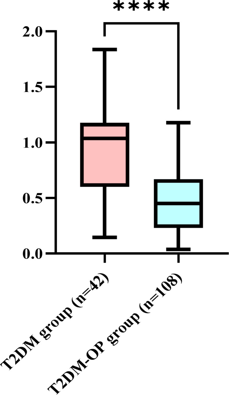
Differential expression of lncRNA H19 between the T2DM group and the T2DM-OP group visualized by box plots. Abbreviations: T2DM, type 2 diabetes mellitus; T2DM-OP, type 2 diabetes mellitus with comorbid osteoporosis; lncRNA H19, long non-coding RNA H19. Notes: The box represents the IQR (25th-75th percentiles) with a midline at the median. Whiskers extend to extreme values within 1.5×IQR. ****P < 0.0001
Bivariate correlations between lncRNA H19 expression and other variables
Bivariate correlation analysis revealed that lncRNA H19 expression was significantly positively correlated with ALB (R = 0.311, P < 0.001; Fig. 2(b)) and BMD (R = 0.696, P < 0.001; Fig. 2(e)). Conversely, it was significantly negatively correlated with age (R = -0.514, P < 0.001; Fig. 2(a)), HbA1c (R = -0.272, P < 0.001; Fig. 2(c)), FBG (R = -0.275, P < 0.001; Fig. 2(d)), and TGF-β1 (R = -0.392, P < 0.001; Fig. 2(f)).
Fig. 2.

Bivariate correlations between lncRNA H19 expression and other variables. Abbreviations: lncRNA H19, long non-coding RNA H19; ALB, albumin; HbA1c, glycated hemoglobin A1c; FBG, fasting blood glucose; BMD, bone mineral density; TGF-β1, transforming growth factor-β1
Logistic regression analysis of the influencing factors for postmenopausal T2DM-OP
After adjusting for covariates, age (odds ratios (OR) = 1.479, 95% confidence interval (CI): 1.088–2.011, P = 0.012), BMI (OR = 0.360, 95% CI: 0.167–0.779, P = 0.010), HbA1c (OR = 2.591, 95% CI: 1.245–5.390, P = 0.011), TGF-β1 (OR = 1.178, 95% CI: 1.057–1.312, P = 0.003), and lncRNA H19 (OR = 0.008, 95% CI: 0.000-0.415, P = 0.017) were identified as factors significantly associated with postmenopausal T2DM-OP (Table 2). Due to the lack of a significant correlation, ALB (OR = 1.510, 95% CI: 0.662–3.444, P = 0.328) and FBG (OR = 1.359, 95% CI: 0.591–3.126, P = 0.470) were excluded.
Table 2.
Logistic regression analysis of the influencing factors for postmenopausal T2DM-OP
| Factors | B | SE | Wald χ2 | P value | OR | 95%CI for OR |
|---|---|---|---|---|---|---|
| Age | 0.391 | 0.157 | 6.244 | 0.012 | 1.479 | 1.088–2.011 |
| BMI | -1.021 | 0.394 | 6.723 | 0.010 | 0.360 | 0.167–0.779 |
| ALB | 0.412 | 0.421 | 0.958 | 0.328 | 1.510 | 0.662–3.444 |
| HbA1c | 0.952 | 0.374 | 6.487 | 0.011 | 2.591 | 1.245–5.390 |
| FBG | 0.307 | 0.425 | 0.521 | 0.470 | 1.359 | 0.591–3.126 |
| TGF-β1 | 0.164 | 0.055 | 8.839 | 0.003 | 1.178 | 1.057–1.312 |
| LncRNA H19 | -4.865 | 2.033 | 5.726 | 0.017 | 0.008 | 0.000-0.415 |
| Constant | -39.746 | 23.116 | 2.956 | 0.086 | 0.000 |
Abbreviations: SE, standard error; OR, odds ratios; CI, confidence interval; BMI, body mass index; ALB, albumin; HbA1c, glycated hemoglobin A1c; FBG, fasting blood glucose; TGF-β1, transforming growth factor-β1; lncRNA H19, long non-coding RNA H19
ROC curve analysis: diagnostic performance of lncRNA H19 for T2DM-OP
ROC curve analysis demonstrated that AUC of lncRNA H19 for diagnosing T2DM-OP was 0.833 (95% CI: 0.748–0.919), with an optimal cutoff value of 0.88, a sensitivity of 71.4%, and specificity of 97.2%. These findings confirm its significant diagnostic value for postmenopausal T2DM-OP (Fig. 3).
Fig. 3.

ROC curve analysis: diagnostic performance of lncRNA H19 for T2DM-OP. Abbreviations: lncRNA H19, long non-coding RNA H19; AUC, area under the curve; CI, confidence interval; ROC, receiver operating characteristic
Multiple linear regression analysis of influencing factors for lncRNA H19
Multiple linear regression analysis was performed with lncRNA H19 as the dependent variable and age, BMI, ALB, HbA1c, FBG, TGF-β1, and BMD as independent variables (Table 3). The regression model fit well (F = 41.711, P < 0.001). The analysis results showed that age (P = 0.005), HbA1c (P = 0.010), FBG (P < 0.001), TGF-β1 (P = 0.032), and BMD (P < 0.001) were influencing factors for lncRNA H19 expression.
Table 3.
Multiple linear regression analysis of influencing factors for LncRNA H19
| Factors | B (unstandardized coefficient) | SE | Beta (standardized coefficient) | t | P value |
|---|---|---|---|---|---|
| Constant | -0.564 | 0.566 | -0.996 | 0.321 | |
| Age | -0.011 | 0.004 | -0.205 | -2.855 | 0.005 |
| BMI | -0.001 | 0.007 | -0.004 | -0.073 | 0.942 |
| ALB | 0.008 | 0.009 | 0.053 | 0.889 | 0.375 |
| HbA1c | 0.036 | 0.014 | 0.175 | 2.622 | 0.010 |
| FBG | -0.050 | 0.011 | -0.293 | -4.378 | < 0.001 |
| TGF-β1 | 0.003 | 0.001 | 0.150 | 2.171 | 0.032 |
| BMD | 1.758 | 0.191 | 0.725 | 9.218 | < 0.001 |
| R2 | 0.673 | ||||
| F | 41.711*** | ||||
Abbreviations: lncRNA H19, long non-coding RNA H19; SE, standard error; BMI, body mass index; ALB, albumin; HbA1c, glycated hemoglobin A1c; FBG, fasting blood glucose; TGF-β1, transforming growth factor-β1; BMD, bone mineral density. Notes: ***P < 0.001
Discussion
Postmenopausal T2DM-OP is defined as a condition characterized by increased bone fragility and decreased bone mass in the skeletal system of postmenopausal T2DM patients [22]. As age advances, islet dysfunction and estrogen depletion jointly elevate the risk of fractures in postmenopausal T2DM patients, significantly impacting their lifespan and quality of life [23]. This has imposed a substantial economic burden on patients’ families and healthcare systems and has emerged as an urgent public health concern that requires immediate attention in contemporary society. It is estimated that the global prevalence of diabetes mellitus among adults aged 20 to 79 years is 10.5%, and it is projected to rise to 12.2% by 2045 [24]. Epidemiological data in China indicates that the prevalence rate of OP among individuals over 65 years old is 32%, and both T1DM and T2DM can increase the susceptibility to fractures [25]. Therefore, it is especially necessary to actively manage diabetic OP in clinical practice. Currently, there is still a dearth of effective biomarkers for detecting diabetic OP and grading the severity of the disease. Although bone turnover markers are associated with BMD and fracture risk and can predict therapeutic responses and adverse events in postmenopausal OP [26, 27], their specific application value and effectiveness in postmenopausal T2DM-OP still require further validation. In this context, this study explores the role and clinical relevance of lncRNA H19 in the pathogenesis of postmenopausal T2DM-OP, offering important references for clinical diagnosis and treatment.
LncRNAs are transcripts exceeding 200 nucleotides in length without protein-coding capacity. They play a crucial role in gene expression regulation and cell physiology and are anticipated to become biomarkers for numerous diseases, including diabetes complications [28]. LncRNA H19 is located in the telomere region of human chromosome 11p15.5, with a transcript length of 2.3 kb. This imprinted gene is only expressed in maternally inherited alleles [29]. As a pivotal regulator of osteogenic differentiation, lncRNA H19 is involved in mediating bone regeneration and regulating skeletal metabolic pathways [30].
The precise coupling between osteoblastic bone formation and osteoclastic resorption serves as the foundation for maintaining skeletal homeostasis, and the imbalance of this coupling relationship represents the core pathophysiological mechanism of OP [31]. The results of this study demonstrated that, compared with the T2DM group, patients in the T2DM-OP group exhibited significantly older age, elevated levels of HbA1c, FBG, and TGF-β1, while BMI, ALB, BMD, and lncRNA H19 expression levels decreased. After adjusting for covariates such as age, BMI, ALB, FBG, HbA1c, TGF-β1, and lncRNA H19, age, BMI, HbA1c, TGF-β1, and lncRNA H19 remained influencing factors for postmenopausal T2DM-OP, aligning with previous research findings [32–38].
The incidence rate of OP exhibits a positive correlation with age. Fragility fractures are highly prevalent among the elderly population and are closely associated with age-related OP [39]. As age advances, the bone-forming ability of osteoblasts declines, while simultaneously, the resorptive function of osteoclasts is relatively enhanced, resulting in an imbalance in bone metabolism, where bone resorption surpasses bone formation. This imbalance leads to a net loss of bone mass and an increase in skeletal fragility [38]. The incidence of OP in postmenopausal women has increased significantly, characterized by a marked decrease in estrogen levels. Estrogen deficiency can accelerate bone resorption, leading to thinning or discontinuity of bone trabeculae, rendering postmenopausal women more prone to OP [8]. Furthermore, persistent calcium loss and alterations in immune cell function collectively heighten the risk of OP in postmenopausal women [40, 41].
BMI represents a significant risk factor for OP. Studies indicated that the BMI of postmenopausal women with T2DM-OP was low [32, 42]. A higher BMI elevates the mechanical loading on bone tissue, stimulates osteoblast activity more strongly, enhances BMD, and decelerates the progression of OP [43]. The Osteoporosis Self-assessment Tool for Asians (OSTA) calculates OP risk scores using the following formula: OSTA score = [body weight (kg) - age (years)] × 0.2, suggesting that weight gain aids in reducing the risk of age-related OP [44]. A recent cross-sectional study further validated the diagnostic value of weight, BMI, and age in screening for OP and proposed new cutoffs for identifying high-risk women (weight < 57.4 kg, BMI < 23.8 kg/m², and age ≥ 72 years) [45]. However, extant evidence indicates that both low and high BMI elevate OP risk [32, 33]. Unfortunately, due to the limited sample size, it is currently infeasible to conduct BMI subgroup analysis to determine the diagnostic cutoff point values for OP.
HbA1c can reflect the stability of a patient’s blood glucose level. Hyperglycemia can inhibit the differentiation of osteoblasts and facilitate osteoclastogenesis [46]. Additionally, factors such as heightened levels of oxidative stress, accumulation of AGEs, reduced levels of insulin-like growth factor-1 (IGF-1), hormonal imbalances, and immune dysfunction collectively constitute the pathophysiological mechanism of OP [34].
TGF-β1 synthesized by osteoblasts exerts a positive regulatory effect on osteoblasts and a bidirectional regulatory effect on osteoclasts [47–50]. As a pivotal factor in maintaining bone metabolic balance, TGF-β1 plays a key role in the proliferation and differentiation of osteoblasts [51]. Research has demonstrated that high concentrations of TGF-β1 can promote osteoclast apoptosis, whereas low concentrations of TGF-β1 induce osteoclast maturation [52]. Xiao et al. [35] reported that the level of TGF-β1 in the serum of patients with T2DM was notably higher than that of healthy individuals. Animal studies by Liu et al. [36] demonstrated that the level of TGF-β1 secreted by senescent cells in the callus of elderly mice increased, which inhibited fracture healing in these mice. This implies that elevated levels of TGF-β1 may foster the occurrence and development of OP.
Previous study by Liang et al. [37] demonstrated that lncRNA H19 can function as a microRNA (miRNA) sponge, reducing the expression levels of miR-141 and miR-22, upregulating runt-related transcription factor 2 (RUNX2), and activating the Wnt/β-catenin signaling pathway to facilitate osteoblast differentiation. However, the association between lncRNA H19 and TGF-β1 in postmenopausal women with T2DM-OP remains unclear. This study revealed that, when compared with the T2DM group, the expression level of lncRNA H19 in PBMCs of patients in the T2DM-OP group was significantly decreased, whereas the level of TGF-β1 was elevated. After adjusting for factors including age, BMI, HbA1c, TGF-β1, and lncRNA H19, lncRNA H19 and TGF-β1 remained significant influencing factors associated with postmenopausal T2DM-OP. This finding implies that lncRNA H19 and TGF-β1 may collaborate in the development of postmenopausal T2DM-OP. A reduction in lncRNA H19 levels, when accompanied by an increase in TGF-β1 levels, may indicate an elevated susceptibility to postmenopausal T2DM-OP.
Further analysis of the correlations among lncRNA H19, TGF-β1, and other clinical parameters in the T2DM group and the T2DM-OP group revealed that the expression level of lncRNA H19 was positively correlated with ALB and BMD, yet negatively correlated with age, HbA1c, FBG, and TGF-β1. Additionally, age, HbA1c, FBG, TGF-β1, and BMD have been recognized as significant factors influencing the expression of lncRNA H19. These findings imply that the downregulation of lncRNA H19 expression may contribute to the pathogenesis of postmenopausal T2DM-OP, potentially facilitating an elevation in TGF-β1 expression via the lncRNA H19/TGF-β1 pathway. Huang et al. [16] verified that the lncRNA H19/miR-675 axis regulated osteoblast differentiation via the TGF-β1/Smad3/histone deacetylase (HDAC) signaling pathway, suggesting its participation in bone metabolism. Furthermore, Cipriani et al. [34] demonstrated that insulin resistance upregulated the expression of factors related to the TGF-β pathway via chronic inflammation and hyperglycemia, consequently impairing bone microstructure and heightening the risk of fractures. In this study, significant associations among lncRNA H19, blood glucose indicators (HbA1c and FBG), TGF-β1, and BMD suggest that lncRNA H19 may be implicated in insulin resistance-related bone metabolism regulation; however, its specific mechanism requires further verification. Therefore, the preliminary results of this study indicate a potential association between lncRNA H19 and TGF-β1 in the pathogenesis of postmenopausal T2DM-OP. To clarify the causal relationship, future longitudinal studies are warranted for further validation.
Mounting evidence suggests that non-coding RNAs (ncRNAs) play a crucial role in musculoskeletal disorders. Recent research findings have particularly emphasized their regulatory potential in OP [53–58]. After analyzing 14 in vitro and in vivo studies, the Gargano team [57] discovered that small interfering RNAs (siRNAs) can specifically silence key genes related to bone metabolism, thereby restoring the dynamic balance between osteoblasts and osteoclasts. Subsequently, the team [58] reviewed 17 studies on circular RNAs (circRNAs) and confirmed that molecules such as circular Forkhead box protein P1 (circFOXP1) and hsa_circ_0006215 can promote the osteogenic differentiation of BMSCs by sponging miRNAs and upregulating RUNX2. These findings are consistent with the phenomenon we observed, where the expression of lncRNA H19 is reduced and the level of TGF-β1 is increased. This implies that different types of ncRNA molecules (siRNA, circRNA, and lncRNA) may play synergistic or antagonistic roles at different nodes of the same pathological network, thus providing a theoretical basis for the targeted combination therapy for postmenopausal T2DM-OP.
The study yielded several key findings. Firstly, we reported for the first time the association between lncRNA H19 levels and postmenopausal T2DM-OP. In comparison with the T2DM group, the expression of lncRNA H19 was significantly lower in the T2DM-OP group. Secondly, bivariate correlation analysis demonstrated that the expression of lncRNA H19 was positively correlated with ALB and BMD, and negatively correlated with age, HbA1c, FBG, and TGF-β1. Thirdly, logistic regression analysis identified age, BMI, HbA1c, TGF-β1, and lncRNA H19 as independent influencing factors for postmenopausal T2DM-OP. Finally, linear regression analysis revealed that age, HbA1c, FBG, TGF-β1, and BMD were independent determinants of the expression of lncRNA H19. Collectively, these results indicate that lncRNA H19 may participate in the pathogenesis of postmenopausal T2DM-OP through mechanisms related to glucose metabolism and the lncRNA H19/TGF-β1 signaling pathway. It is noteworthy that the ROC analysis in this study demonstrated that the AUC of lncRNA H19 in PBMCs for diagnosing postmenopausal T2DM-OP was 0.833, with a sensitivity of 71.4% and specificity of 97.2%, indicating its considerable diagnostic value. This finding suggests that lncRNA H19 may serve as a novel non-invasive biomarker for postmenopausal T2DM-OP.
However, this study has several limitations. Firstly, as a cross-sectional investigation, it is impossible to establish causal relationships. Further animal experiments and in vitro studies are warranted to validate the causal role of lncRNA H19 and its potential regulatory effect on TGF-β1 in the pathogenesis of postmenopausal T2DM-OP through the lncRNA H19/TGF-β1 signaling pathway. Secondly, the sample size was relatively limited and the data were non-normally distributed. This necessitates future studies to adopt previous non-parametric sample size estimation methods during the design phase and conduct large-scale multicenter cohorts to verify the clinical relevance, expression patterns, and mechanisms of action of lncRNA H19. Thirdly, the absence of a healthy control group and the failure to fully control confounding factors related to diabetes make it difficult to comprehensively explore the pathogenesis of T2DM-specific OP, which limits the analysis of dynamic changes of relevant parameters in the “healthy-T2DM-T2DM-OP” spectrum. Fourthly, the lack of measurement of insulin resistance indicators and inflammatory markers makes it impossible to directly elucidate the upstream role of insulin resistance in the lncRNA H19/TGF-β1 axis. Therefore, future research needs to supplement relevant data to improve understanding of its mechanism of action. It is important to note that subsequent studies will include healthy controls for triple comparisons to clarify the specific expression of lncRNA H19 and related indicators in the disease.
Conclusions
In conclusion, the expression of lncRNA H19 in PBMCs of postmenopausal T2DM-OP patients may contribute to the onset and progression of the disease via pathways related to glucose metabolism and transforming growth factors. Therefore, monitoring the changes in lncRNA H19 levels is highly valuable for the early prediction of postmenopausal T2DM-OP.
Acknowledgements
Not applicable.
Abbreviations
- AGEs
Advanced glycation end products
- ALB
Albumin
- AUC
Area under the curve
- BMD
Bone mineral density
- BMI
Body mass index
- BMSCs
Bone marrow mesenchymal stem cells
- cDNA
Complementary DNA
- CI
Confidence interval
- circFOXP1
Circular Forkhead box protein P1
- circRNAs
Circular RNAs
- DM
Diabetes mellitus
- DXA
Dual-energy x-ray absorptiometry
- EDTA-K2
Ethylenediaminetetraacetic acid dipotassium
- ELISA
Enzyme-linked immunosorbent assay
- FBG
Fasting blood glucose
- GAPDH
Glyceraldehyde-3-phosphate dehydrogenase
- HbA1c
Glycated hemoglobin A1c
- HCY
Homocysteine
- HDAC
Histone deacetylase
- HDL-C
High-density lipoprotein cholesterol
- HPLC
High-performance liquid chromatography
- IGF-1
Insulin-like growth factor-1
- IQR
Interquartile range
- LDL-C
Low-density lipoprotein cholesterol
- lncRNAs
Long non-coding RNAs
- lncRNA H19
Long non-coding RNA H19
- miRNA
microRNA
- ncRNAs
Non-coding RNAs
- OGTT
Oral glucose tolerance test
- OP
Osteoporosis
- OR
Odds ratios
- OSTA
Osteoporosis self-assessment tool for Asians
- PBMCs
Peripheral blood mononuclear cells
- qRT-PCR
Quantitative reverse transcription polymerase chain reaction
- ROC
Receiver operating characteristic
- RUNX2
Runt-related transcription factor 2
- SD
Standard deviation
- SE
Standard error
- siRNAs
Small interfering RNAs
- TC
Total cholesterol
- T1DM
Type 1 diabetes mellitus
- T2DM
Type 2 diabetes mellitus
- T2DM-OP
Type 2 diabetes mellitus with comorbid osteoporosis
- TG
Triglycerides
- TGF-β1
Transforming growth factor-β1
- UA
Uric acid
Author contributions
TY and XF were responsible for data curation, formal analysis, and original draft preparation. NW, TZ, and ZZ contributed to patient enrollment and data collection. GW oversaw project administration, manuscript review, and editing. All authors read and approved the final manuscript.
Funding
This study was funded by 2024 Healthcare Quality (Evidence-Based) Management Research Project (YLZLXZ24G069), 2024 Lianyungang Association for Science and Technology Soft Science Research Project (Lkxyb24160), 2023 Lianyungang Sixth Phase “521” Scientific Research Funding Project (LYG06521202372), 2022 Lianyungang Health and Medical Technology Project (202206) and 2021 Lianyungang Elderly health research project (L202105).
Data availability
The datasets used and/or analysed during the current study are available from the corresponding author on reasonable request.
Declarations
Ethics approval and consent to participate
The study protocol was approved by the Ethics Committee of Lianyungang First People’s Hospital (KY-20210712001-01) and registered with the Chinese Clinical Trial Registry (ChiCTR2300074807). Written informed consent was obtained from all participants in accordance with the Declaration of Helsinki.
Consent for publication
Not applicable.
Competing interests
The authors declare no competing interests.
Footnotes
Publisher’s note
Springer Nature remains neutral with regard to jurisdictional claims in published maps and institutional affiliations.
Teng Yang and Xiuli Feng contributed equally to this work.
References
- 1.Mlonga J, Damian D. Lipid profile abnormalities in type 2 diabetes mellitus patients at Mbeya zonal referral hospital, tanzania: a retrospective study. J Diabetes Res. 2025;2025:9966933. 10.1155/jdr/9966933. [DOI] [PMC free article] [PubMed]
- 2.Farooqui KJ, Mithal A, Kerwen AK, Chandran M. Type 2 diabetes and bone fragility- an under-recognized association. Diabetes Metab Syndr. 2021;15(3):927–35. 10.1016/j.dsx.2021.04.017. [DOI] [PubMed] [Google Scholar]
- 3.Bao X, Liu C, Liu H, Wang Y, Xue P, Li Y. Association between polymorphisms of glucagon-like peptide-1 receptor gene and susceptibility to osteoporosis in Chinese postmenopausal women. J Orthop Surg Res. 2024;19(1):869. 10.1186/s13018-024-05361-z. [DOI] [PMC free article] [PubMed] [Google Scholar]
- 4.Migliorini F, Maffulli N, Colarossi G, Eschweiler J, Tingart M, Betsch M. Effect of drugs on bone mineral density in postmenopausal osteoporosis: a bayesian network meta-analysis. J Orthop Surg Res. 2021;16(1):533. 10.1186/s13018-021-02678-x. [DOI] [PMC free article] [PubMed] [Google Scholar]
- 5.Conti V, Russomanno G, Corbi G, et al. A polymorphism at the translation start site of the vitamin D receptor gene is associated with the response to anti-osteoporotic therapy in postmenopausal women from Southern Italy. Int J Mol Sci. 2015;16(3):5452–66. 10.3390/ijms16035452. [DOI] [PMC free article] [PubMed] [Google Scholar]
- 6.Park SY, Choi KH, Jun JE, Chung HY. Effects of advanced glycation end products on differentiation and function of osteoblasts and osteoclasts. J Korean Med Sci. 2021;36(37):e239. 10.3346/jkms.2021.36.e239. [DOI] [PMC free article] [PubMed] [Google Scholar]
- 7.Huang F, Wang Y, Liu J, Cheng Y, Zhang X, Jiang H. Asperuloside alleviates osteoporosis by promoting autophagy and regulating Nrf2 activation. J Orthop Surg Res. 2024;19(1):855. 10.1186/s13018-024-05320-8. [DOI] [PMC free article] [PubMed] [Google Scholar]
- 8.Li J, Chen X, Lu L, Yu X. The relationship between bone marrow adipose tissue and bone metabolism in postmenopausal osteoporosis. Cytokine Growth Factor Rev. 2020;52:88–98. 10.1016/j.cytogfr.2020.02.003. [DOI] [PubMed] [Google Scholar]
- 9.Sun W, Xing Y, Zhou F, Ma Y, Wan X, Ma H. Association analysis of triglyceride Glucose-Body mass index and bone turnover markers in patients with type 2 diabetes mellitus. Diabetes Metab Syndr Obes. 2023;16:1435–47. 10.2147/DMSO.S406849. [DOI] [PMC free article] [PubMed] [Google Scholar]
- 10.Ouyang X, Li S, Ding Y, Xin F, Liu M. Mechanism of miRNA-31 regulating Wnt/β-catenin signaling pathway by targeting Satb2 in the osteogenic differentiation of human bone Marrow-Derived mesenchymal stem cells. J Musculoskelet Neuronal Interact. 2023;23(3):346–54. [PMC free article] [PubMed] [Google Scholar]
- 11.Migliorini F, Colarossi G, Baroncini A, Eschweiler J, Tingart M, Maffulli N. Pharmacological management of postmenopausal osteoporosis: a level I evidence Based - Expert opinion. Expert Rev Clin Pharmacol. 2021;14(1):105–19. 10.1080/17512433.2021.1851192. [DOI] [PubMed] [Google Scholar]
- 12.Andersen MØ, Andresen AK, Hartvigsen J, Hermann AP, Sørensen J, Carreon LY. Vertebroplasty for painful osteoporotic vertebral compression fractures: a protocol for a single-center doubled-blind randomized sham-controlled clinical trial. VOPE2. J Orthop Surg Res. 2024;19(1):813. 10.1186/s13018-024-05301-x. [DOI] [PMC free article] [PubMed] [Google Scholar]
- 13.Migliorini F, Colarossi G, Eschweiler J, Oliva F, Driessen A, Maffulli N. Antiresorptive treatments for corticosteroid-induced osteoporosis: a bayesian network meta-analysis. Br Med Bull. 2022;143(1):46–56. 10.1093/bmb/ldac017. [DOI] [PMC free article] [PubMed] [Google Scholar]
- 14.Shen L, Yang H, Zhou F, Jiang T, Jiang Z. Risk factors of short-term residual low back pain after PKP for the first thoracolumbar osteoporotic vertebral compression fracture. J Orthop Surg Res. 2024;19(1):792. 10.1186/s13018-024-05295-6. [DOI] [PMC free article] [PubMed] [Google Scholar]
- 15.Dimai HP. Use of dual-energy X-ray absorptiometry (DXA) for diagnosis and fracture risk assessment; WHO-criteria, T- and Z-score, and reference databases. Bone. 2017;104:39–43. 10.1016/j.bone.2016.12.016. [DOI] [PubMed] [Google Scholar]
- 16.Huang Y, Zheng Y, Jia L, Li W, Long Noncoding. RNA H19 promotes osteoblast differentiation via TGF-β1/Smad3/HDAC signaling pathway by deriving miR-675. Stem Cells. 2015;33(12):3481–92. 10.1002/stem.2225. [DOI] [PubMed] [Google Scholar]
- 17.Zhou P, Li Y, Di R, et al. H19 and Foxc2 synergistically promotes osteogenic differentiation of BMSCs via wnt-β-catenin pathway. J Cell Physiol. 2019;234(8):13799–806. 10.1002/jcp.28060. [DOI] [PMC free article] [PubMed] [Google Scholar]
- 18.Zhang P, Zhang H, Lin J, et al. Insulin impedes osteogenesis of BMSCs by inhibiting autophagy and promoting premature senescence via the TGF-β1 pathway. Aging. 2020;12(3):2084–100. 10.18632/aging.102723. [DOI] [PMC free article] [PubMed] [Google Scholar]
- 19.ElSayed NA, Aleppo G, Aroda VR, et al. 2. Classification and diagnosis of diabetes: standards of care in diabetes-2023. Diabetes Care. 2023;46(Suppl 1):S19–40. 10.2337/dc23-S002. [DOI] [PMC free article] [PubMed] [Google Scholar]
- 20.Kanis JA, Cooper C, Rizzoli R, Reginster JY, Scientific Advisory Board of the European Society for Clinical and Economic Aspects of Osteoporosis (ESCEO) and the Committees of Scientific Advisors and National Societies of the International Osteoporosis Foundation (IOF). European guidance for the diagnosis and management of osteoporosis in postmenopausal women. Osteoporos Int. 2019;30(1):3–44. 10.1007/s00198-018-4704-5. [DOI] [PMC free article] [PubMed] [Google Scholar]
- 21.Obuchowski NA. Sample size tables for receiver operating characteristic studies. AJR Am J Roentgenol. 2000;175(3):603–8. 10.2214/ajr.175.3.1750603. [DOI] [PubMed] [Google Scholar]
- 22.Chen Y, Mei X, Liang X, et al. Application of magnetic resonance image compilation (MAGiC) in the diagnosis of middle-aged and elderly women with osteoporosis. BMC Med Imaging. 2023;23(1):63. 10.1186/s12880-023-01010-9. [DOI] [PMC free article] [PubMed] [Google Scholar]
- 23.Liang Y, Zhu B, Li S, et al. Curcumin protects bone Biomechanical properties and microarchitecture in type 2 diabetic rats with osteoporosis via the TGFβ/Smad2/3 pathway. Exp Ther Med. 2020;20(3):2200–8. 10.3892/etm.2020.8943. [DOI] [PMC free article] [PubMed] [Google Scholar]
- 24.Sun H, Saeedi P, Karuranga S, et al. IDF diabetes atlas: global, regional and country-level diabetes prevalence estimates for 2021 and projections for 2045. Diabetes Res Clin Pract. 2022;183:109119. 10.1016/j.diabres.2021.109119. [DOI] [PMC free article] [PubMed] [Google Scholar]
- 25.Yu F, Xia W. The epidemiology of osteoporosis, associated fragility fractures, and management gap in China. Arch Osteoporos. 2019;14(1):32. 10.1007/s11657-018-0549-y. [DOI] [PubMed] [Google Scholar]
- 26.Migliorini F, Maffulli N, Spiezia F, Peretti GM, Tingart M, Giorgino R. Potential of biomarkers during Pharmacological therapy setting for postmenopausal osteoporosis: a systematic review. J Orthop Surg Res. 2021;16(1):351. 10.1186/s13018-021-02497-0. [DOI] [PMC free article] [PubMed] [Google Scholar]
- 27.Migliorini F, Maffulli N, Spiezia F, Tingart M, Maria PG, Riccardo G. Biomarkers as therapy monitoring for postmenopausal osteoporosis: a systematic review. J Orthop Surg Res. 2021;16(1):318. 10.1186/s13018-021-02474-7. [DOI] [PMC free article] [PubMed] [Google Scholar]
- 28.Xu YX, Pu SD, Li X, et al. Exosomal ncrnas: novel therapeutic target and biomarker for diabetic complications. Pharmacol Res. 2022;178:106135. 10.1016/j.phrs.2022.106135. [DOI] [PubMed] [Google Scholar]
- 29.Bitarafan S, Yari M, Broumand MA, et al. Association of increased levels of LncRNA H19 in PBMCs with risk of coronary artery disease. Cell J. 2019;20(4):564–8. 10.22074/cellj.2019.5544. [DOI] [PMC free article] [PubMed] [Google Scholar]
- 30.Wu J, Zhao J, Sun L, Pan Y, Wang H, Zhang WB. Long non-coding RNA H19 mediates mechanical tension-induced osteogenesis of bone marrow mesenchymal stem cells via FAK by sponging miR-138. Bone. 2018;108:62–70. 10.1016/j.bone.2017.12.013. [DOI] [PubMed] [Google Scholar]
- 31.Lin L, He E, Wang H, et al. Intravenous transplantation of human hair follicle-derived mesenchymal stem cells ameliorates trabecular bone loss in osteoporotic mice. Front Cell Dev Biol. 2022;10:814949. 10.3389/fcell.2022.814949. [DOI] [PMC free article] [PubMed] [Google Scholar]
- 32.Lee JH, Kim JH, Hong AR, Kim SW, Shin CS. Optimal body mass index for minimizing the risk for osteoporosis and type 2 diabetes. Korean J Intern Med. 2020;35(6):1432–42. 10.3904/kjim.2018.223. [DOI] [PMC free article] [PubMed] [Google Scholar]
- 33.Gundmi S, Maiya AG, Bhat AK, Hande MH, Kumar AS. Screening for basal metabolic rate and visceral fat among postmenopausal osteoporosis with type 2 diabetes mellitus. Diabetes Metab Syndr. 2019;13(2):981–4. 10.1016/j.dsx.2019.01.003. [DOI] [PubMed] [Google Scholar]
- 34.Cipriani C, Colangelo L, Santori R, et al. The interplay between bone and glucose metabolism. Front Endocrinol (Lausanne). 2020;11:122. 10.3389/fendo.2020.00122. [DOI] [PMC free article] [PubMed] [Google Scholar]
- 35.Xiao Y, Wang Y, Ryu J, et al. Upregulated TGF-β1 contributes to hyperglycaemia in type 2 diabetes by potentiating glucagon signalling. Diabetologia. 2023;66(6):1142–55. 10.1007/s00125-023-05889-5. [DOI] [PubMed] [Google Scholar]
- 36.Liu J, Zhang J, Lin X, Boyce BF, Zhang H, Xing L. Age-associated callus senescent cells produce TGF-β1 that inhibits fracture healing in aged mice. J Clin Invest. 2022;132(8):e148073. 10.1172/JCI148073. [DOI] [PMC free article] [PubMed] [Google Scholar]
- 37.Liang WC, Fu WM, Wang YB, et al. H19 activates Wnt signaling and promotes osteoblast differentiation by functioning as a competing endogenous RNA. Sci Rep. 2016;6:20121. 10.1038/srep20121. [DOI] [PMC free article] [PubMed] [Google Scholar]
- 38.Ding P, Gao C, Gao Y, et al. Osteocytes regulate senescence of bone and bone marrow. Elife. 2022;11:e81480. 10.7554/eLife.81480. [DOI] [PMC free article] [PubMed] [Google Scholar]
- 39.Migliorini F, Giorgino R, Hildebrand F, et al. Fragility fractures: risk factors and management in the elderly. Med (Kaunas). 2021;57(10):1119. 10.3390/medicina57101119. [DOI] [PMC free article] [PubMed] [Google Scholar]
- 40.Rosen CJ. Clinical practice. Postmenopausal osteoporosis. N Engl J Med. 2005;353(6):595–603. 10.1056/NEJMcp043801. [DOI] [PubMed] [Google Scholar]
- 41.Fischer V, Haffner-Luntzer M. Interaction between bone and immune cells: implications for postmenopausal osteoporosis. Semin Cell Dev Biol. 2022;123:14–21. 10.1016/j.semcdb.2021.05.014. [DOI] [PubMed] [Google Scholar]
- 42.Zhao X, Yu X, Zhang X. Association between uric acid and bone mineral density in postmenopausal women with type 2 diabetes mellitus in china: a cross-sectional inpatient study. J Diabetes Res. 2020;2020:3982831. 10.1155/2020/3982831. [DOI] [PMC free article] [PubMed] [Google Scholar]
- 43.Wang D, Zhang Y, He Y, et al. Associations of perchlorate, nitrate, and thiocyanate with bone mineral density in the US general population: a multi-cycle study of NHANES 2011–2018. Nutrients. 2024;16(16):2658. 10.3390/nu16162658. [DOI] [PMC free article] [PubMed] [Google Scholar]
- 44.Fan Z, Li X, Zhang X, Yang Y, Fei Q, Guo A. Comparison of OSTA, FRAX and BMI for predicting postmenopausal osteoporosis in a Han population in beijing: a cross sectional study. Clin Interv Aging. 2020;15:1171–80. 10.2147/CIA.S257166. [DOI] [PMC free article] [PubMed] [Google Scholar]
- 45.Leeyaphan J, Rojjananukulpong K, Intarasompun P, Peerakul Y. Simple clinical predictors for making directive decisions in osteoporosis screening for women: a cross-sectional study. J Orthop Surg Res. 2024;19(1):789. 10.1186/s13018-024-05287-6. [DOI] [PMC free article] [PubMed] [Google Scholar]
- 46.Park YS, Lee BA, Choi SH, Kim YT. Evaluation of failed implants and reimplantation at sites of previous dental implant failure: survival rates and risk factors. J Periodontal Implant Sci. 2022;52(3):230–41. 10.5051/jpis.2105020251. [DOI] [PMC free article] [PubMed] [Google Scholar]
- 47.Janssens K, ten Dijke P, Janssens S, Van Hul W. Transforming growth factor-beta1 to the bone. Endocr Rev. 2005;26(6):743–74. 10.1210/er.2004-0001. [DOI] [PubMed] [Google Scholar]
- 48.Zhang Z, Zhang X, Zhao D, et al. TGF–β1 promotes the osteoinduction of human osteoblasts via the PI3K/AKT/mTOR/S6K1 signalling pathway. Mol Med Rep. 2019;19(5):3505–18. 10.3892/mmr.2019.10051. [DOI] [PMC free article] [PubMed] [Google Scholar]
- 49.Inoue M, Nagai-Yoshioka Y, Yamasaki R, Kawamoto T, Nishihara T, Ariyoshi W. Mechanisms involved in suppression of osteoclast supportive activity by transforming growth factor-β1 via the ubiquitin-proteasome system. PLoS ONE. 2022;17(2):e0262612. 10.1371/journal.pone.0262612. [DOI] [PMC free article] [PubMed] [Google Scholar]
- 50.Gingery A, Bradley EW, Pederson L, Ruan M, Horwood NJ, Oursler MJ. TGF-beta coordinately activates TAK1/MEK/AKT/NFkB and SMAD pathways to promote osteoclast survival. Exp Cell Res. 2008;314(15):2725–38. 10.1016/j.yexcr.2008.06.006. [DOI] [PMC free article] [PubMed] [Google Scholar]
- 51.Steinbrech DS, Mehrara BJ, Rowe NM, et al. Gene expression of TGF-beta, TGF-beta receptor, and extracellular matrix proteins during membranous bone healing in rats. Plast Reconstr Surg. 2000;105(6):2028–38. 10.1097/00006534-200005000-00018. [DOI] [PubMed] [Google Scholar]
- 52.Kasagi S, Chen W. TGF-beta1 on osteoimmunology and the bone component cells. Cell Biosci. 2013;3(1):4. 10.1186/2045-3701-3-4. [DOI] [PMC free article] [PubMed] [Google Scholar]
- 53.Giordano L, Porta GD, Peretti GM, Maffulli N. Therapeutic potential of MicroRNA in tendon injuries. Br Med Bull. 2020;133(1):79–94. 10.1093/bmb/ldaa002. [DOI] [PubMed] [Google Scholar]
- 54.Oliviero A, Della Porta G, Peretti GM, Maffulli N. MicroRNA in osteoarthritis: physiopathology, diagnosis and therapeutic challenge. Br Med Bull. 2019;130(1):137–47. 10.1093/bmb/ldz015. [DOI] [PubMed] [Google Scholar]
- 55.Gargano G, Oliviero A, Oliva F, Maffulli N. Small interfering RNAs in tendon homeostasis. Br Med Bull. 2021;138(1):58–67. 10.1093/bmb/ldaa040. [DOI] [PubMed] [Google Scholar]
- 56.Gargano G, Oliva F, Oliviero A, Maffulli N. Small interfering RNAs in the management of human rheumatoid arthritis. Br Med Bull. 2022;142(1):34–43. 10.1093/bmb/ldac012. [DOI] [PMC free article] [PubMed] [Google Scholar]
- 57.Gargano G, Asparago G, Spiezia F, Oliva F, Maffulli N. Small interfering RNAs in the management of human osteoporosis. Br Med Bull. 2023;148(1):58–69. 10.1093/bmb/ldad023. [DOI] [PMC free article] [PubMed] [Google Scholar]
- 58.Gargano G, Pagano SM, Maffulli N. Circular RNAs in the management of human osteoporosis. Br Med Bull. 2025;153(1):ldae024. 10.1093/bmb/ldae024. [DOI] [PubMed] [Google Scholar]
Associated Data
This section collects any data citations, data availability statements, or supplementary materials included in this article.
Data Availability Statement
The datasets used and/or analysed during the current study are available from the corresponding author on reasonable request.