

Abstract
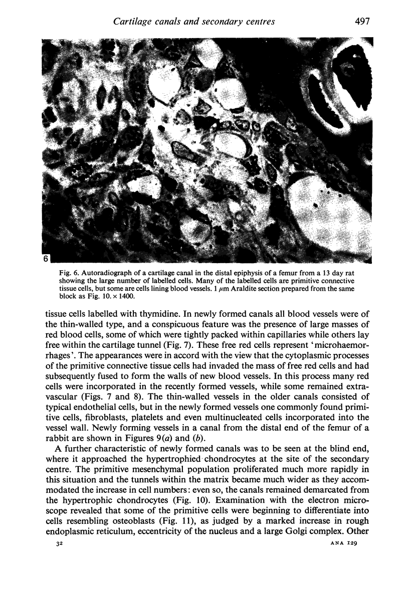
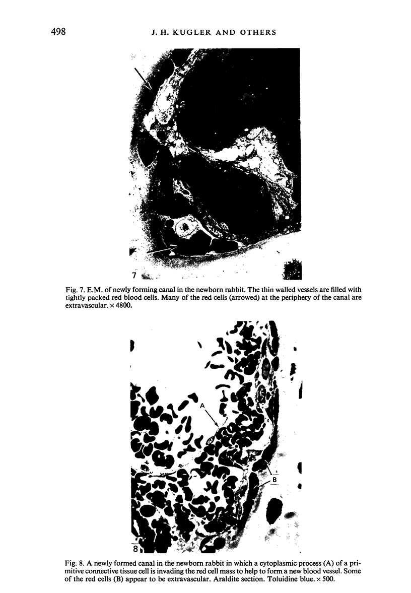
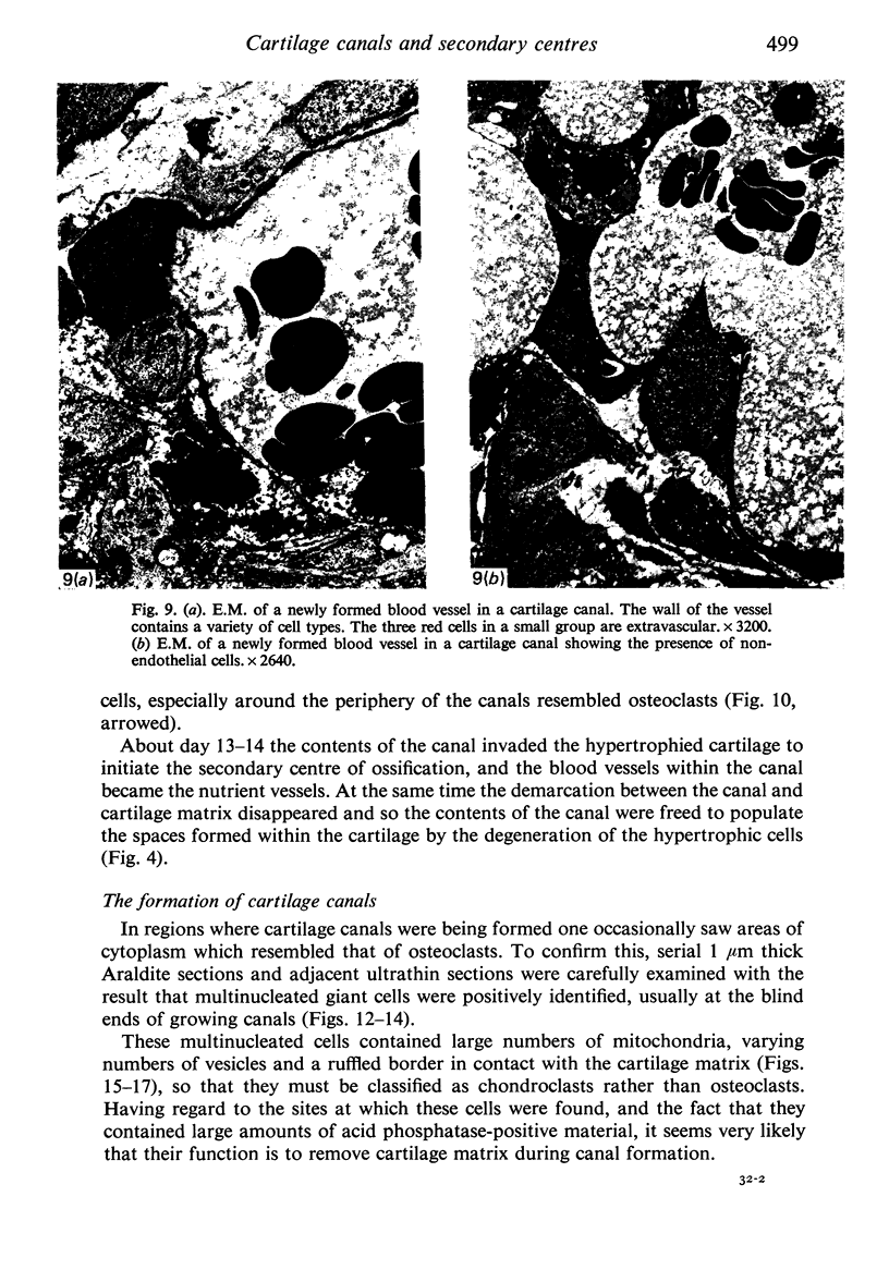
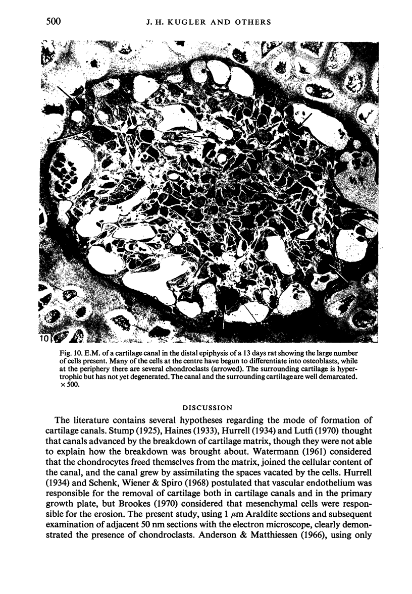
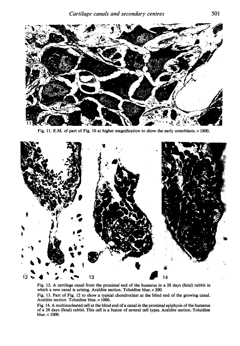
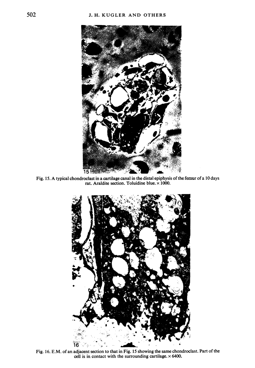
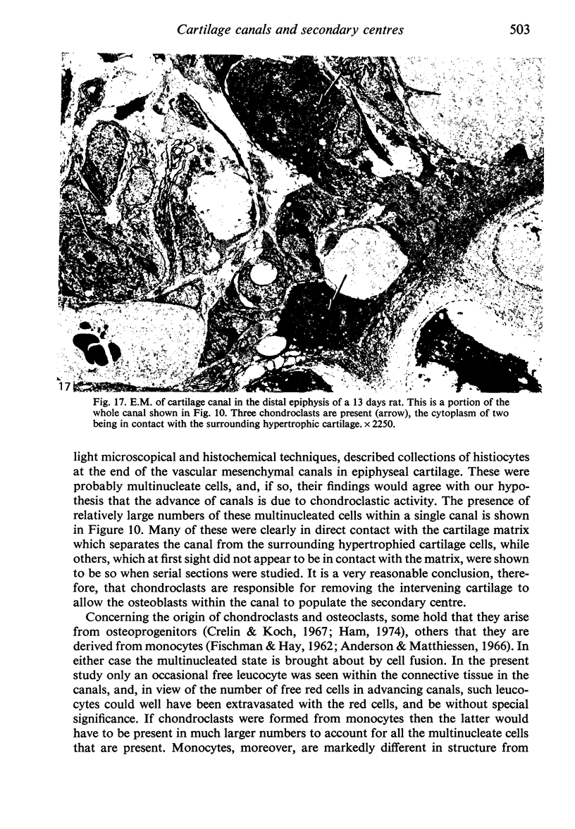
The present studies indicate that in the growth of cartilage canals the cartilage is removed by chondroclasts which stem from the primitive connective tissue cells of the perichondrium. Besides fusing to form multinucleated chondroclasts, these cells also provide the osteoblasts which establish the secondary centre of ossification. The growing tips of the blood vessels within the canals are also fashioned from these primitive connective tissue cells as they invade the microhaemorrhages at the ends of the canals. This is an identical procedure to that found in the vascular invasion of the primary growth plate. The cartilage canals are clearly useful sites in which to study the development of osteoclasts and chondroclasts from more primitive (osteoprogenitor) cells.
Full text
PDF
Images in this article
Selected References
These references are in PubMed. This may not be the complete list of references from this article.
- Andersen H., Matthiessen M. E. The histiocyte in human foetal tissues: its morphology, cytochemistry, origin, function, and fate. Z Zellforsch Mikrosk Anat. 1966;72(2):193–211. doi: 10.1007/BF00334275. [DOI] [PubMed] [Google Scholar]
- BRIDGES J. B., PRITCHARD J. J. Bone and cartilage induction in the rabbit. J Anat. 1958 Jan;92(1):28–38. [PMC free article] [PubMed] [Google Scholar]
- Crelin E. S., Koch W. E. An autoradiographic study of chondrocyte transformation into chondroclasts and osteocytes during bone formation in vitro. Anat Rec. 1967 Aug;158(4):473–483. doi: 10.1002/ar.1091580410. [DOI] [PubMed] [Google Scholar]
- FISCHMAN D. A., HAY E. D. Origin of osteoclasts from mononuclear leucocytes in regenerating newt limbs. Anat Rec. 1962 Aug;143:329–337. doi: 10.1002/ar.1091430402. [DOI] [PubMed] [Google Scholar]
- HARALDSSON S. The vascular pattern of a growing and fullgrown human epiphysis. Acta Anat (Basel) 1962;48:156–167. doi: 10.1159/000141835. [DOI] [PubMed] [Google Scholar]
- Haines R. W. Cartilage Canals. J Anat. 1933 Oct;68(Pt 1):45–64. [PMC free article] [PubMed] [Google Scholar]
- Hurrell D. J. The Vascularisation of Cartilage. J Anat. 1934 Oct;69(Pt 1):47–61. [PMC free article] [PubMed] [Google Scholar]
- LEVENE C. THE PATTERNS OF CARTILAGE CANALS. J Anat. 1964 Oct;98:515–538. [PMC free article] [PubMed] [Google Scholar]
- Lutfi A. M. Mode of growth, fate and functions of cartilage canals. J Anat. 1970 Jan;106(Pt 1):135–145. [PMC free article] [PubMed] [Google Scholar]
- Schenk R. K., Wiener J., Spiro D. Fine structural aspects of vascular invasion of the tibial epiphyseal plate of growing rats. Acta Anat (Basel) 1968;69(1):1–17. doi: 10.1159/000143059. [DOI] [PubMed] [Google Scholar]
- Stockwell R. A. The ultrastructure of cartilage canals and the surrounding cartilage in the sheep fetus. J Anat. 1971 Sep;109(Pt 3):397–410. [PMC free article] [PubMed] [Google Scholar]
- Stump C. W. The Histogenesis of Bone. J Anat. 1925 Jan;59(Pt 2):136–154. [PMC free article] [PubMed] [Google Scholar]
- Wilsman N. J., Van Sickle D. C. The relationship of cartilage canals to the initial osteogenesis of secondary centers of ossification. Anat Rec. 1970 Nov;168(3):381–391. doi: 10.1002/ar.1091680305. [DOI] [PubMed] [Google Scholar]