Abstract
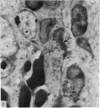
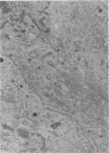
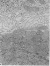
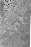
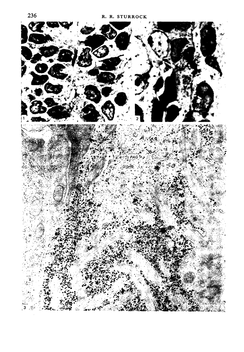
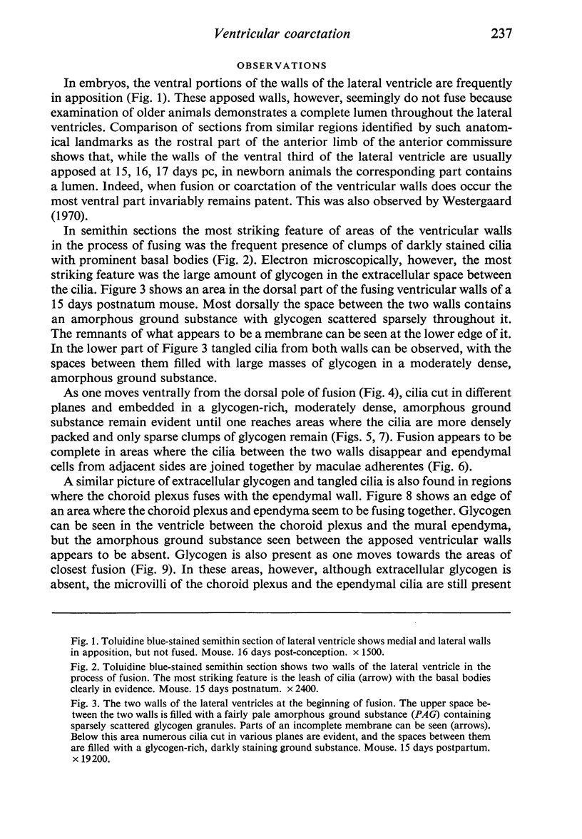
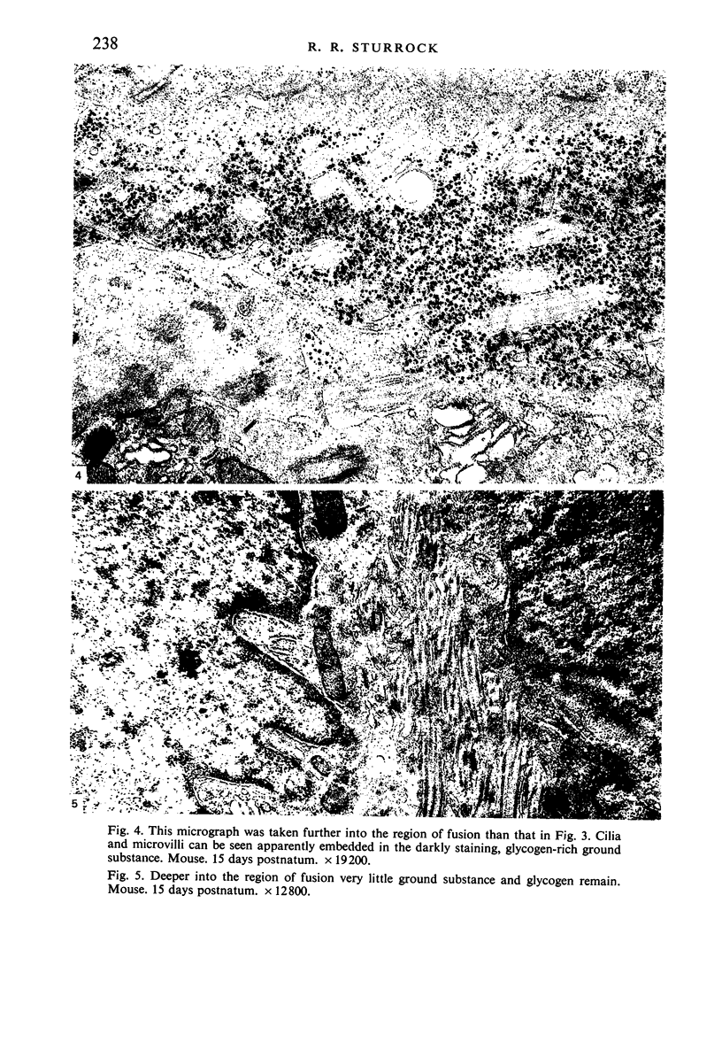
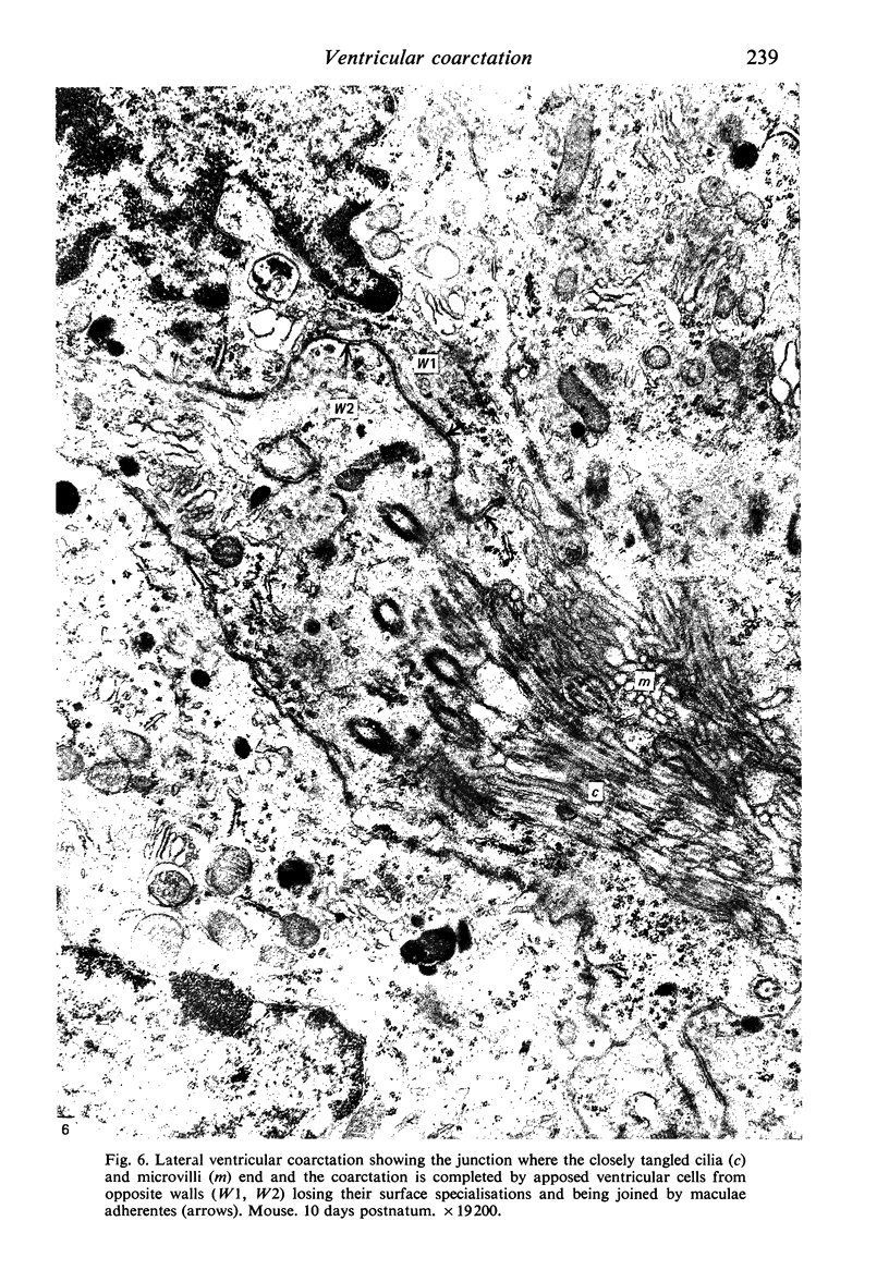
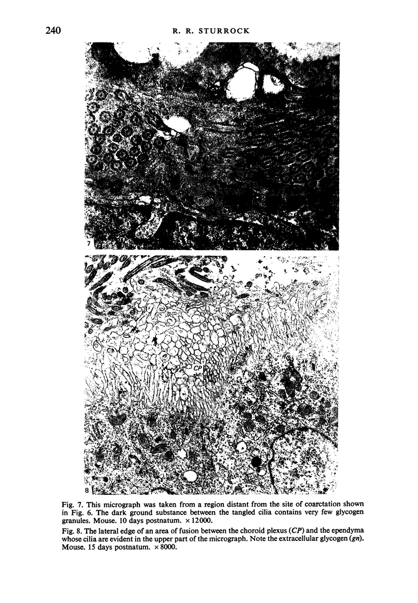
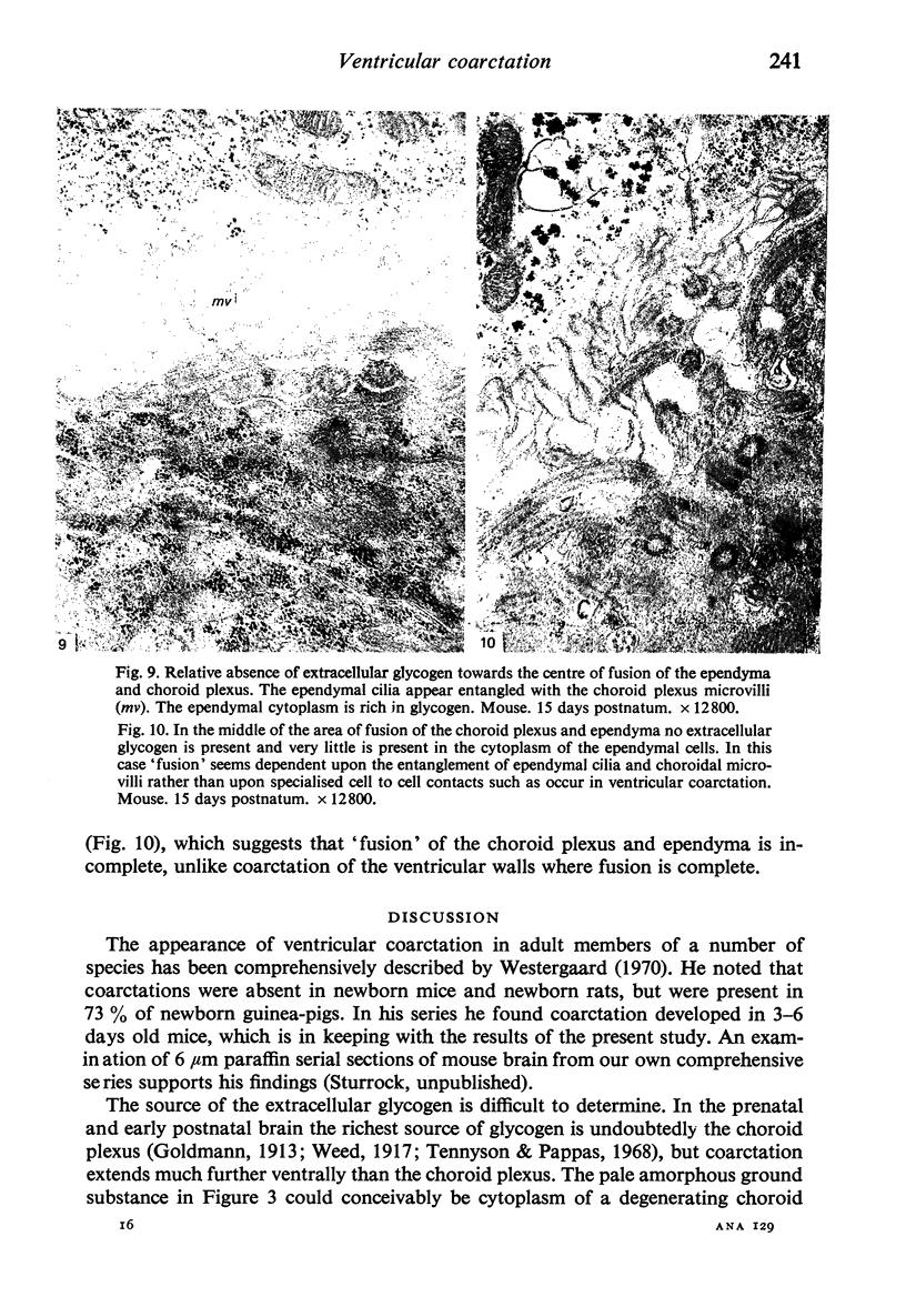
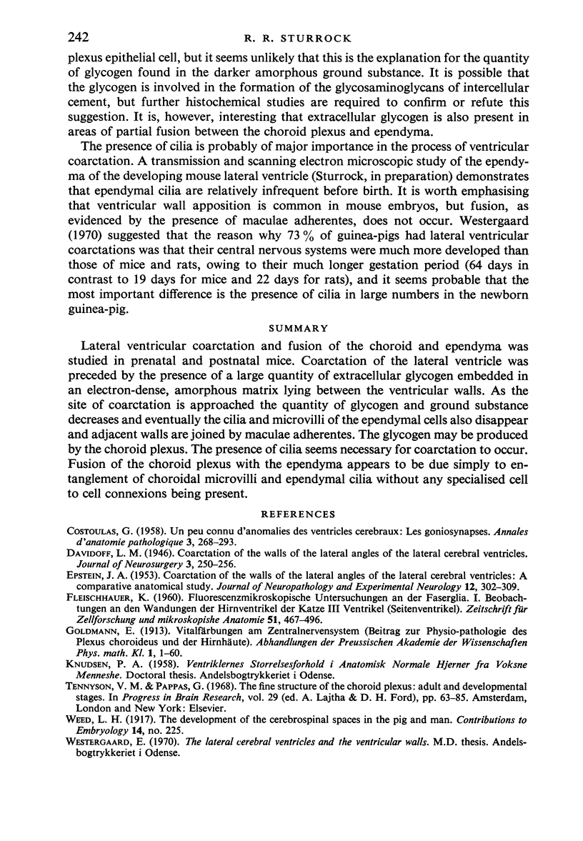
Lateral ventricular coarctation and fusion of the choroid and ependyma was studied in prenatal and postnatal mice. Coarctation of the lateral ventricle was preceded by the presence of a large quantity of extracellular glycogen embedded in an electron-dense, amorphous matrix lying between the ventricular walls. As the site of coarctation is approached the quantity of glycogen and ground substance decreases and eventually the cilia and microvilli of the ependymal cells also disappear and adjacent walls are joined by maculae adherentes. The glycogen may be produced by the choroid plexus. The presence of cilia seems necessary for coarctation to occur. Fusion of the choroid plexus with the ependyma appears to be due simply to entanglement of choroidal microvilli and ependymal cilia without any specialised cell to cell connexions being present.
Full text
PDF
Images in this article
Selected References
These references are in PubMed. This may not be the complete list of references from this article.
- COSTOULAS G. Un type peu connu d'anomalies des ventricules cérébraux: les goniosynapses; étude anatomo-clinique et statistique. Ann Anat Pathol (Paris) 1958 Apr-Jun;3(2):268–293. [PubMed] [Google Scholar]
- EPSTEIN J. A. Coarctation of the walls of the lateral angles of the lateral cerebral ventricles; a comparative anatomical study. J Neuropathol Exp Neurol. 1953 Jul;12(3):302–309. doi: 10.1097/00005072-195307000-00010. [DOI] [PubMed] [Google Scholar]
- Tennyson V. M., Appas G. D. The fine structure of the choroid plexus adult and developmental stages. Prog Brain Res. 1968;29:63–85. doi: 10.1016/s0079-6123(08)64149-7. [DOI] [PubMed] [Google Scholar]


