

Abstract
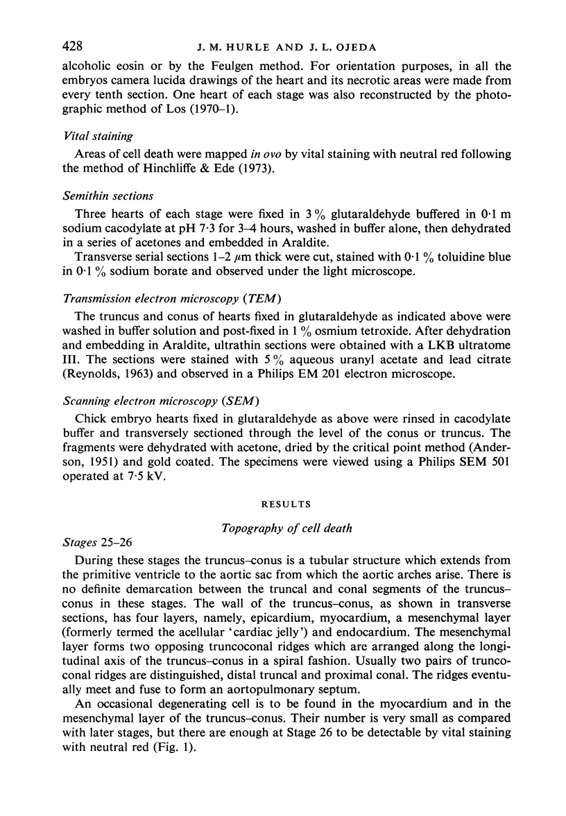
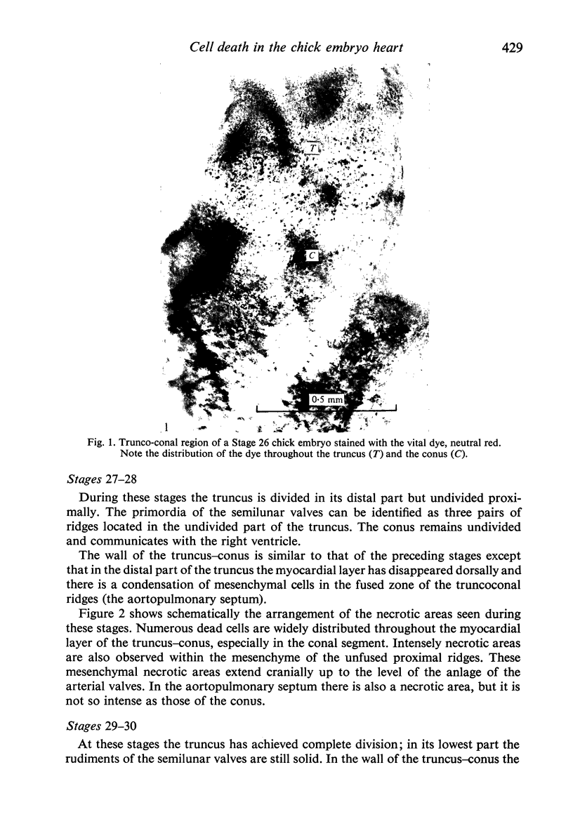
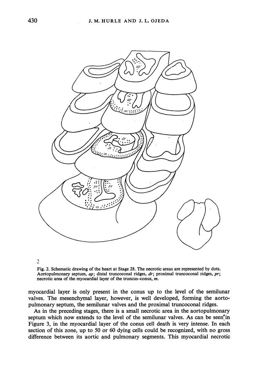
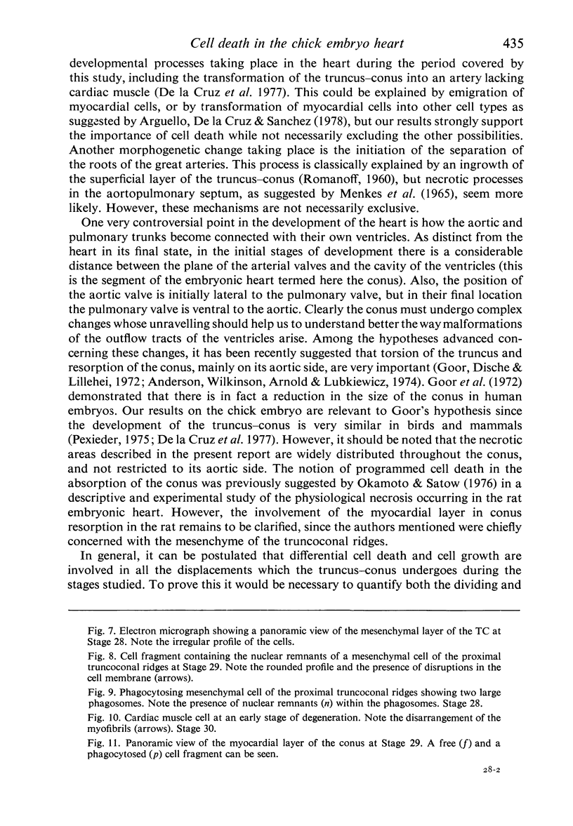
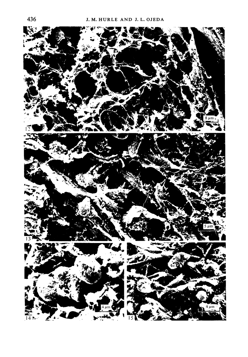
The presence of cell death in the walls of the truncus and conus of the developing chick heart was investigated by a variety of light and electron microscopic techniques. Necrotic areas were observed in the myocardial layer of the truncus and conus and within the mesenchymal cells of the truncoconal ridges and aortopulmonary septum. These necrotic zones appeared first at Stage 25-26 and reached their maximum extent at Stages 29-32 undergoing later progressive disappearance. The morphological changes of the degenerating cells detectable under both transmission and scanning electron microscopy are also reported. The possible role of cell death in the morphogenesis of the truncus and conus is discussed.
Full text
PDF
Images in this article
Selected References
These references are in PubMed. This may not be the complete list of references from this article.
- Anderson R. H., Wilkinson J. L., Arnold R., Lubkiewicz K. Morphogenesis of bulboventricular malformations. I. Consideration of embryogenesis in the normal heart. Br Heart J. 1974 Mar;36(3):242–255. doi: 10.1136/hrt.36.3.242. [DOI] [PMC free article] [PubMed] [Google Scholar]
- Argüello C., De La Cruz M. V., Sánchez C. Ultrastructural and experimental evidence of myocardial cell differentiation into connective tissue cells in embryonic chick heart. J Mol Cell Cardiol. 1978 Apr;10(4):307–315. doi: 10.1016/0022-2828(78)90380-2. [DOI] [PubMed] [Google Scholar]
- Goor D. A., Dische R., Lillehei C. W. The conotruncus. I. Its normal inversion and conus absorption. Circulation. 1972 Aug;46(2):375–384. doi: 10.1161/01.cir.46.2.375. [DOI] [PubMed] [Google Scholar]
- Hay D. A., Low F. N. The fusion of dorsal and ventral endocardial cushions in the embryonic chick heart: a study in fine structure. Am J Anat. 1972 Jan;133(1):1–23. doi: 10.1002/aja.1001330102. [DOI] [PubMed] [Google Scholar]
- Hendrix M. J., Morse D. E. Atrial septation. I. Scanning electron microscopy in the chick. Dev Biol. 1977 Jun;57(2):345–363. doi: 10.1016/0012-1606(77)90220-2. [DOI] [PubMed] [Google Scholar]
- Hinchliffe J. R., Ede D. A. Cell death and the development of limb form and skeletal pattern in normal and wingless (ws) chick embryos. J Embryol Exp Morphol. 1973 Dec;30(3):753–772. [PubMed] [Google Scholar]
- Hinchliffe J. R., Thorogood P. V. Genetic inhibition of mesenchymal cell death and the development of form and skeletal pattern in the limbs of talpid3 (ta3) mutant chick embryos. J Embryol Exp Morphol. 1974 Jun;31(3):747–760. [PubMed] [Google Scholar]
- Hurle J. M., Lafarga M., Ojeda J. L. Cytological and cytochemical studies of the necrotic area of the bulbus of the chick embryo heart: phagocytosis by developing myocardial cells. J Embryol Exp Morphol. 1977 Oct;41:161–173. [PubMed] [Google Scholar]
- Hurle J. M., Lafarga M., Ojeda J. L. In vivo phagocytosis by developing myocardial cells: an ultrastructural study. J Cell Sci. 1978 Oct;33:363–369. doi: 10.1242/jcs.33.1.363. [DOI] [PubMed] [Google Scholar]
- Hurle J., Hinchcliffe J. R. Cell death in the posterior necrotic zone (PNZ) of the chick wing-bud: a stereoscan and ultrastructural survey of autolysis and cell fragmentation. J Embryol Exp Morphol. 1978 Feb;43:123–136. [PubMed] [Google Scholar]
- Kerr J. F., Wyllie A. H., Currie A. R. Apoptosis: a basic biological phenomenon with wide-ranging implications in tissue kinetics. Br J Cancer. 1972 Aug;26(4):239–257. doi: 10.1038/bjc.1972.33. [DOI] [PMC free article] [PubMed] [Google Scholar]
- Krstić R., Pexieder T. Ultrastructure of cell death in bulbar cushions of chick embryo heart. Z Anat Entwicklungsgesch. 1973 Aug 30;140(3):337–350. doi: 10.1007/BF00525060. [DOI] [PubMed] [Google Scholar]
- Lemanski L. F. Heart development in the Mexican salamander, Ambystoma Mexicanum. II. Ultrastructure. Am J Anat. 1973 Apr;136(4):487–525. doi: 10.1002/aja.1001360408. [DOI] [PubMed] [Google Scholar]
- Los J. A. A new method of three-dimensional reconstruction of microscopical structures based on photographic techniques. Acta Morphol Neerl Scand. 1971 May;8(4):273–279. [PubMed] [Google Scholar]
- Los J. A., van Eijndthoven E. The fusion of the endocardial cushions in the heart of the chick embryo. A light-microscopical and electron-microscopical study. Z Anat Entwicklungsgesch. 1973;141(1):55–75. doi: 10.1007/BF00523365. [DOI] [PubMed] [Google Scholar]
- Manasek F. J. Myocardial cell death in the embryonic chick ventricle. J Embryol Exp Morphol. 1969 Apr;21(2):271–284. [PubMed] [Google Scholar]
- Ojeda J. L., Hurle J. M. Cell death during the formation of tubular heart of the chick embryo. J Embryol Exp Morphol. 1975 Jun;33(3):523–534. [PubMed] [Google Scholar]
- Pexieder T. The tissue dynamics of heart morphogenesis. I. The phenomena of cell death. B. Topography. Z Anat Entwicklungsgesch. 1972;138(3):241–253. doi: 10.1007/BF00520705. [DOI] [PubMed] [Google Scholar]
- REYNOLDS E. S. The use of lead citrate at high pH as an electron-opaque stain in electron microscopy. J Cell Biol. 1963 Apr;17:208–212. doi: 10.1083/jcb.17.1.208. [DOI] [PMC free article] [PubMed] [Google Scholar]
- Saunders J. W., Jr Death in embryonic systems. Science. 1966 Nov 4;154(3749):604–612. doi: 10.1126/science.154.3749.604. [DOI] [PubMed] [Google Scholar]
- Schlüter G. Ultrastructural observations on cell necrosis during formation of the neural tube in mouse embryos. Z Anat Entwicklungsgesch. 1973;141(3):251–264. doi: 10.1007/BF00519046. [DOI] [PubMed] [Google Scholar]
- Shapiro B. L., Sweney L. Electron microscopic and histochemical examination of oral epithelial-mesenchymal interaction (programmed cell death). J Dent Res. 1969 Sep-Oct;48(5):652–660. doi: 10.1177/00220345690480050801. [DOI] [PubMed] [Google Scholar]
- Waterman R. E., Meller S. M. Alterations in the epithelial surface of human palatal shelves prior to and during fusion: a scanning electron microscopic study. Anat Rec. 1974 Sep;180(1):111–135. doi: 10.1002/ar.1091800111. [DOI] [PubMed] [Google Scholar]
- de la Cruz M. V., Sánchez Gómez C., Arteaga M. M., Argüello C. Experimental study of the development of the truncus and the conus in the chick embryo. J Anat. 1977 Jul;123(Pt 3):661–686. [PMC free article] [PubMed] [Google Scholar]