Abstract
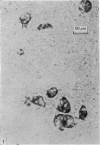
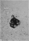
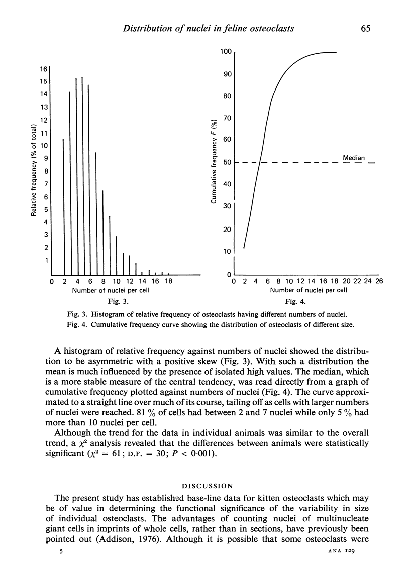
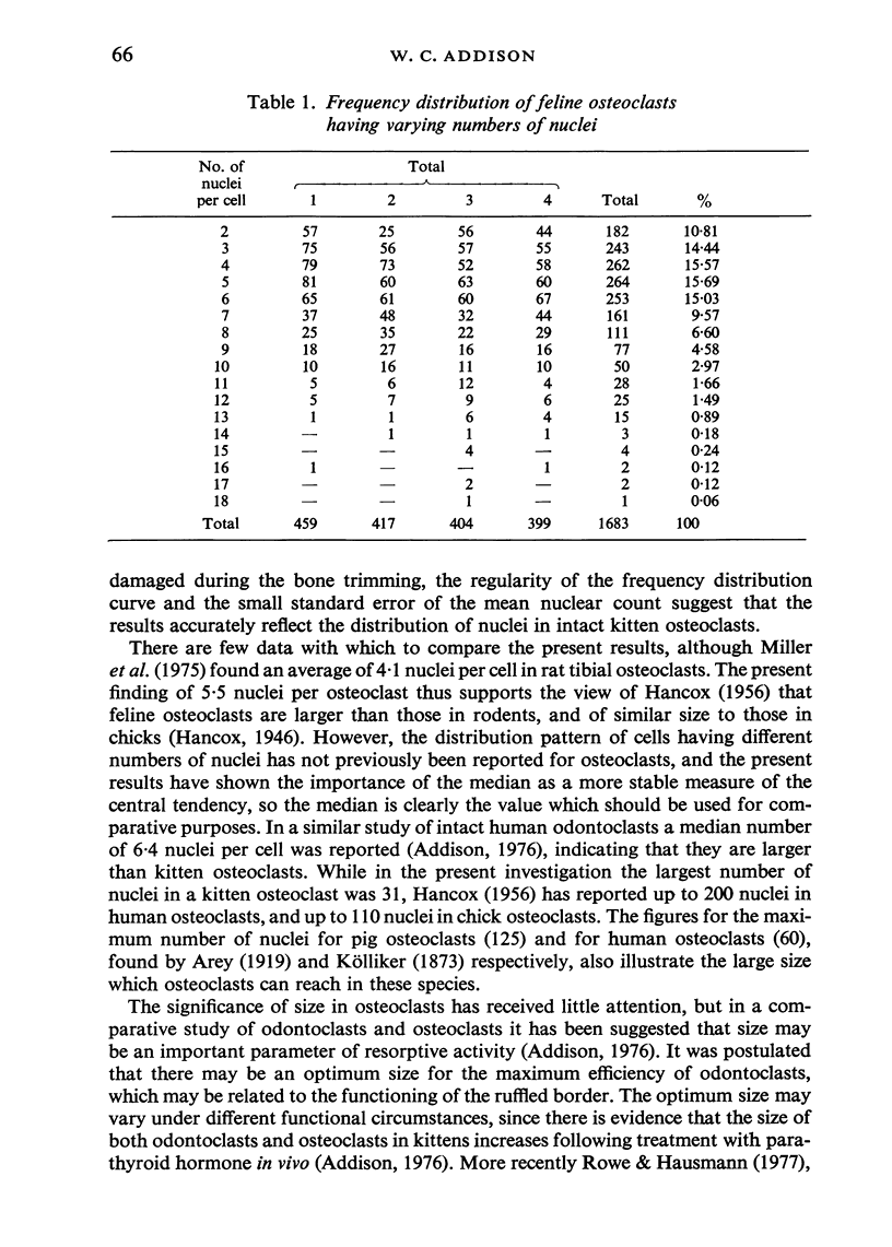
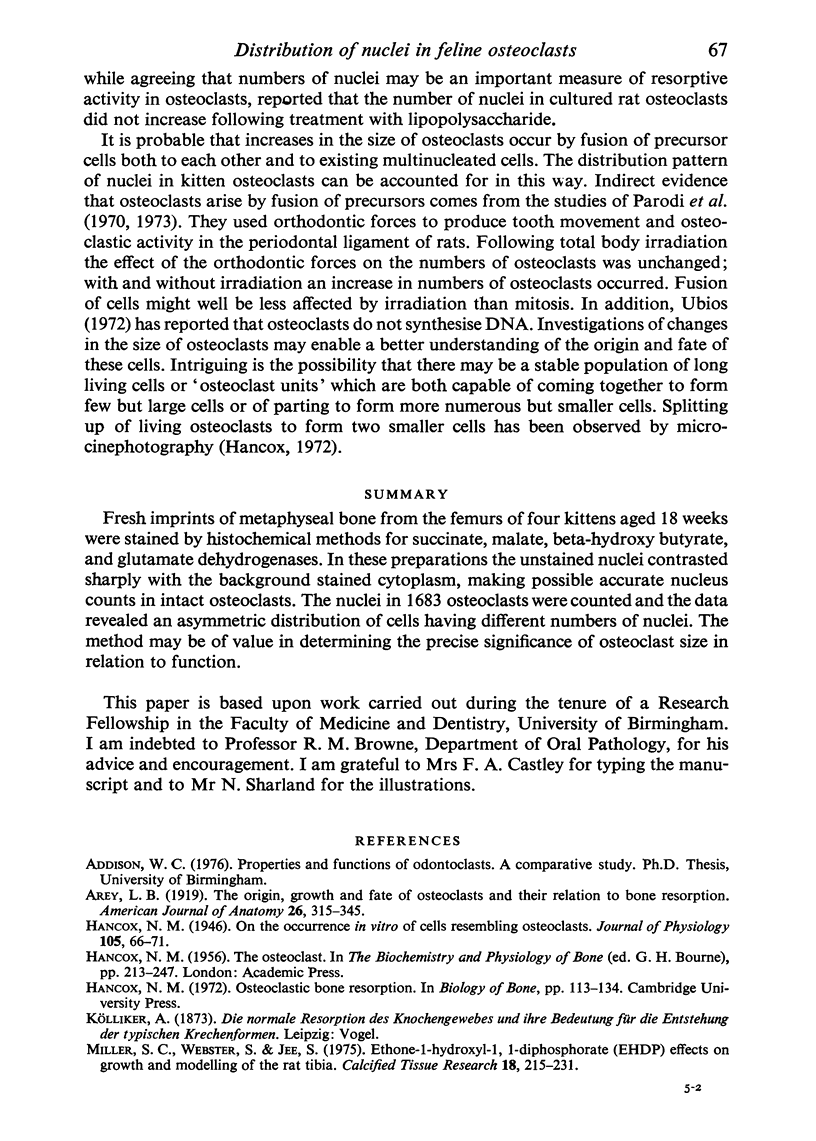
Fresh imprints of metaphyseal bone from the femurs of four kittens aged 18 weeks were stained by histochemical methods for succinate, malate, beta-hydroxy butyrate, and glutamate dehydrogenases. In these preparations the unstained nuclei contrasted sharply with the background stained cytoplasm, making possible accurate nucleus counts in intact osteoclasts. The nuclei in 1683 osteoclasts were counted and the data revealed an asymmetric distribution of cells having different numbers of nuclei. The method may be of value in determining the precise significance of osteoclast size in relation to function.
Full text
PDF

Images in this article
Selected References
These references are in PubMed. This may not be the complete list of references from this article.
- Hancox N. M. On the occurrence in vitro of cells resembling osteoclasts. J Physiol. 1946 Jul 15;105(1):66–71. [PMC free article] [PubMed] [Google Scholar]
- Miller S. C., Jee W. S. Ethane-1-hydroxy-1, 1-diphosphonate (EHDP). Effects on growth and modeling of the rat tibia. Calcif Tissue Res. 1975 Sep 5;18(3):215–231. doi: 10.1007/BF02546241. [DOI] [PubMed] [Google Scholar]
- Parodi R. J., Ubios A. M., Mayo J., Cabrini R. L. Total body irradiation effects on the bone resorption mechanism in rats subjected to orthodontic movements. J Oral Pathol. 1973;2(1):1–6. doi: 10.1111/j.1600-0714.1973.tb01668.x. [DOI] [PubMed] [Google Scholar]
- Rowe D. J., Hausmann E. Quantitative analyses of osteoclast changes in resorbing bone organ cultures. Calcif Tissue Res. 1977 Oct 20;23(3):283–289. doi: 10.1007/BF02012798. [DOI] [PubMed] [Google Scholar]


