

Abstract
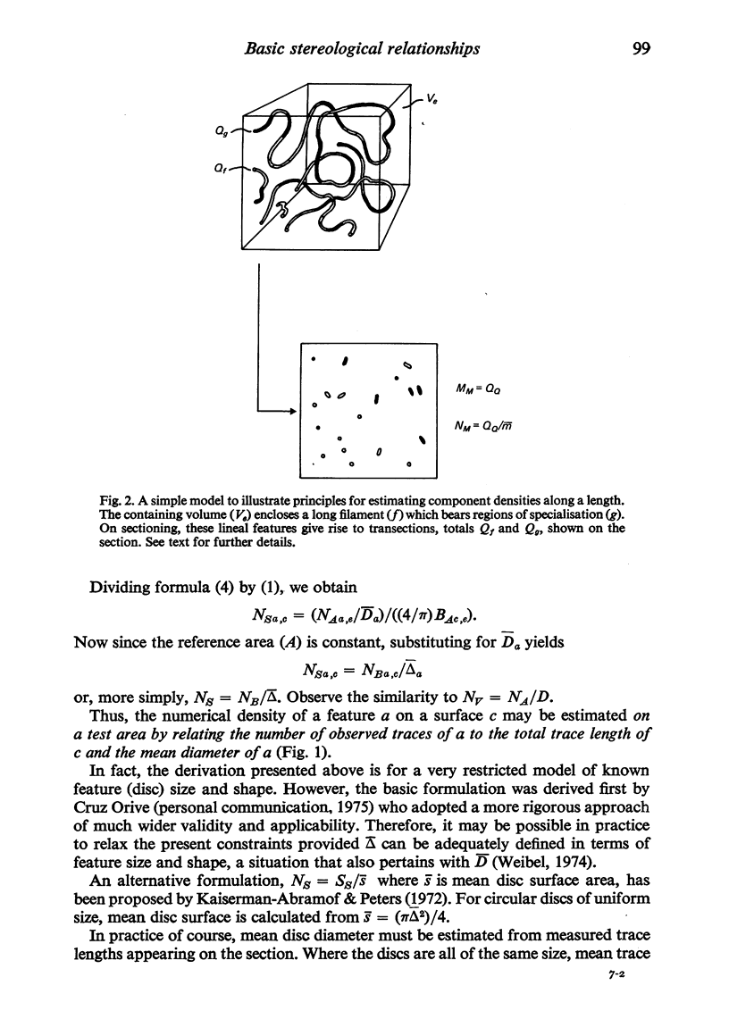
There exists in the literature a core of formulations regarded as 'the basic stereological principles' for quantifying cell and tissue morphology. They may be used to obtain information relating component volume, surface area, length and number to a specified containing volume (the so-called component densities in a volume: VV, SV, MV and NV). However, principles may also be formulated for relating these component dimensions to a containing surface (SS, MS and NS), containing length (MM and NM) and a containing number (NN). Methods for estimating these previously neglected stereological relations are presented. Possible biological applications of the principles are also discussed.
Full text
PDF

Selected References
These references are in PubMed. This may not be the complete list of references from this article.
- Anker R. L., Cragg B. G. Estimation of the number of synapses in a volume of nervous tissue from counts in thin sections by electron microscopy. J Neurocytol. 1974 Dec;3(6):725–735. doi: 10.1007/BF01097194. [DOI] [PubMed] [Google Scholar]
- BLACKSTAD T. W., DAHL H. A. Quantitative evaluation of structures in contact with neuronal somata. An electron microscopic study on the fascia dentata of the rat. Acta Morphol Neerl Scand. 1962;4:329–343. [PubMed] [Google Scholar]
- Blouin A., Bolender R. P., Weibel E. R. Distribution of organelles and membranes between hepatocytes and nonhepatocytes in the rat liver parenchyma. A stereological study. J Cell Biol. 1977 Feb;72(2):441–455. doi: 10.1083/jcb.72.2.441. [DOI] [PMC free article] [PubMed] [Google Scholar]
- Conradi S. Ultrastructure and distribution of neuronal and glial elements on the motoneuron surface in the lumbosacral spinal cord of the adult cat. Acta Physiol Scand Suppl. 1969;332:5–48. [PubMed] [Google Scholar]
- Elias H., Hennig A., Schwartz D. E. Stereology: applications to biomedicalresearch. Physiol Rev. 1971 Jan;51(1):158–200. doi: 10.1152/physrev.1971.51.1.158. [DOI] [PubMed] [Google Scholar]
- FARQUHAR M. G., PALADE G. E. Junctional complexes in various epithelia. J Cell Biol. 1963 May;17:375–412. doi: 10.1083/jcb.17.2.375. [DOI] [PMC free article] [PubMed] [Google Scholar]
- Farquhar M. G., Palade G. E. Cell junctions in amphibian skin. J Cell Biol. 1965 Jul;26(1):263–291. doi: 10.1083/jcb.26.1.263. [DOI] [PMC free article] [PubMed] [Google Scholar]
- Gabella G. Quantitative morphological study of smooth muscle cells of the guinea-pig taenia coli. Cell Tissue Res. 1976 Jul 26;170(2):161–186. doi: 10.1007/BF00224297. [DOI] [PubMed] [Google Scholar]
- Kaiserman-Abramof I. R., Peters A. Some aspects of the morphology of Betz cells in the cerebral cortex of the cat. Brain Res. 1972 Aug 25;43(2):527–546. doi: 10.1016/0006-8993(72)90406-4. [DOI] [PubMed] [Google Scholar]
- Karlsson U. Three-dimensional studies of neurons in the lateral geniculate nucleus of the rat. II. Environment of perikarya and proximal parts of their branches. J Ultrastruct Res. 1966 Dec;16(5):482–504. doi: 10.1016/s0022-5320(66)80002-3. [DOI] [PubMed] [Google Scholar]
- Loud A. V. A quantitative stereological description of the ultrastructure of normal rat liver parenchymal cells. J Cell Biol. 1968 Apr;37(1):27–46. doi: 10.1083/jcb.37.1.27. [DOI] [PMC free article] [PubMed] [Google Scholar]
- Mayhew T. M., Momoh C. K. Stereological description of the anterior horn cervical cord of the adult rat. A quantitative study using the optical microscope. J Comp Neurol. 1974 Jul 1;156(1):107–121. doi: 10.1002/cne.901560109. [DOI] [PubMed] [Google Scholar]
- Weibel E. R. Selection of the best method in stereology. J Microsc. 1974 Apr;100(3):261–269. doi: 10.1111/j.1365-2818.1974.tb03938.x. [DOI] [PubMed] [Google Scholar]
- Weibel E. R. Stereological principles for morphometry in electron microscopic cytology. Int Rev Cytol. 1969;26:235–302. doi: 10.1016/s0074-7696(08)61637-x. [DOI] [PubMed] [Google Scholar]