

Abstract
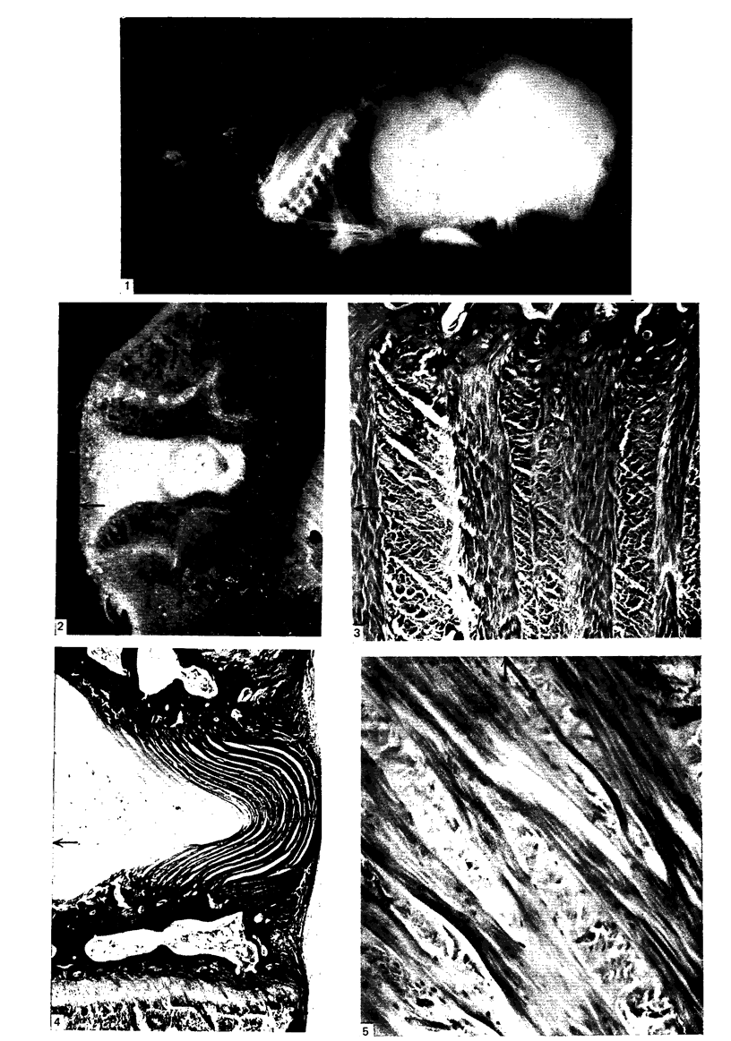
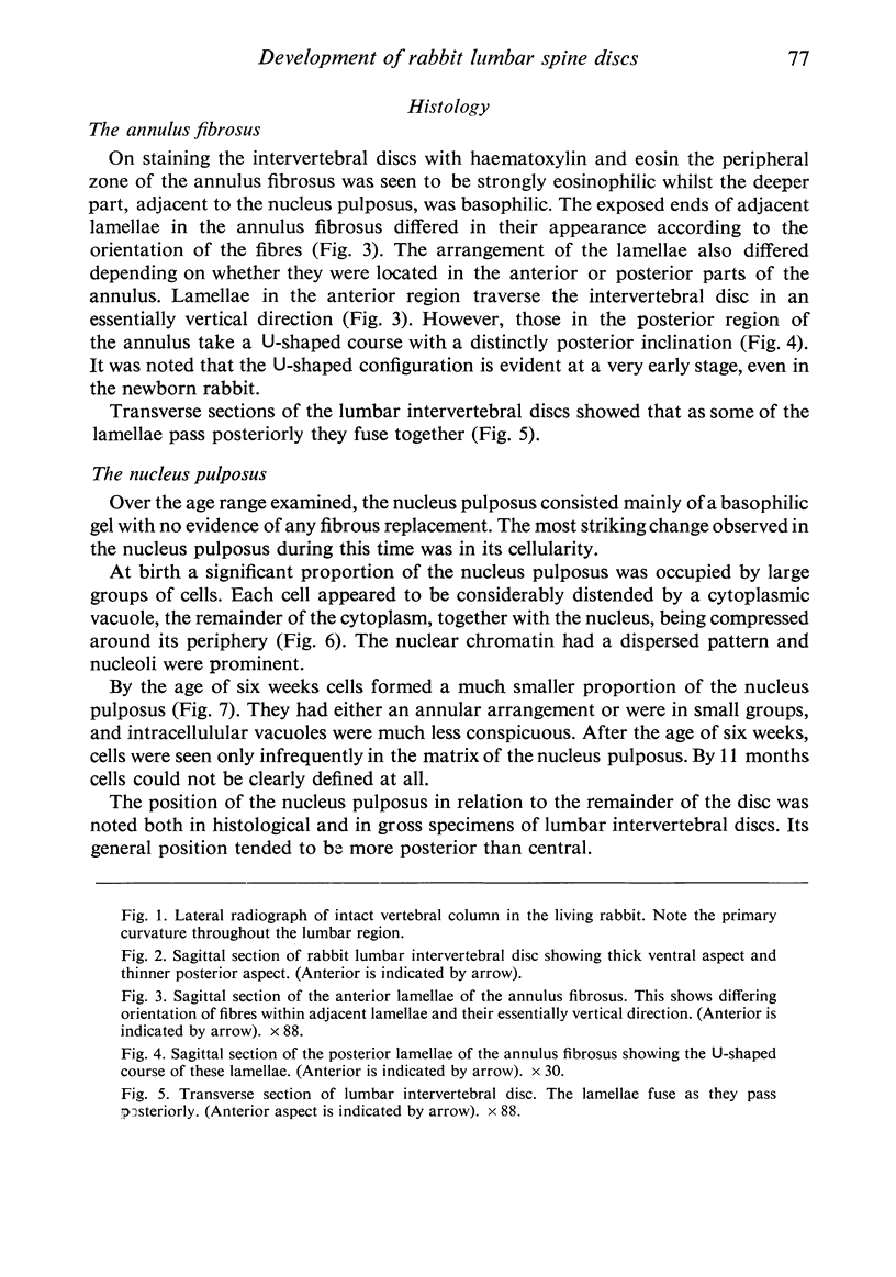
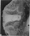
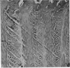
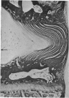
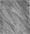
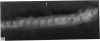
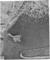
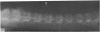
Some basic features in the development of the structure of the annulus fibrosus and nucleus pulposus in the rabbit, as described by previous workers, have been confirmed in the present study. However, the greater thickness of the anterior part of the disc, as compared with the posterior region, and the distinctive arrangement of lamellae in the posterior part of the disc, cannot be attributed, as conventionally claimed from studies of the human spine, to a secondary curvature in the lumbar spine associated with an upright posture: for these features are present in the lumbar spine of the quadrupedal rabbit with its primary curvature. Secondary ossification produces a plate-like epiphysis separating the growth cartilage from the intervertebral disc. A distinct cartilaginous plate, limiting the nucleus pulposus in the rabbit intervertebral disc, only becomes apparent when collagen fibres cease to traverse the area above and below the nucleus pulposus.
Full text
PDF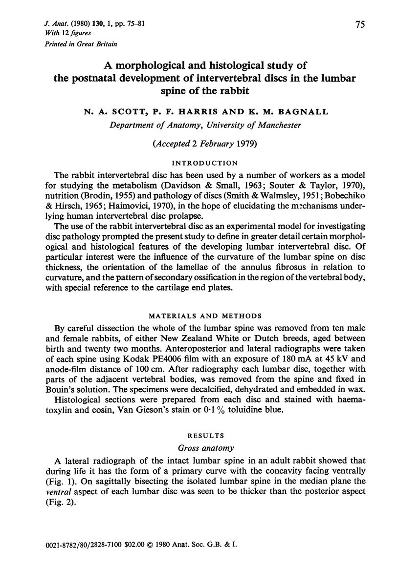
Images in this article
Selected References
These references are in PubMed. This may not be the complete list of references from this article.
- BICK E. M., COPEL J. W. The ring apophysis of the human vertebra; contribution to human osteogeny. II. J Bone Joint Surg Am. 1951 Jul;33-A(3):783–787. [PubMed] [Google Scholar]
- BOBECHKO W. P., HIRSCH C. AUTO-IMMUNE RESPONSE TO NUCLEUS PULPOSUS IN THE RABBIT. J Bone Joint Surg Br. 1965 Aug;47:574–580. [PubMed] [Google Scholar]
- BRODIN H. Paths of nutrition in articular cartilage and intervertebral discs. Acta Orthop Scand. 1955;24(3):177–183. doi: 10.3109/17453675408988561. [DOI] [PubMed] [Google Scholar]
- DAVIDSON E. A., SMALL W. Metabolism in vivo of connective-tissue mucopolysaccharides. I. Chondroitin sulfate C and keratosulfate of nucleus pulposus. Biochim Biophys Acta. 1963 Mar 5;69:445–452. doi: 10.1016/0006-3002(63)91292-7. [DOI] [PubMed] [Google Scholar]
- Haimovici E. H. Experimental disc lesion in rabbits. The effect of repeated ACTH administration. Acta Orthop Scand. 1970;41(5):505–521. doi: 10.3109/17453677008991539. [DOI] [PubMed] [Google Scholar]
- Inoue H. Three-dimensional observation of collagen framework of intervertebral discs in rats, dogs and humans. Arch Histol Jpn. 1973 Sep;36(1):39–56. doi: 10.1679/aohc1950.36.39. [DOI] [PubMed] [Google Scholar]
- LEESON T. S., LEESON C. R. Observations on the histochemistry and fine structure of the notochord in rabbit embryos. J Anat. 1958 Apr;92(2):278–285. [PMC free article] [PubMed] [Google Scholar]
- PEACOCK A. Observations on the postnatal structure of the intervertebral disc in man. J Anat. 1952 Apr;86(2):162–179. [PMC free article] [PubMed] [Google Scholar]
- SMITH J. W., WALMSLEY R. Experimental incision of the intervertebral disc. J Bone Joint Surg Br. 1951 Nov;33-B(4):612–625. doi: 10.1302/0301-620X.33B4.612. [DOI] [PubMed] [Google Scholar]
- Souter W. A., Taylor T. K. Sulphated acid mucopolysaccharide metabolism in the rabbit intervertebral disc. J Bone Joint Surg Br. 1970 May;52(2):371–384. [PubMed] [Google Scholar]
- WALMSLEY R. The development and growth of the intervertebral disc. Edinb Med J. 1953 Aug;60(8):341–364. [PMC free article] [PubMed] [Google Scholar]