Abstract
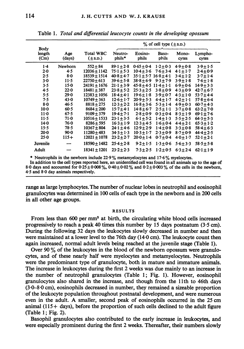
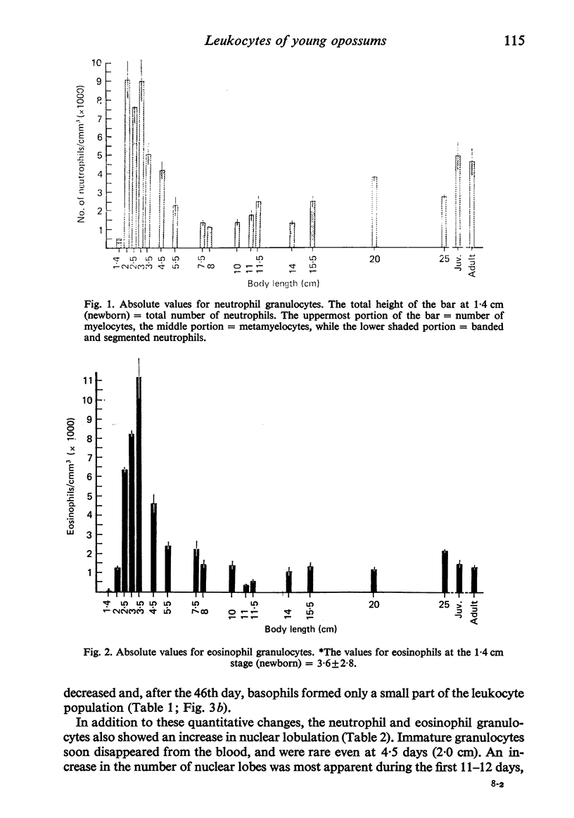
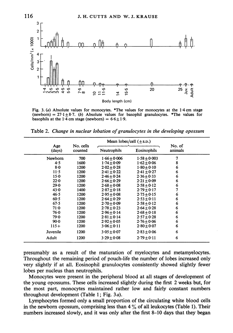
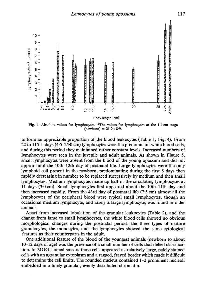
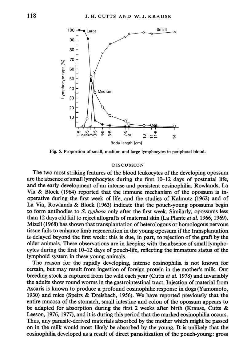
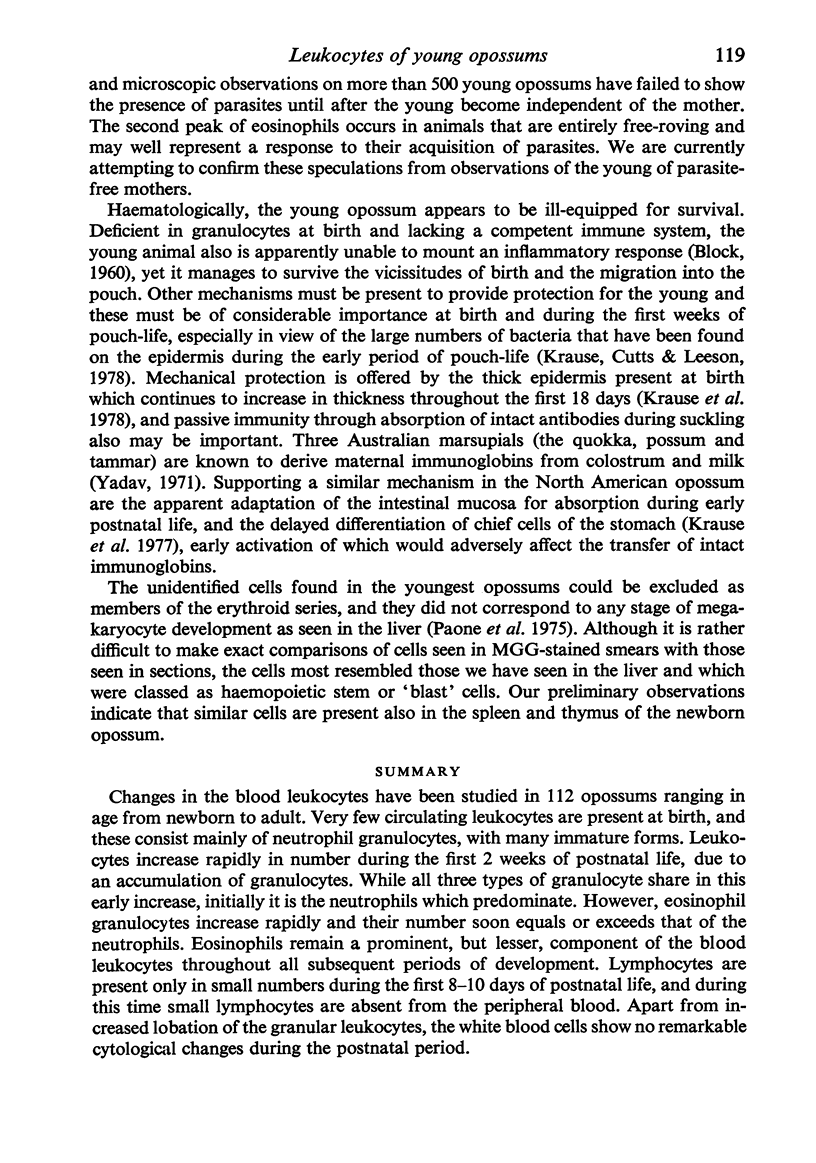
Changes in the blood leukocytes have been studied in 112 opossums ranging in age from newborn to adult. Very few circulating leukocytes are present at birth, and these consist mainly of neutrophil granulocytes, with many immature forms. Leukocytes increase rapidly in number during the first 2 weeks of postnatal life, due to an accumulation of granulocytes. While all three types of granulocyte share in this early increase, initially it is the neutrophils which predominate. However, eosinophil granulocytes increase rapidly and their number soon equals or exceeds that of the neutrophils. Eosinophils remain a prominent, but lesser, component of the blood leukocytes throughout all subsequent periods of development. Lymphocytes are present only in small numbers during the first 8--10 days of postnatal life, and during this time small lymphocytes are absent from the peripheral blood. Apart from increased lobation of the granular leukocytes, the white blood cells show no remarkable cytological changes during the postnatal period.
Full text
PDF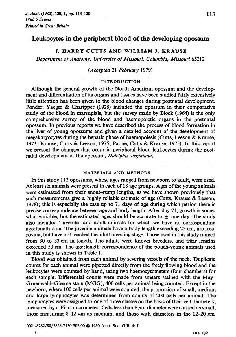
Selected References
These references are in PubMed. This may not be the complete list of references from this article.
- BLOCK M. THE BLOOD FORMING TISSUES AND BLOOD OF THE NEWBORN OPOSSUM (DIDELPHYS VIRGINIANA). I. NORMAL DEVELOPMENT THROUGH ABOUT THE ONE HUNDREDTH DAY OF LIFE. Ergeb Anat Entwicklungsgesch. 1964;37:237–366. [PubMed] [Google Scholar]
- BLOCK M. Wound healing in the new-born opossum (Didelphis virginianam). Nature. 1960 Jul 23;187:340–341. doi: 10.1038/187340a0. [DOI] [PubMed] [Google Scholar]
- Cutts J. H., Krause W. J., Leeson C. R. General observations on the growth and development of the young pouch opossum, Didelphis virginiana. Biol Neonate. 1978;33(5-6):264–272. doi: 10.1159/000241082. [DOI] [PubMed] [Google Scholar]
- Cutts J. H., Leeson C. R., Krause W. J. The postnatal development of the liver in a marsupial, Didelphis virginiana. 1. Light microscopy. J Anat. 1973 Sep;115(Pt 3):327–346. [PMC free article] [PubMed] [Google Scholar]
- KALMUTZ S. E. Antibody production in the opossum embryo. Nature. 1962 Mar 3;193:851–853. doi: 10.1038/193851a0. [DOI] [PubMed] [Google Scholar]
- Krause W. J., Cutts J. H., Leeson C. R. Postnatal development of the epidermis in a marsupial, Didelphis virginiana. J Anat. 1978 Jan;125(Pt 1):85–99. [PMC free article] [PubMed] [Google Scholar]
- Krause W. J., Cutts J. H., Leeson C. R. The postnatal development of the alimentary canal in the opossum. II. Stomach. J Anat. 1976 Dec;122(Pt 3):499–519. [PMC free article] [PubMed] [Google Scholar]
- Krause W. J., Cutts J. H., Leeson C. R. The postnatal development of the alimentary canal in the opossum. III. Small intestine and colon. J Anat. 1977 Feb;123(Pt 1):21–45. [PMC free article] [PubMed] [Google Scholar]
- Krause W. J., Cutts J. H., Leeson C. R. The postnatal development of the liver in a marsupial, Didelphis virginiana. 2. Electron microscopy. J Anat. 1975 Sep;120(Pt 1):191–205. [PMC free article] [PubMed] [Google Scholar]
- La Via M. F., Rowlands D. T., Jr, Block M. Antibody Formation in Embryos. Science. 1963 Jun 14;140(3572):1219–1220. doi: 10.1126/science.140.3572.1219. [DOI] [PubMed] [Google Scholar]
- LaPlante E. S., Burrell R. G., Watne A. L., Zimmermann B. Permanent survival of skin allografts applied to the pouch young of the opossum. Surg Forum. 1966;17:200–201. [PubMed] [Google Scholar]
- LaPlante E. S., Burrell R., Watne A. L., Taylor D. L., Zimmermann B. Skin allograft studies in the pouch young of the opossum. Transplantation. 1969 Jan;7(1):67–72. doi: 10.1097/00007890-196901000-00006. [DOI] [PubMed] [Google Scholar]
- Mizell M. Limb regeneration: induction in the newborn opossum. Science. 1968 Jul 19;161(3838):283–286. doi: 10.1126/science.161.3838.283. [DOI] [PubMed] [Google Scholar]
- Paone D. B., Cutts J. H., Krause W. J. Megakaryocytopoiesis in the liver of the developing opossum (Didelphis virginiana). J Anat. 1975 Nov;120(Pt 2):239–252. [PMC free article] [PubMed] [Google Scholar]
- ROWLANDS D. T., Jr, LAVIA M. F., BLOCK M. H. THE BLOOD FORMING TISSUES AND BLOOD OF THE NEWBORN OPOSSUM (DIDELPHYS VIRGINIANA). II. ONTOGENESIS OF ANTIBODY FORMATION TO FLAGELLA OF SALMONELLA TYPHI. J Immunol. 1964 Jul;93:157–164. [PubMed] [Google Scholar]
- SPEIRS R. S., DREISBACH M. E. Quantitative studies of the cellular responses to antigen injections in normal mice; technic for determining cells in the peritoneal fluid. Blood. 1956 Jan;11(1):44–55. [PubMed] [Google Scholar]
- Yadav M. The transmissions of antibodies across the gut of pouch-young marsupials. Immunology. 1971 Nov;21(5):839–851. [PMC free article] [PubMed] [Google Scholar]


