Abstract
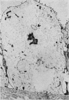
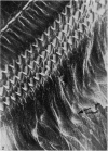
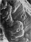
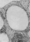
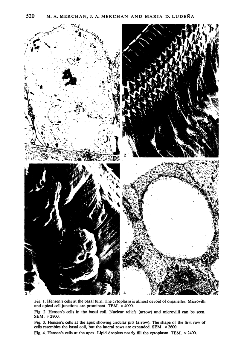
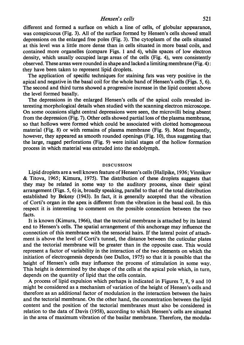
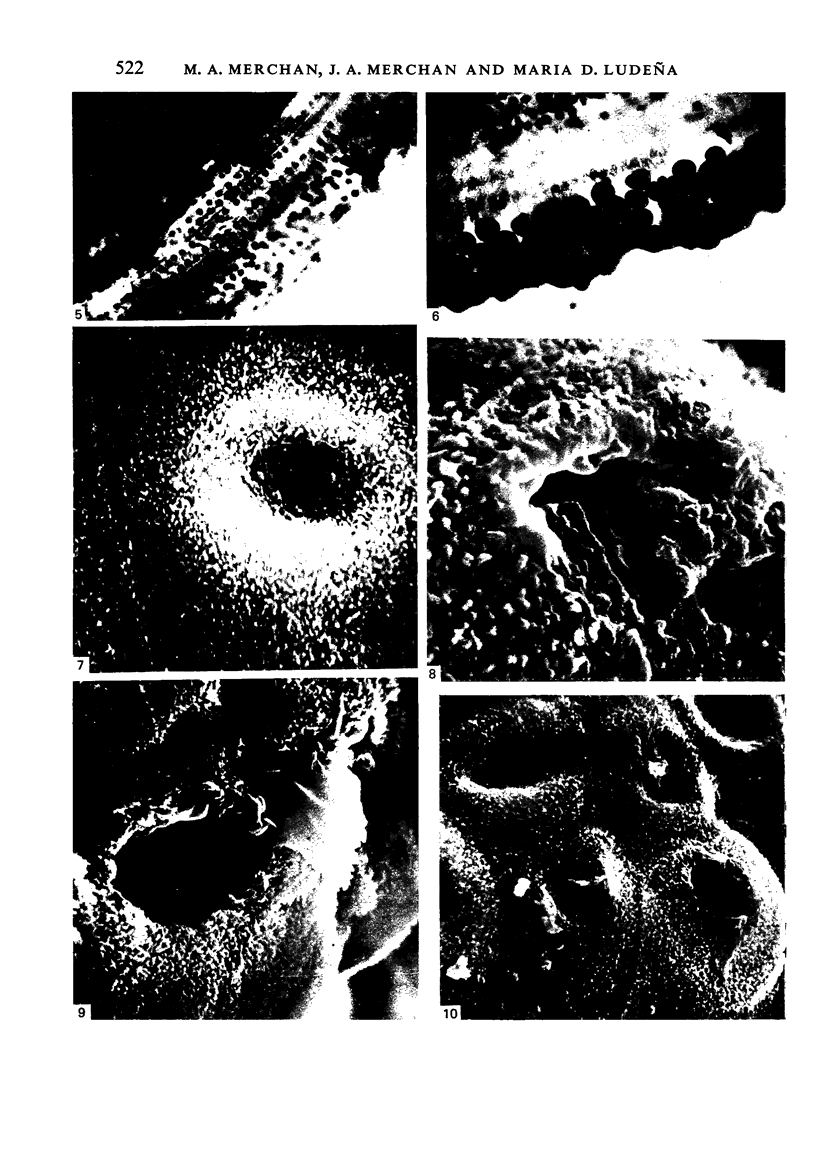
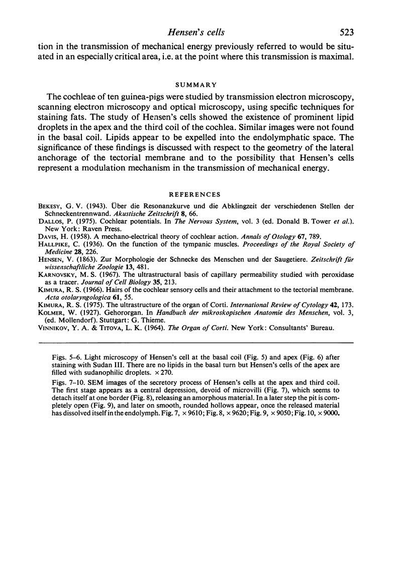
The cochleae of ten guinea-pigs were studied by transmission electron microscopy, scanning electron microscopy and optical microscopy, using specific techniques for staining fats. The study of Hensen's cells showed the existence of prominent lipid droplets in the apex and the third coil of the cochlea. Similar images were not found in the basal coil. Lipids appear to be expelled into the endolymphatic space. The significance of these findings is discussed with respect to the geometry of the lateral anchorage of the tectorial membrane and to the possibility that Hensen's cells represent a modulation mechanism in the transmission of mechanical energy.
Full text
PDF
Images in this article
Selected References
These references are in PubMed. This may not be the complete list of references from this article.
- DAVIS H. [A mechano-electrical theory of cochlear action]. Ann Otol Rhinol Laryngol. 1958 Sep;67(3):789–801. doi: 10.1177/000348945806700315. [DOI] [PubMed] [Google Scholar]
- Karnovsky M. J. The ultrastructural basis of capillary permeability studied with peroxidase as a tracer. J Cell Biol. 1967 Oct;35(1):213–236. doi: 10.1083/jcb.35.1.213. [DOI] [PMC free article] [PubMed] [Google Scholar]
- Kimura R. S. The ultrastructure of the organ of Corti. Int Rev Cytol. 1975;42:173–222. doi: 10.1016/s0074-7696(08)60981-x. [DOI] [PubMed] [Google Scholar]


