

Abstract
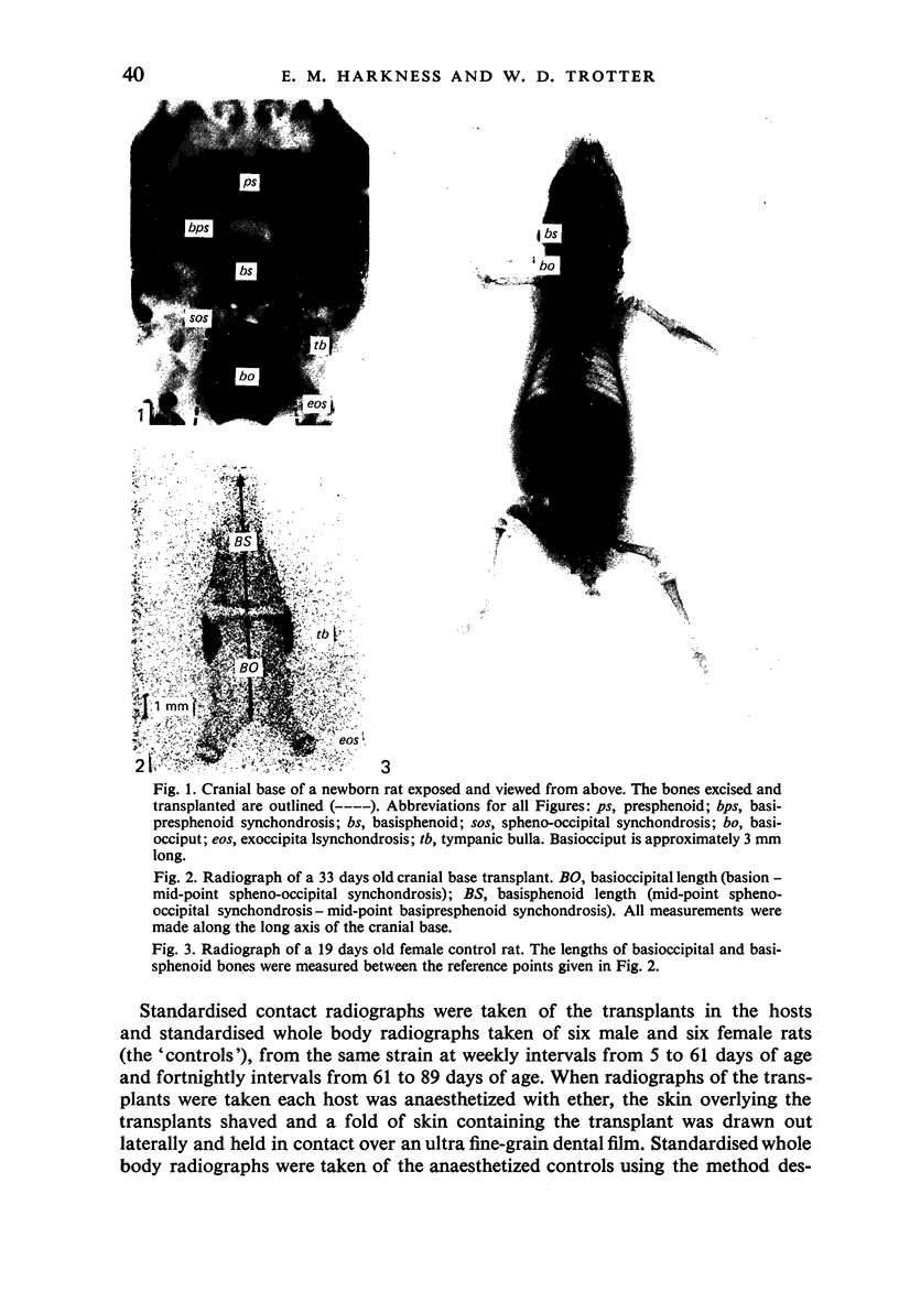
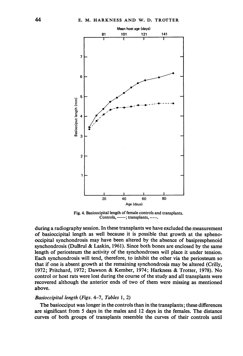
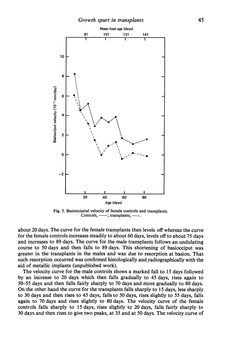
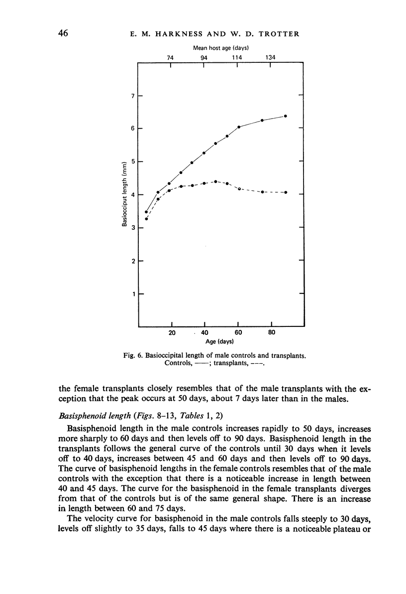
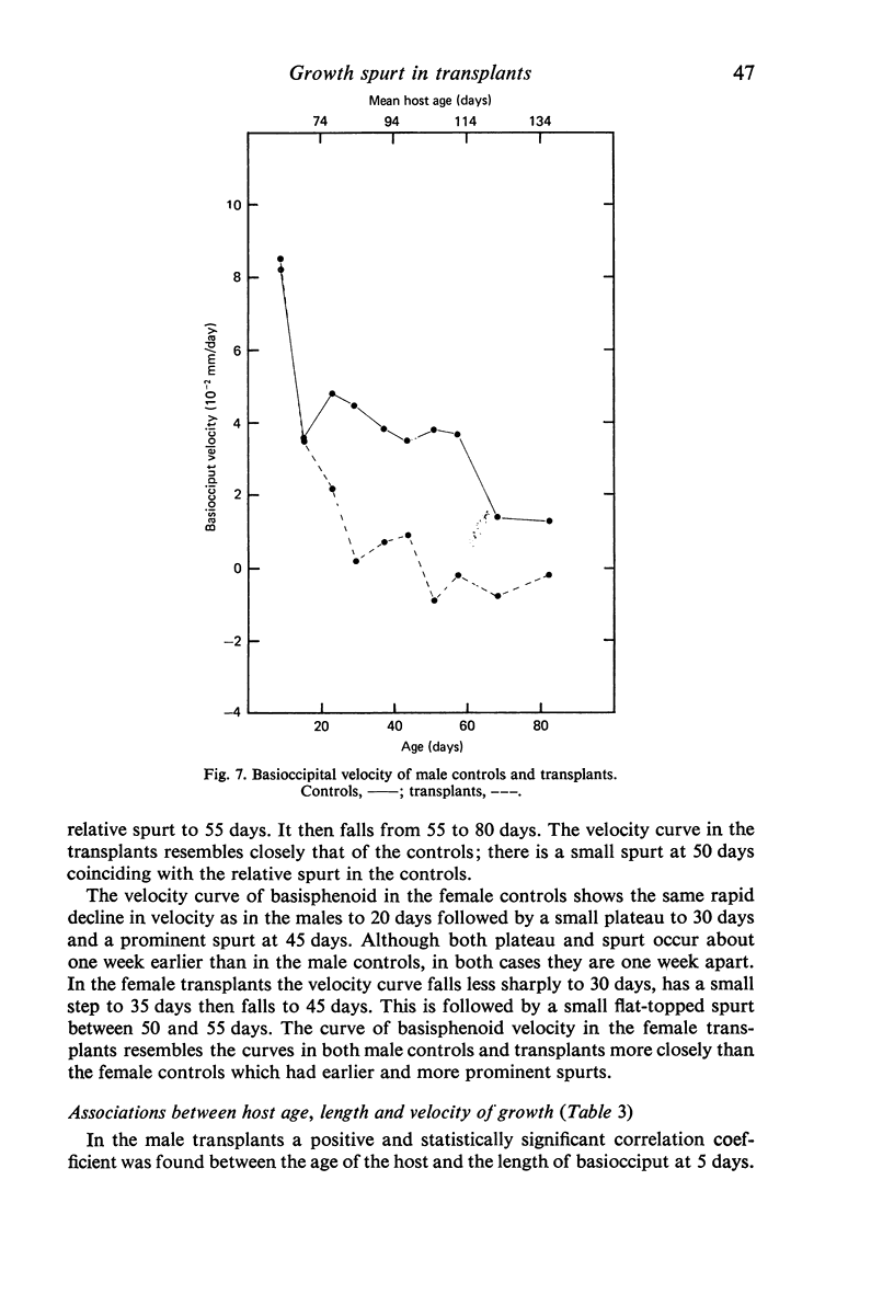
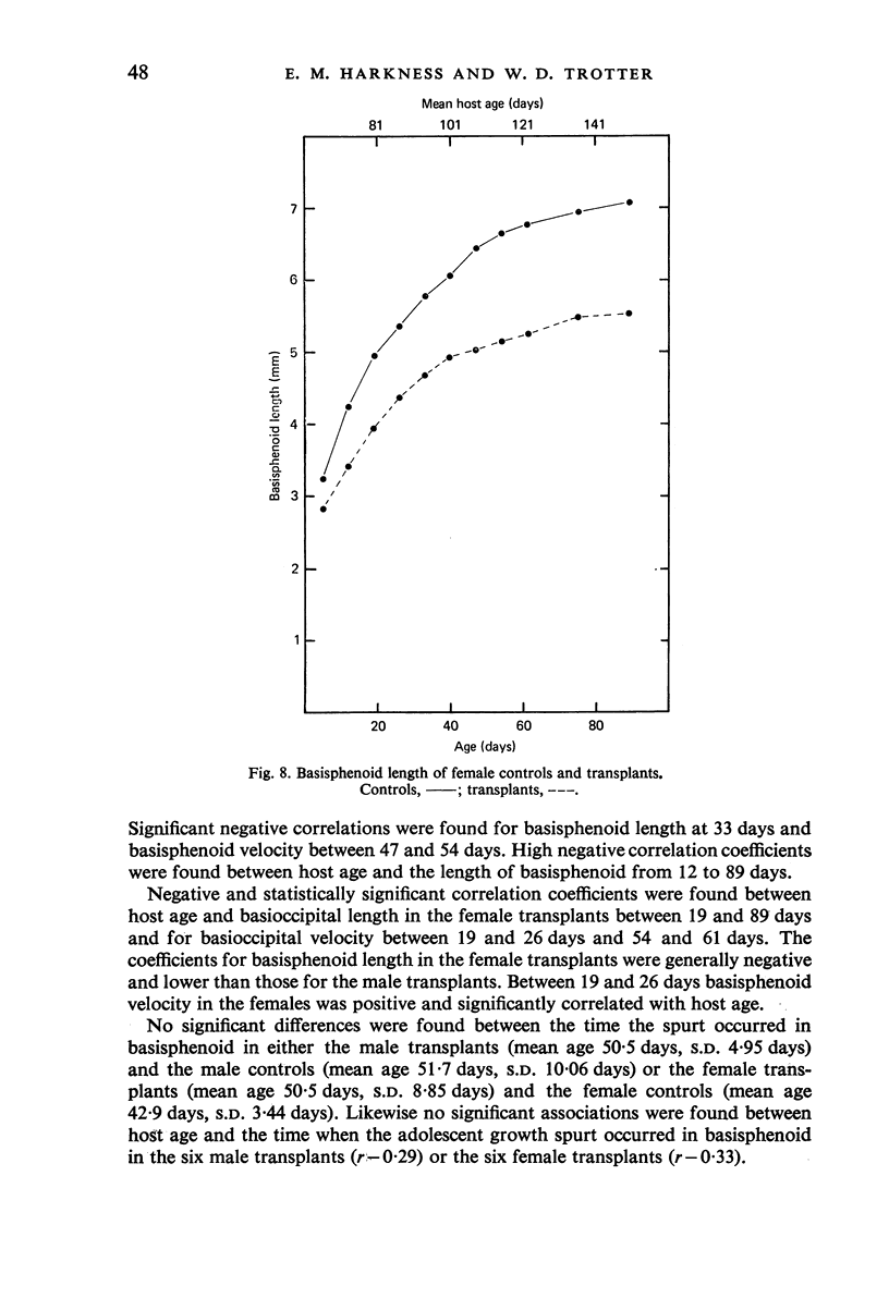
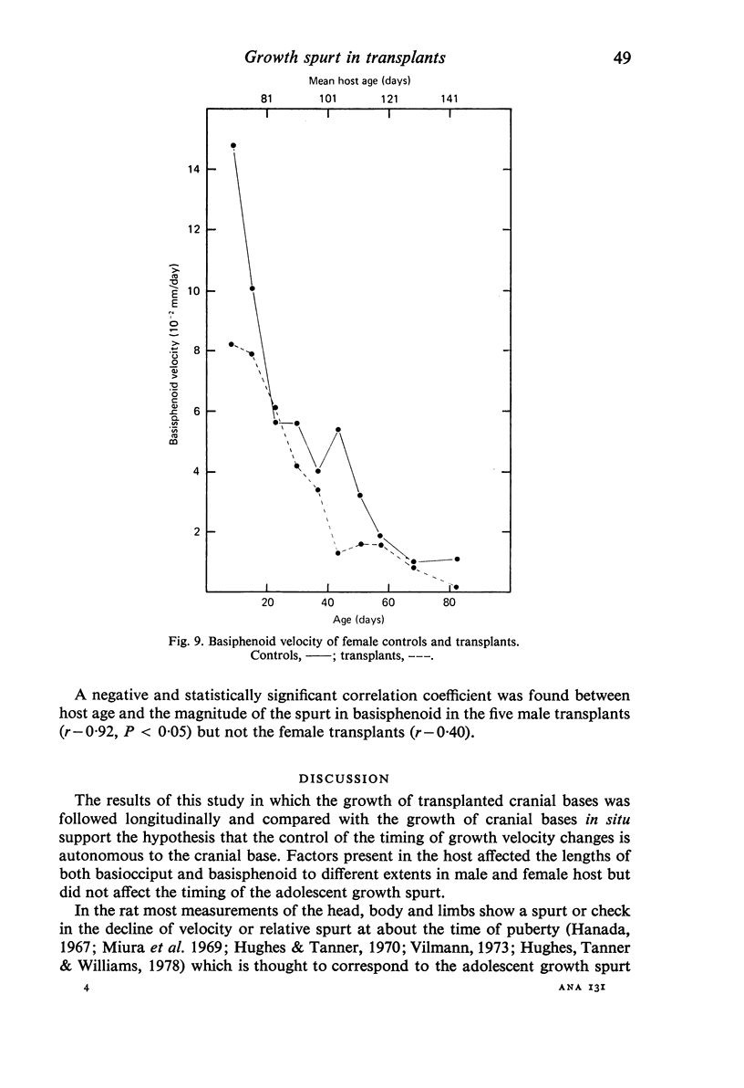
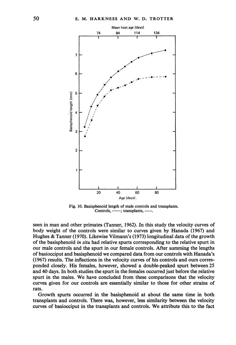
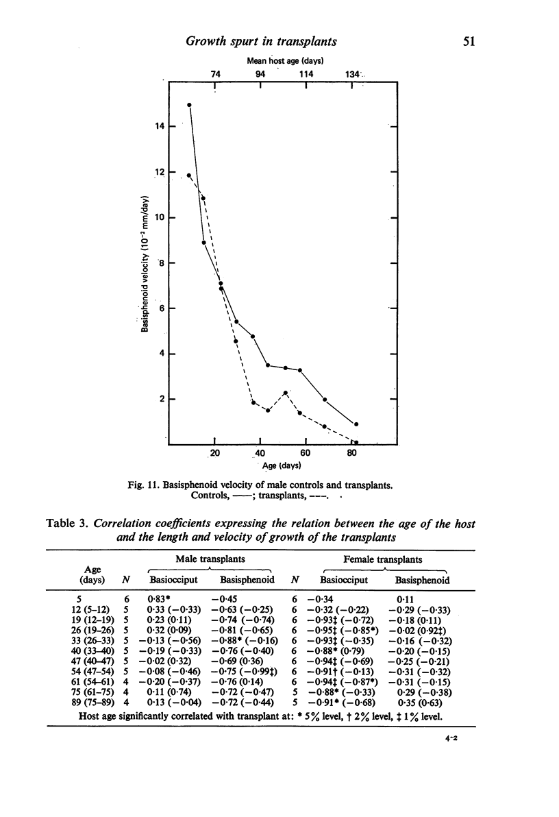
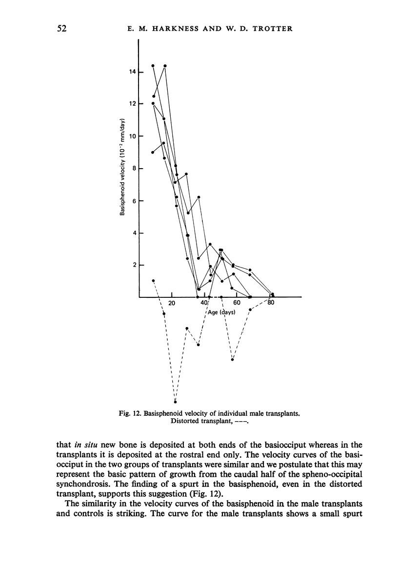
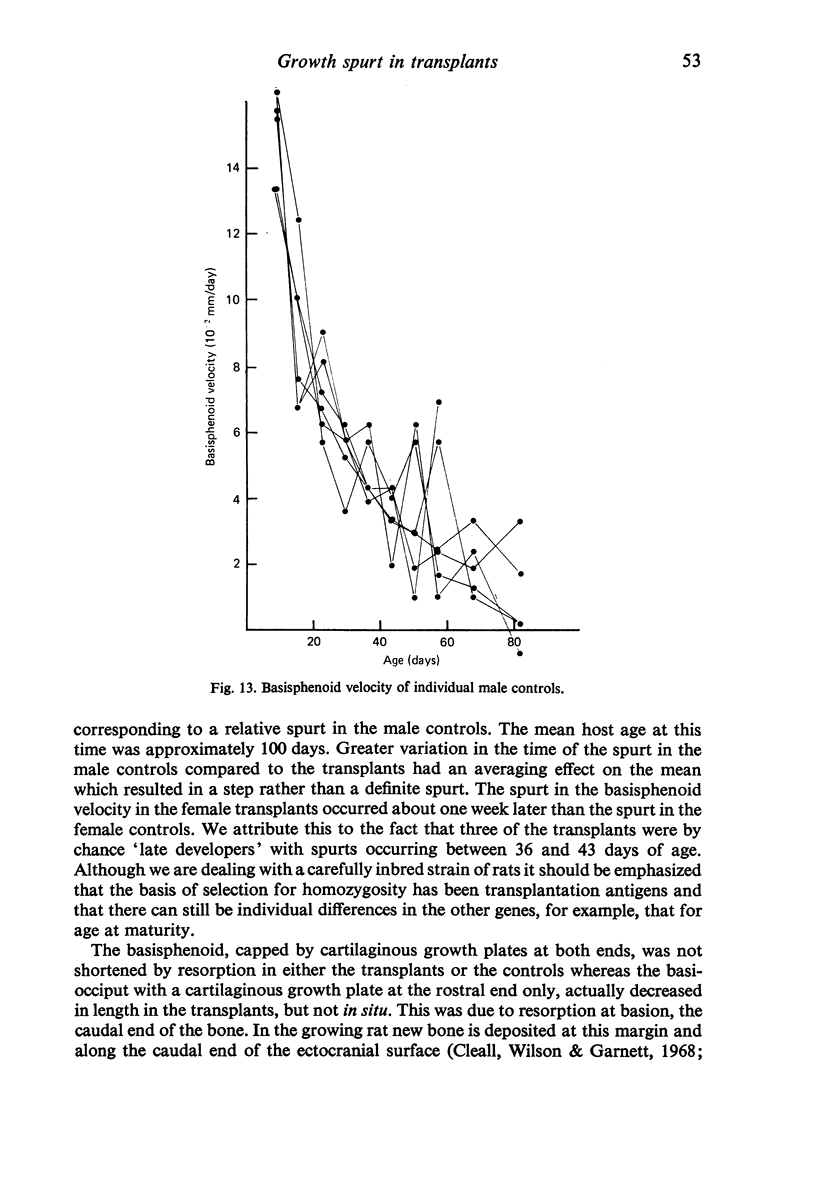
The growth of young rat cranial bases, consisting of the basioccipital, basisphenoid and a fragment of presphenoid bones, transplanted to older sex-matched isohistogenic hosts has been studied longitudinally. Detailed figures are given for absolute growth and velocity of growth both in transplants and in situ. Similarities between the pattern of growth of basisphenoid and in particular the timing of the so-called adolescent growth spurt in the transplanted bones and in situ were found. There was less similarity in the pattern of velocity changes in basiocciput. This was attributed to resorption of basion in the transplants. Significant negative correlations were found between host age and basioccipital length in the females and the velocity of growth of the basisphenoid in the males between 47 and 54 days. There was also a trend for host age to be negatively correlated with basisphenoid length in the males. These findings support the view that intrinsic, presumably genetic, factors regulate the pattern of timing of growth velocity changes in the cranial base.
Full text
PDF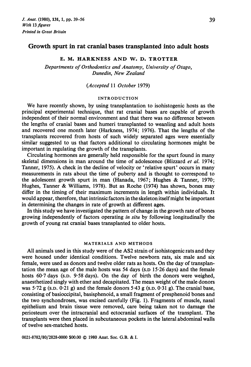

Images in this article
Selected References
These references are in PubMed. This may not be the complete list of references from this article.
- BJORK A., SOLOW B. Measurement on radiographs. J Dent Res. 1962 May-Jun;41:672–683. doi: 10.1177/00220345620410032101. [DOI] [PubMed] [Google Scholar]
- BUDY A. M., URIST M. R., MCLEAN F. C. The effect of estrogens on the growth apparatus of the bones of immature rats. Am J Pathol. 1952 Nov-Dec;28(6):1143–1167. [PMC free article] [PubMed] [Google Scholar]
- Caputo C. B., Meadows D., Raisz L. G. Failure of estrogens and androgens to inhibit bone resorption in tissue culture. Endocrinology. 1976 Apr;98(4):1065–1068. doi: 10.1210/endo-98-4-1065. [DOI] [PubMed] [Google Scholar]
- Cleall J. F., Wilson G. W., Garnett D. S. Normal craniofacial skeletal growth of the rat. Am J Phys Anthropol. 1968 Sep;29(2):225–242. doi: 10.1002/ajpa.1330290216. [DOI] [PubMed] [Google Scholar]
- Crilly R. G. Longitudinal overgrowth of chicken radius. J Anat. 1972 May;112(Pt 1):11–18. [PMC free article] [PubMed] [Google Scholar]
- DUBRUL E. L., LASKIN D. M. Preadaptive potentialities of the mammalian skull: an experiment in growth and form. Am J Anat. 1961 Sep;109:117–132. doi: 10.1002/aja.1001090203. [DOI] [PubMed] [Google Scholar]
- Dawson A., Kember N. F. Compensatory growth in the rat tibia. Cell Tissue Kinet. 1974 May;7(3):285–291. doi: 10.1111/j.1365-2184.1974.tb00908.x. [DOI] [PubMed] [Google Scholar]
- Döhler K. D., Wuttke W. Changes with age in levels of serum gonadotropins, prolactin and gonadal steroids in prepubertal male and female rats. Endocrinology. 1975 Oct;97(4):898–907. doi: 10.1210/endo-97-4-898. [DOI] [PubMed] [Google Scholar]
- Germain B. J., Campbell P. S., Anderson J. N. Role of the serum estrogen-binding protein in the control of tissue estradiol levels during postnatal development of the female rat. Endocrinology. 1978 Oct;103(4):1401–1410. doi: 10.1210/endo-103-4-1401. [DOI] [PubMed] [Google Scholar]
- Hamada K. [A study on growth and development of the dentofacial complex of the living rat by means of longitudinal roentgenographic cephalometrics]. Kokubyo Gakkai Zasshi. 1967 Mar;34(1):18–74. [PubMed] [Google Scholar]
- Harkness E. M., Trotter W. D. Growth of transplants of rat humerus following circumferential division of the periosteum. J Anat. 1978 Jun;126(Pt 2):275–289. [PMC free article] [PubMed] [Google Scholar]
- Harkness M. Growth of transplants of the rat cranial base. J Dent Res. 1976 Nov-Dec;55(6):1134–1134. doi: 10.1177/00220345760550062801. [DOI] [PubMed] [Google Scholar]
- Harkness M. Influence of host age on the growth of rat cranial base and humerus transplants. J Dent Res. 1974 Jul-Aug;53(4):943–943. doi: 10.1177/00220345740530043601. [DOI] [PubMed] [Google Scholar]
- Hughes P. C., Tanner J. M. A longitudinal study of the growth of the black-hooded rat: methods of measurement and rates of growth for skull, limbs, pelvis, nose-rump and tail lengths. J Anat. 1970 Mar;106(Pt 2):349–370. [PMC free article] [PubMed] [Google Scholar]
- Hughes P. C., Tanner J. M., Williams J. P. A longitudinal radiographic study of the growth of the rat skull. J Anat. 1978 Sep;127(Pt 1):83–91. [PMC free article] [PubMed] [Google Scholar]
- LINDQUIST B., BUDY A. M., MCLEAN F. C., HOWARD J. L. Skeletal metabolism in estrogen-treated rats studied by means of Ca45. Endocrinology. 1960 Jan;66:100–111. doi: 10.1210/endo-66-1-100. [DOI] [PubMed] [Google Scholar]
- Miura F., Nunota E., Hanada K., Oyama K., Noguchi K. Effect of growth hormone on growth and development of the dentofacial complex in the young rat. A study by means of longitudinal roentgenographic cephalometrics. Bull Tokyo Med Dent Univ. 1969 Jun;16(2):109–122. [PubMed] [Google Scholar]
- Pritchard J. J. The control or trigger mechanism induced by mechanical forces which causes responses of mesenchymal cells in general and bone apposition and resorption in particular. Acta Morphol Neerl Scand. 1972 Oct;10(1):63–69. [PubMed] [Google Scholar]
- Raisz L. G., Canalis E. M., Dietrich J. W., Kream B. E., Gworek S. C. Hormonal regulation of bone formation. Recent Prog Horm Res. 1978;34:335–356. doi: 10.1016/b978-0-12-571134-0.50013-2. [DOI] [PubMed] [Google Scholar]
- Roche A. F. Differential timing of maximum length increments among bones within individuals. Hum Biol. 1974 Feb;46(1):145–157. [PubMed] [Google Scholar]