Abstract
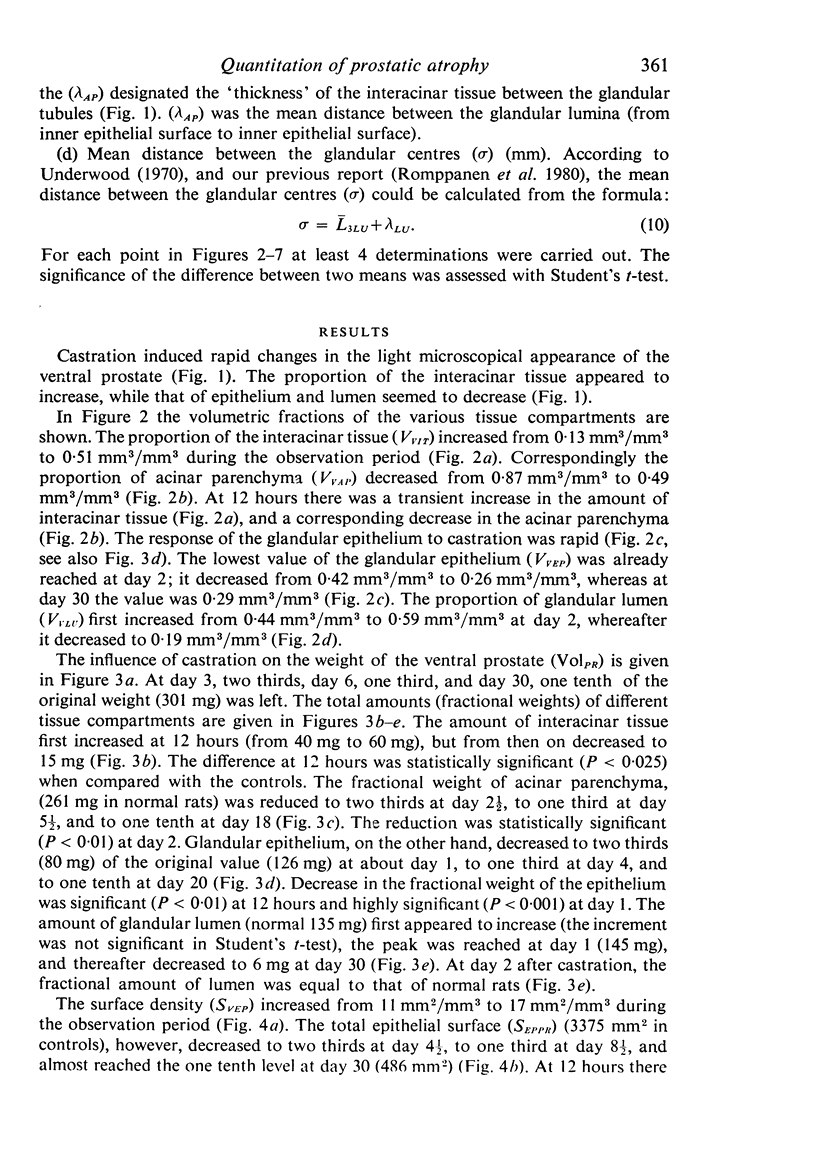
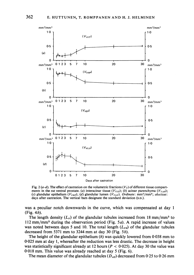
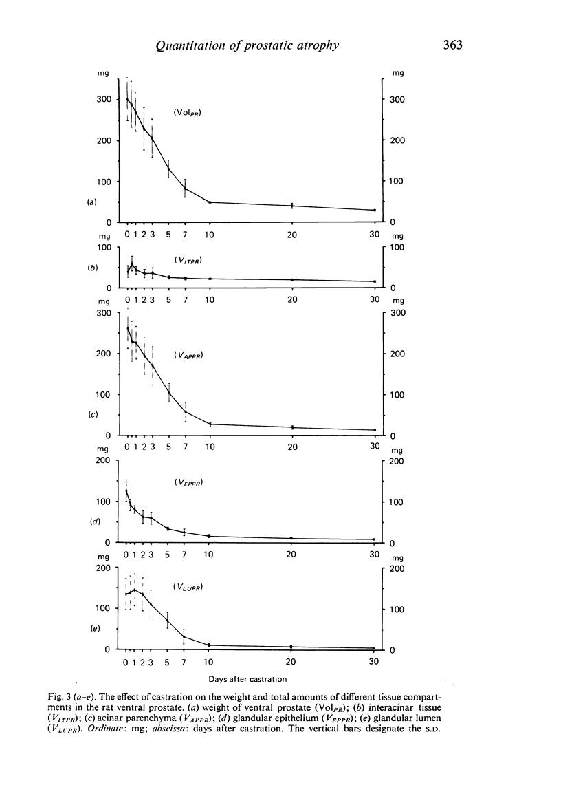
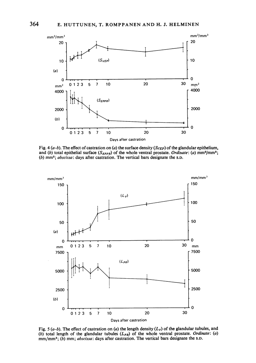
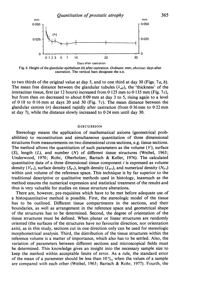
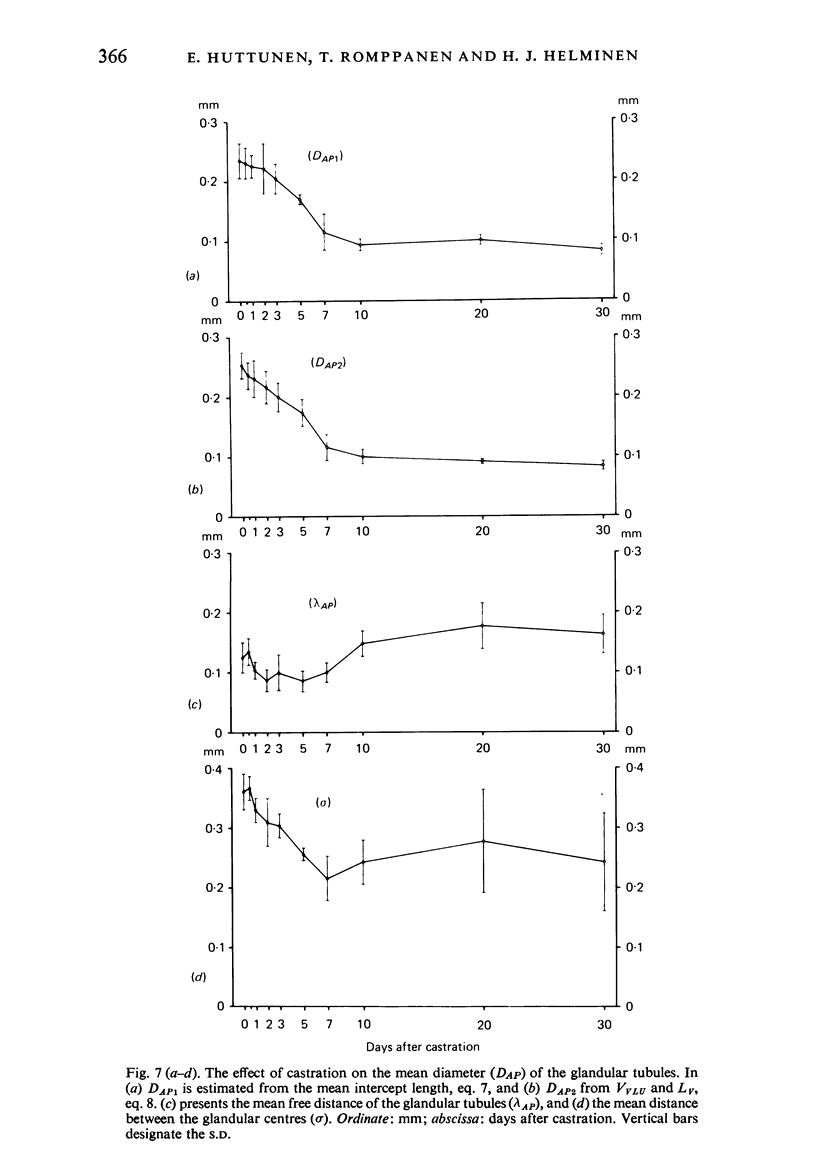
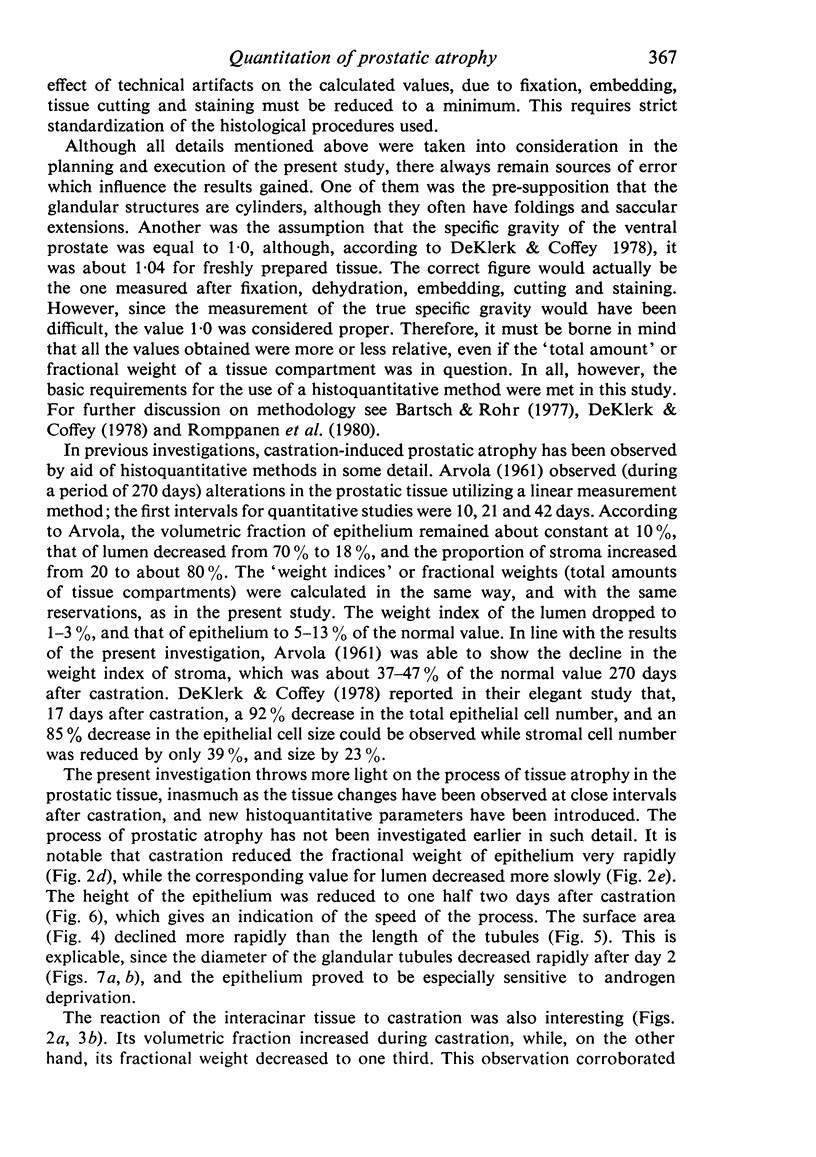
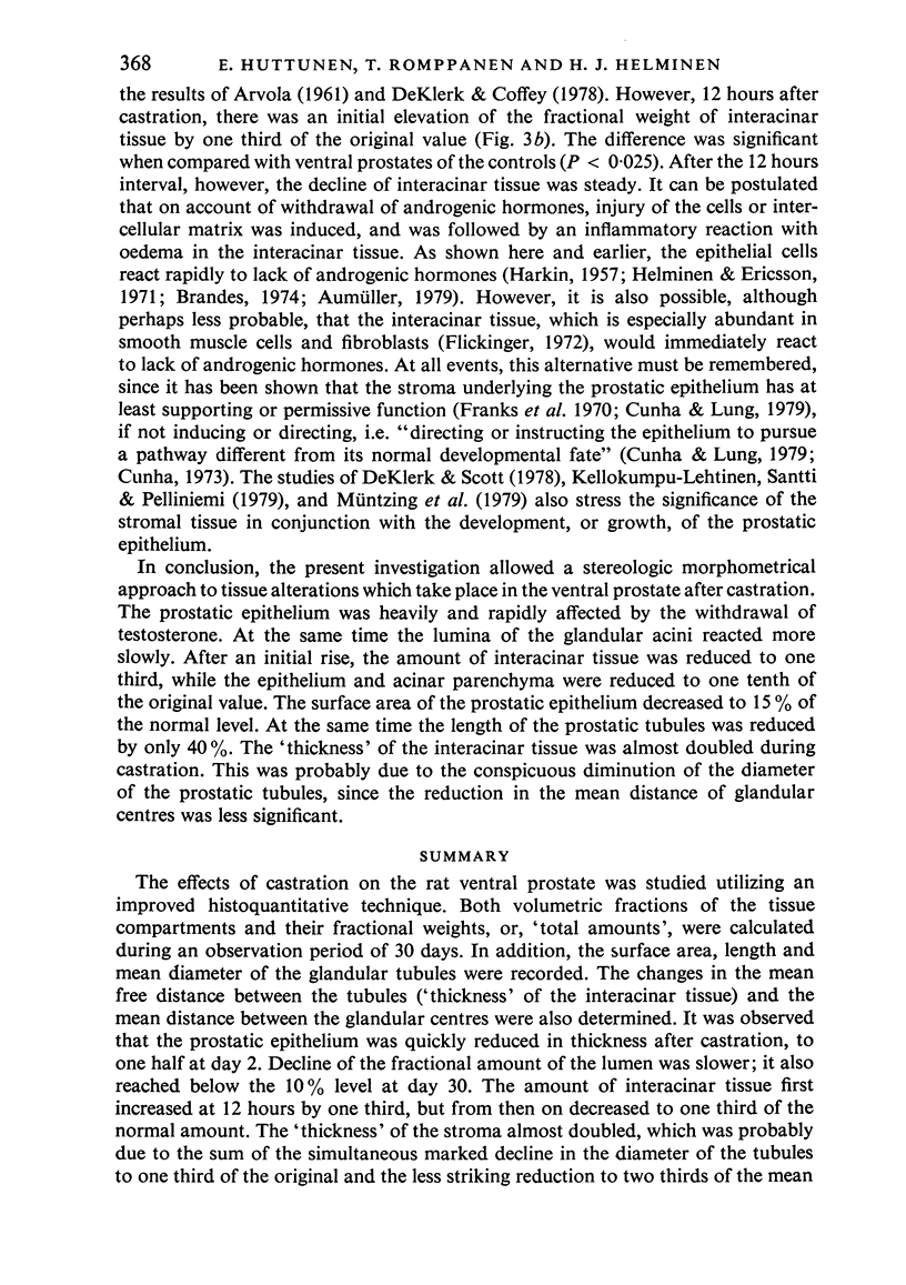
The effects of castration on the rat ventral prostate was studied utilizing an improved histoquantitative technique. Both volumetric fractions of the tissue compartments and their fractional weights, or, 'total amounts', were calculated during an observation period of 30 days. In addition, the surface area, length and mean diameter of the glandular tubules were recorded. The changes in the mean free distance between the tubules were recorded. The changes in the mean free distance between the tubules ('thickness' of the interacinar tissue) and the mean distance between the glandular centres were also determined. It was observed that the prostatic epithelium was quickly reduced in thickness after castration, to one half at day 2. Decline of the fractional amount of the lumen was slower; it also reached below the 10% level at day 30. The amount of interacinar tissue first increased at 12 hours by one third, but from then on decreased to one third of the normal amount. The 'thickness' of the stroma almost doubled, which was probably due to the sum of the simultaneous marked decline in the diameter of the tubules to one third of the original and the less striking reduction to two thirds of the mean distance between the glandular centres. The morphometrical method ensured the acquisition of a quantitative insight into the tissue processes involved in prostatic atrophy. Calculation of the fractional weights was regarded as especially invaluable, inasmuch as a growing body of evidence has been accumulated in favour of the crucial role of stromal-epithelial interactions in the differentiation, and growth, of the prostate.
Full text
PDF
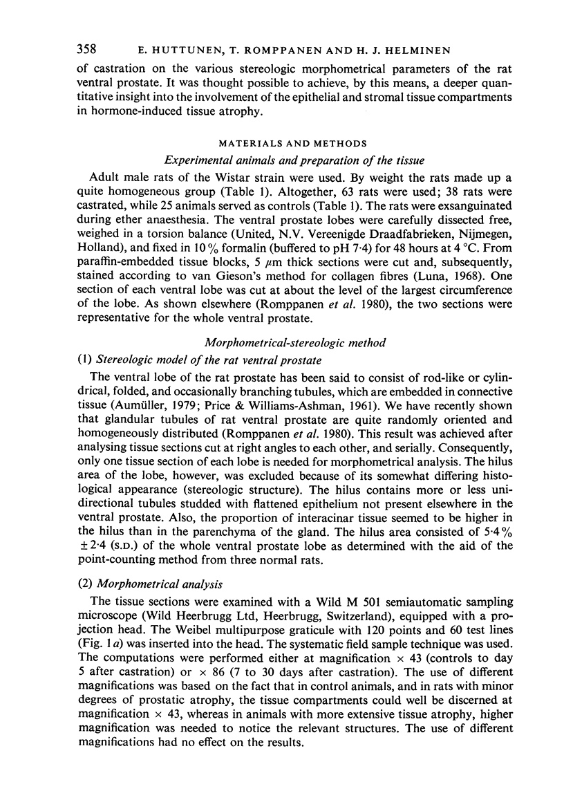
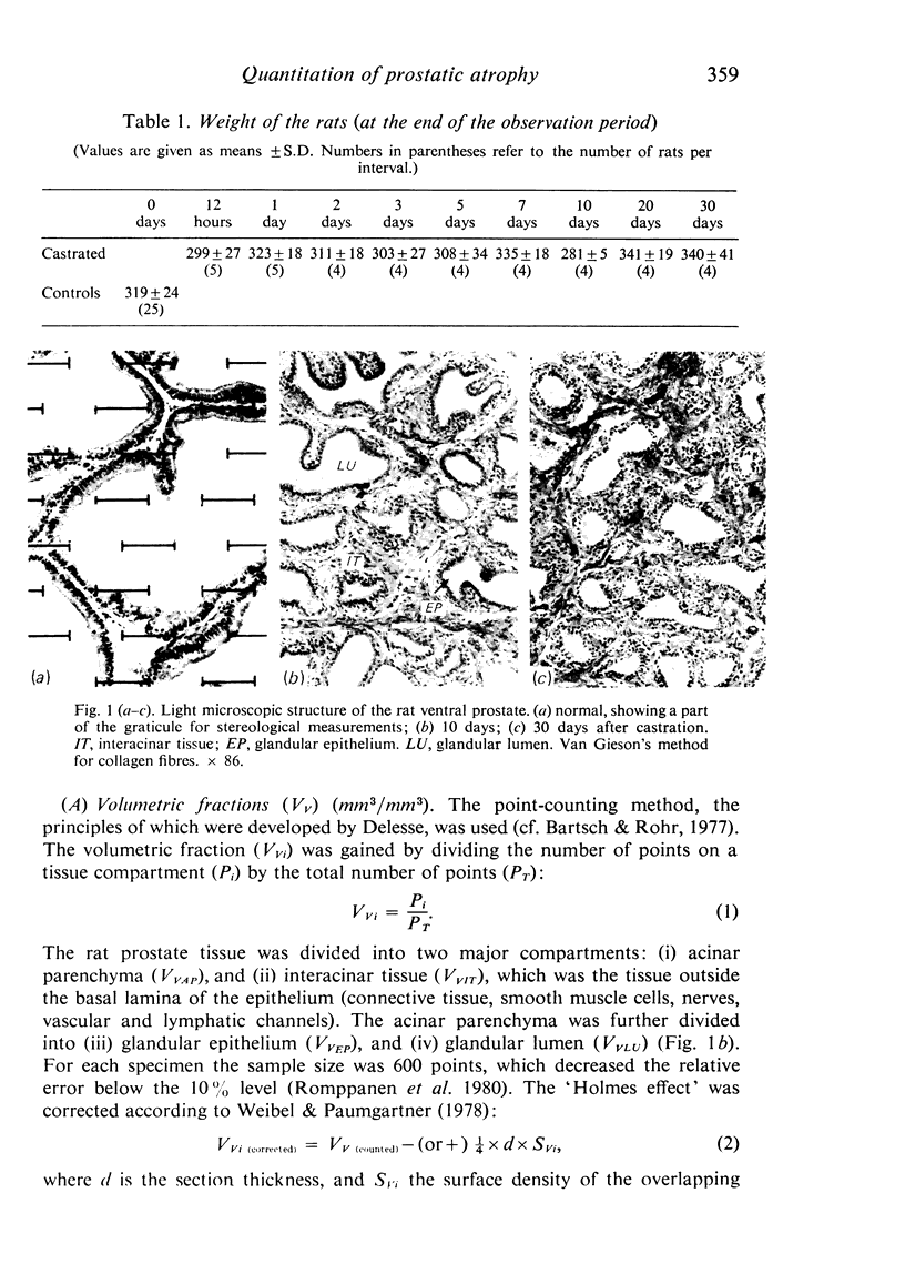
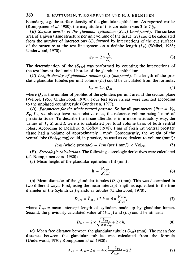
Images in this article
Selected References
These references are in PubMed. This may not be the complete list of references from this article.
- ARVOLA I. The hormonal control of the amounts of the tissue components of the prostate. A histoquantitative investigation performed on rats. Ann Chir Gynaecol Fenn Suppl. 1961;50(102):1–120. [PubMed] [Google Scholar]
- Bartsch G., Frick J., Rüegg I., Bucher M., Holliger O., Oberholzer M., Rohr H. P. Electron microscopic stereological analysis of the normal human prostate and of benign prostatic hyperplasia. J Urol. 1979 Oct;122(4):481–486. doi: 10.1016/s0022-5347(17)56475-7. [DOI] [PubMed] [Google Scholar]
- Bartsch G., Müller H. R., Oberholzer M., Rohr H. P. Light microscopic stereological analysis of the normal human prostate and of benign prostatic hyperplasia. J Urol. 1979 Oct;122(4):487–491. doi: 10.1016/s0022-5347(17)56476-9. [DOI] [PubMed] [Google Scholar]
- Bartsch G., Rohr H. P. Ultrastructural stereology: a new approach to the study of prostatic function. Invest Urol. 1977 Jan;14(4):301–306. [PubMed] [Google Scholar]
- Bartsch G. Stereology, a new quantitative morphological approach to study prostatic function and disease. Eur Urol. 1977;3(2):85–95. doi: 10.1159/000472066. [DOI] [PubMed] [Google Scholar]
- Cunha G. R., Lung B. The importance of stroma in morphogenesis and functional activity of urogenital epithelium. In Vitro. 1979 Jan;15(1):50–71. doi: 10.1007/BF02627079. [DOI] [PubMed] [Google Scholar]
- Cunha G. R. The role of androgens in the epithelio-mesenchymal interactions involved in prostatic morphogenesis in embryonic mice. Anat Rec. 1973 Jan;175(1):87–96. doi: 10.1002/ar.1091750108. [DOI] [PubMed] [Google Scholar]
- DeKlerk D. P., Coffey D. S. Quantitative determination of prostatic epithelial and stromal hyperplasia by a new technique. Biomorphometrics. Invest Urol. 1978 Nov;16(3):240–245. [PubMed] [Google Scholar]
- Deklerk D. P., Scott W. W. Prostatic maldevelopment in the prune belly syndrome: a defect in prostatic stromal-epithelial interaction. J Urol. 1978 Sep;120(3):341–344. doi: 10.1016/s0022-5347(17)57168-2. [DOI] [PubMed] [Google Scholar]
- Flickinger C. J. The fine structure of the interstitial tissue of the rat prostate. Am J Anat. 1972 May;134(1):107–125. doi: 10.1002/aja.1001340109. [DOI] [PubMed] [Google Scholar]
- Franks L. M., Riddle P. N., Carbonell A. W., Gey G. O. A comparative study of the ultrastructure and lack of growth capacity of adult human prostate epithelium mechanically separated from its stroma. J Pathol. 1970 Feb;100(2):113–119. doi: 10.1002/path.1711000206. [DOI] [PubMed] [Google Scholar]
- Helminen H. J., Ericsson J. L. Ultrastructural studies on prostatic involution in the rat. Mechanism of autophagy in epithelial cells, with special reference to the rough-surfaced endoplasmic reticulum. J Ultrastruct Res. 1971 Sep;36(5):708–724. doi: 10.1016/s0022-5320(71)90025-6. [DOI] [PubMed] [Google Scholar]
- Kellokumpu-Lehtinen P., Santti R., Pelliniemi L. J. Early cytodifferentiation of human prostatic urethra and Leydig cells. Anat Rec. 1979 Jul;194(3):429–443. doi: 10.1002/ar.1091940309. [DOI] [PubMed] [Google Scholar]
- Müntzing J., Liljekvist J., Murphy G. P. Chalones and stroma as possible growth-limiting factors in the rat ventral prostate. Invest Urol. 1979 Mar;16(5):399–402. [PubMed] [Google Scholar]
- Rohr H., Oberholzer M., Bartsch G., Keller M. Morphometry in experimental pathology: methods, baseline data and applications. Int Rev Exp Pathol. 1976;15:233–325. [PubMed] [Google Scholar]
- Romppanen T., Huttunen E., Helminen H. J. An improved light microscopical histoquantitative method for the stereological analysis of the rat ventral prostate lobe. Invest Urol. 1980 Jul;18(1):59–65. [PubMed] [Google Scholar]
- WEIBEL E. R. Principles and methods for the morphometric study of the lung and other organs. Lab Invest. 1963 Feb;12:131–155. [PubMed] [Google Scholar]
- Weibel E. R., Paumgartner D. Integrated stereological and biochemical studies on hepatocytic membranes. II. Correction of section thickness effect on volume and surface density estimates. J Cell Biol. 1978 May;77(2):584–597. doi: 10.1083/jcb.77.2.584. [DOI] [PMC free article] [PubMed] [Google Scholar]


