

Abstract
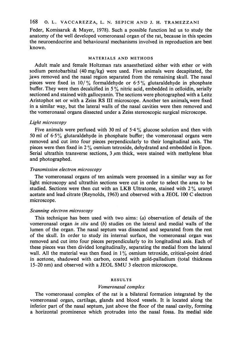
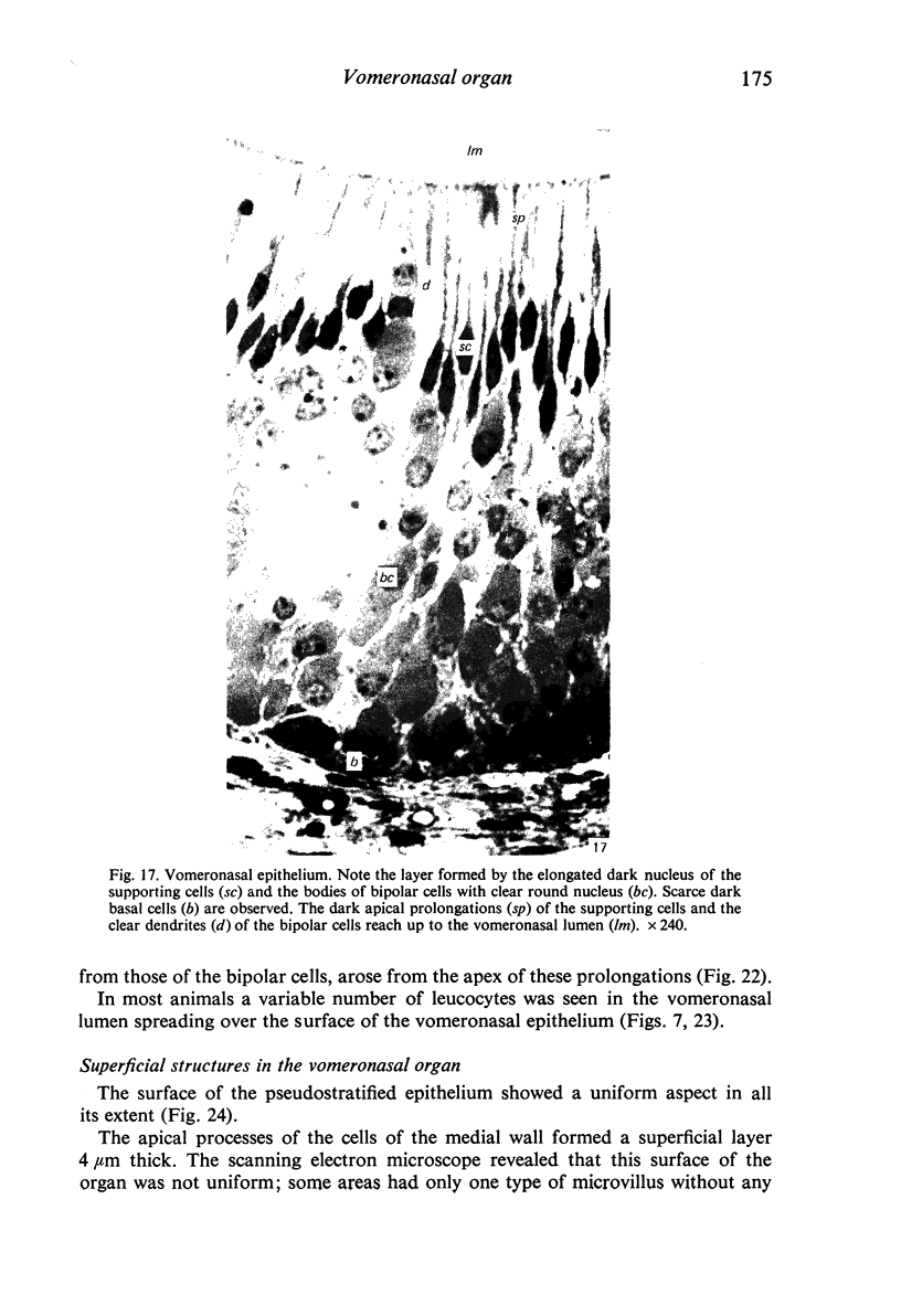
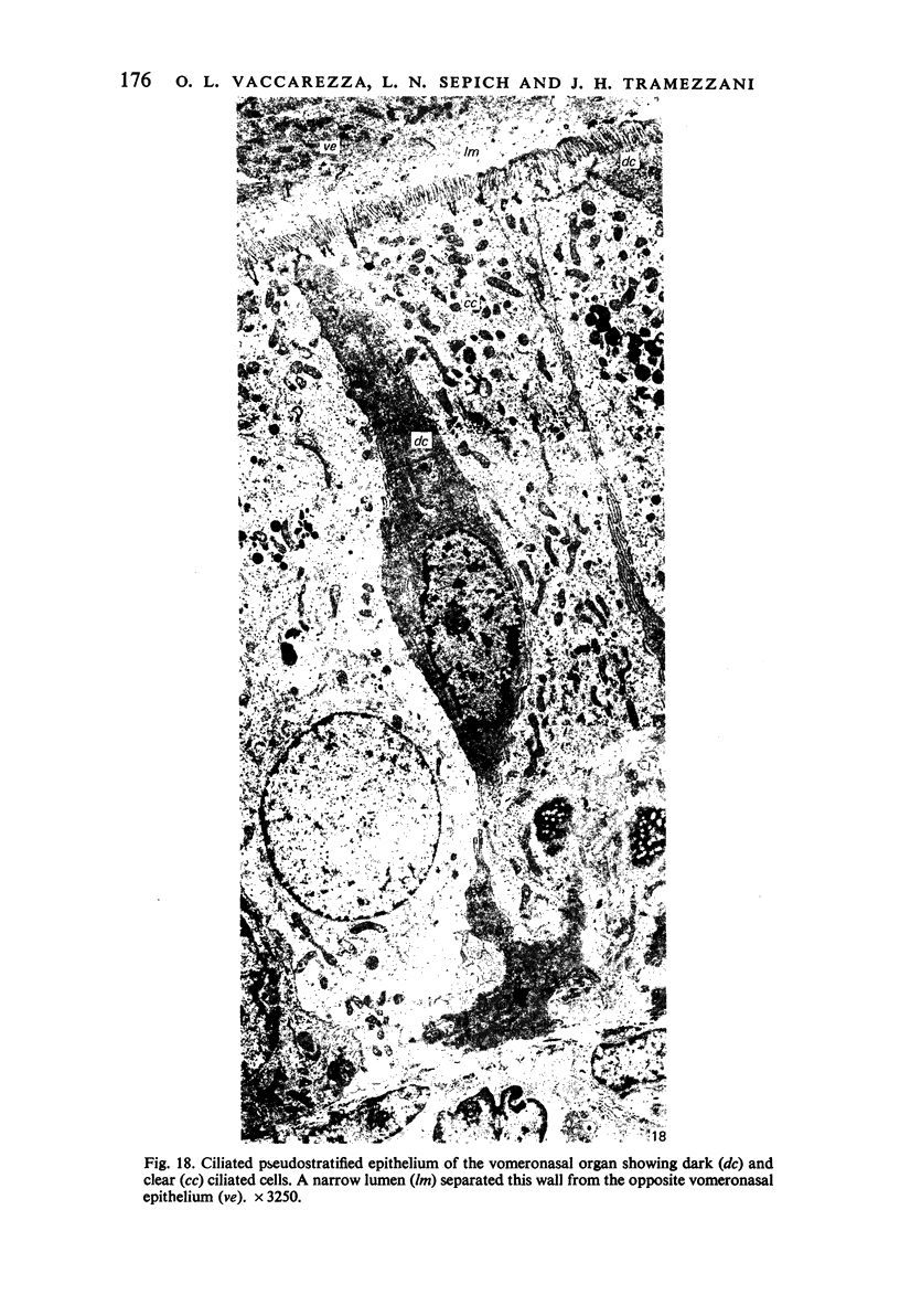
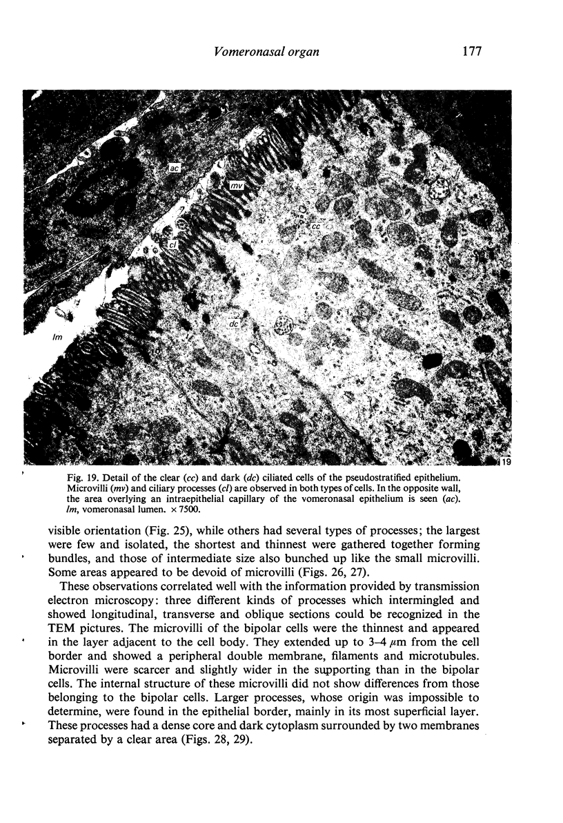
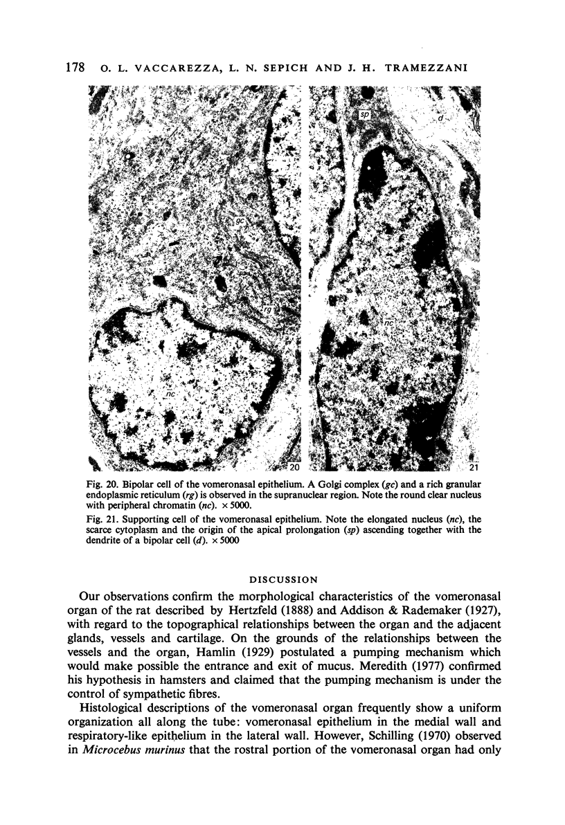
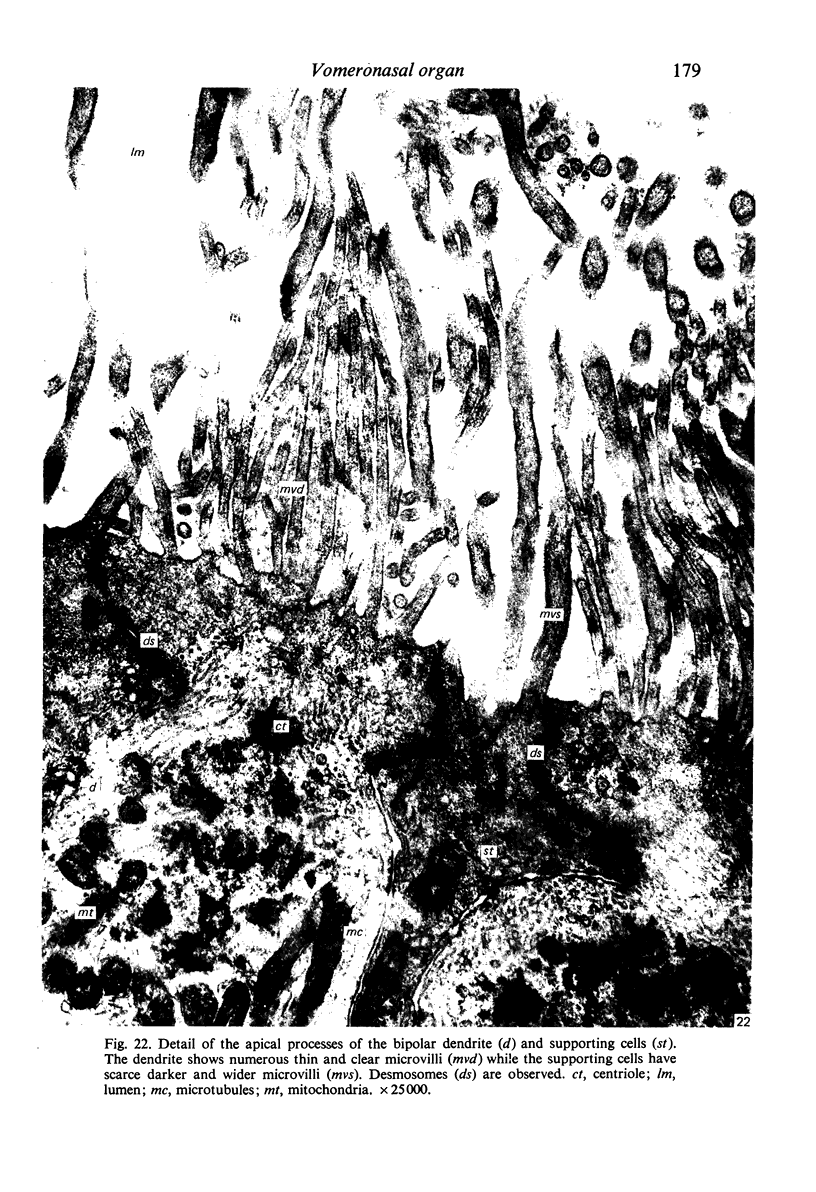
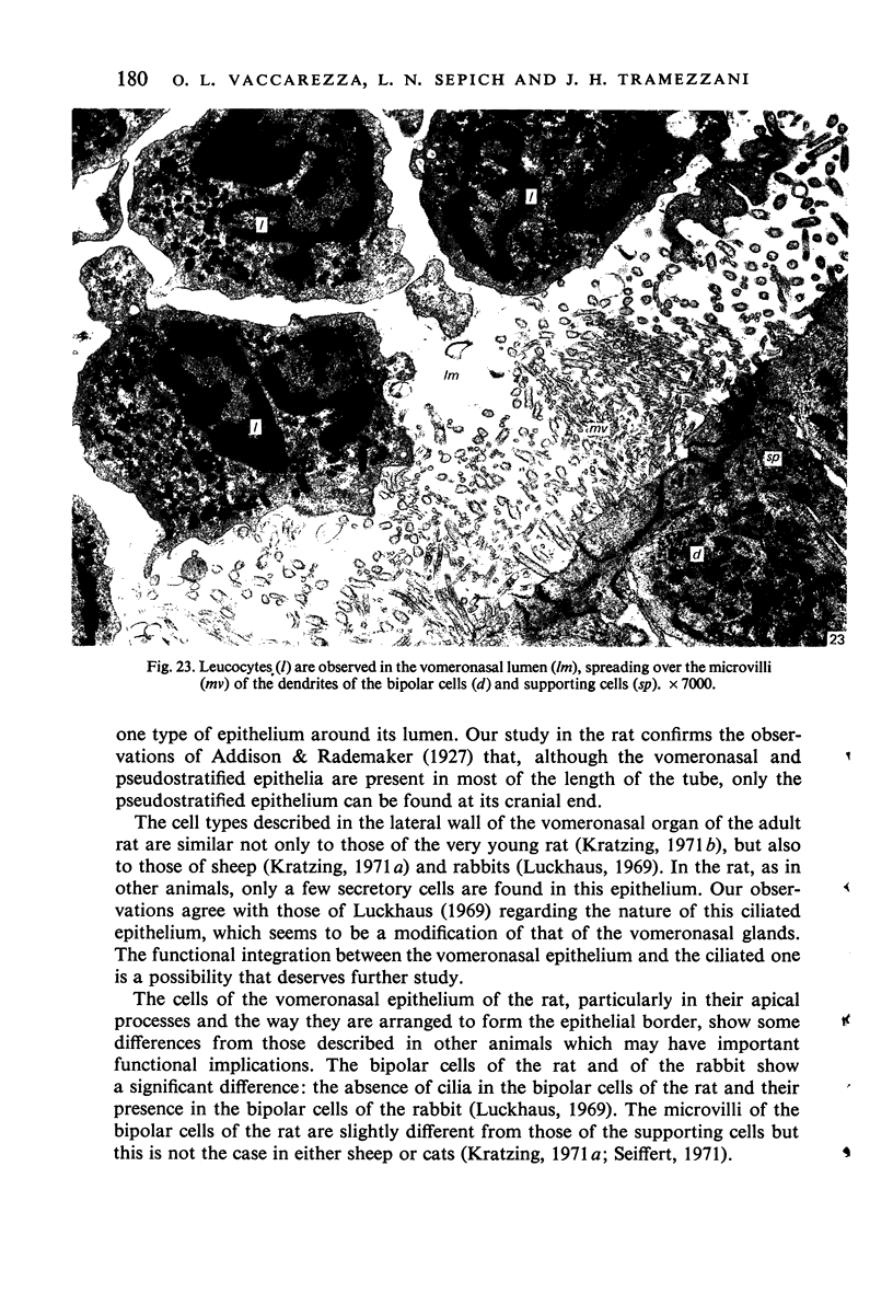
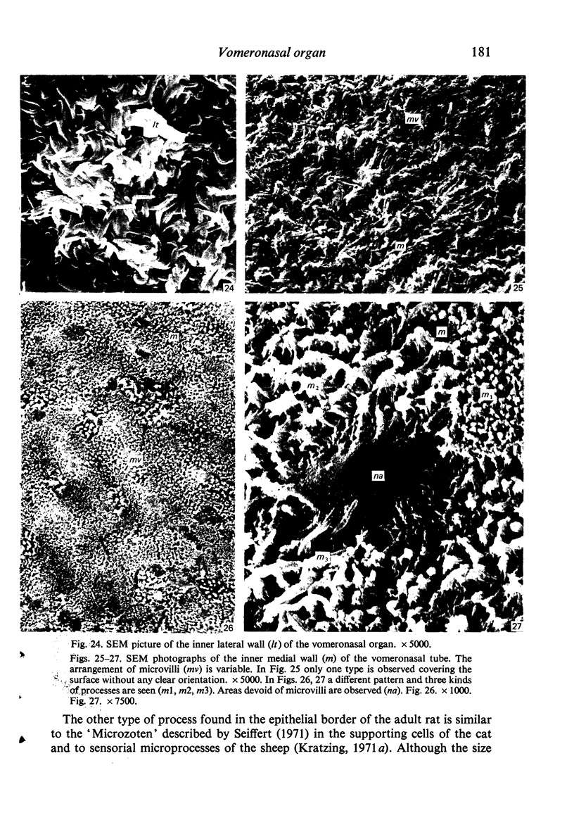
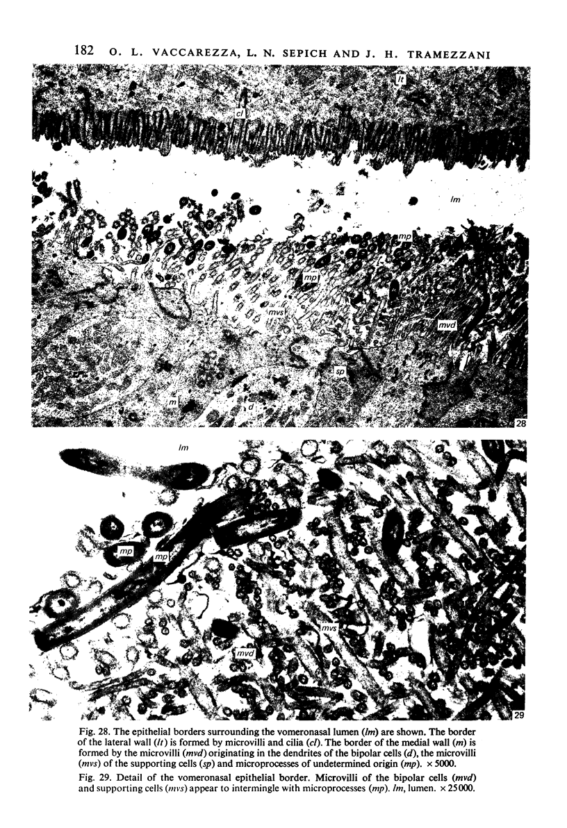
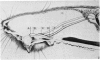
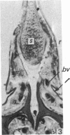
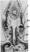
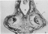
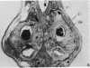
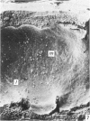
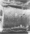
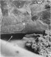
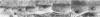
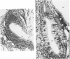
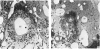
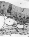
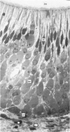
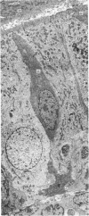
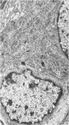
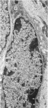
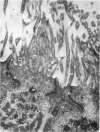
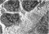
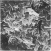
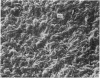
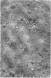
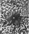
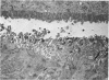
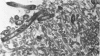
The anatomical organization of the vomeronasal complex of the rat (vomeronasal organ blood vessels, cartilage and glands) and the structure of the vomeronasal organ were studied. This organ is a tubular formation that shows different characteristics along its longitudinal axis. In its rostral portion it has a lateral flattened shape but caudally the organ acquires a typical crescent shape and a greater size. The organ is rotated along its longitudinal axis, the medial wall becoming inferior and the lateral one, superior. In its most caudal portion the organ decreases in size and ends in glandular branches. Three histological segments were recognized in the vomeronasal organ. The rostral one shows a pseudostratified epithelium surrounding all the lumen. The middle segment presents in one of its walls a similar epithelium and, in the other wall, the vomeronasal epithelium. The caudalmost segment shows a simple columnar epithelium that continues with that of glandular ducts. The vomeronasal epithelial border is formed by three types of cellular processes which intermingle, each one showing particular features: (a) microvilli originating from dendrites of bipolar cells; (b) microvilli from supporting cells and (c) micro-processes of undetermined origin. The surface of the vomeronasal epithelium shows an irregular distribution and arrangement of these processes.
Full text
PDF
Images in this article

Selected References
These references are in PubMed. This may not be the complete list of references from this article.
- Barber P. C., Raisman G. Cell division in the vomeronasal organ of the adult mouse. Brain Res. 1978 Feb 3;141(1):57–66. doi: 10.1016/0006-8993(78)90616-9. [DOI] [PubMed] [Google Scholar]
- Barber P. C., Raisman G. Replacement of receptor neurones after section of the vomeronasal nerves in the adult mouse. Brain Res. 1978 May 26;147(2):297–313. doi: 10.1016/0006-8993(78)90841-7. [DOI] [PubMed] [Google Scholar]
- Johns M. A., Feder H. H., Komisaruk B. R., Mayer A. D. Urine-induced reflex ovulation in anovulatory rats may be a vomeronasal effect. Nature. 1978 Mar 30;272(5652):446–448. doi: 10.1038/272446a0. [DOI] [PubMed] [Google Scholar]
- Kolnberger I. Vergleichende Untersuchungen am Riechepithel, insbesondere des Jacobsonschen Organs von Amphibien, Reptilien und Säugetieren. Z Zellforsch Mikrosk Anat. 1971;122(1):53–67. [PubMed] [Google Scholar]
- Kratzing J. E. The fine structure of the sensory epithelium of the vomeronasal organ in suckling rats. Aust J Biol Sci. 1971 Aug;24(4):787–796. [PubMed] [Google Scholar]
- Kratzing J. The structure of the vomeronasal organ in the sheep. J Anat. 1971 Feb;108(Pt 2):247–260. [PMC free article] [PubMed] [Google Scholar]
- Luckhaus G. Licht- und elektronemikroskopixhe Befunde an der Lamina epithelialis des Vomeronasalorgans vom Kaninchen. Anat Anz. 1969;124(5):477–489. [PubMed] [Google Scholar]
- NEGUS V. E. The organ of Jacobson. J Anat. 1956 Oct;90(4):515–519. [PMC free article] [PubMed] [Google Scholar]
- PLANEL H. Etudes sur la physiologie de l'organe de Jacobson. Arch Anat Histol Embryol. 1953;36(4-8):197–205. [PubMed] [Google Scholar]
- REYNOLDS E. S. The use of lead citrate at high pH as an electron-opaque stain in electron microscopy. J Cell Biol. 1963 Apr;17:208–212. doi: 10.1083/jcb.17.1.208. [DOI] [PMC free article] [PubMed] [Google Scholar]
- Winans S. S., Powers J. B. Olfactory and vomeronasal deafferentation of male hamsters: histological and behavioral analyses. Brain Res. 1977 May 6;126(2):325–344. doi: 10.1016/0006-8993(77)90729-6. [DOI] [PubMed] [Google Scholar]