

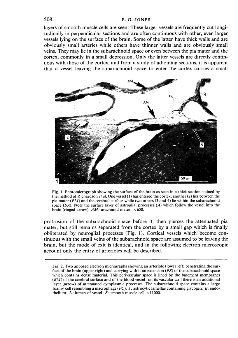
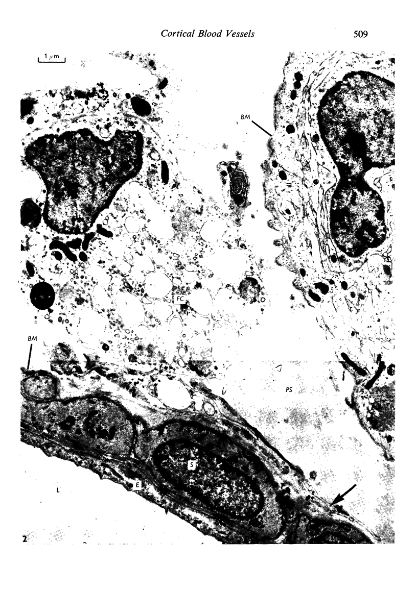
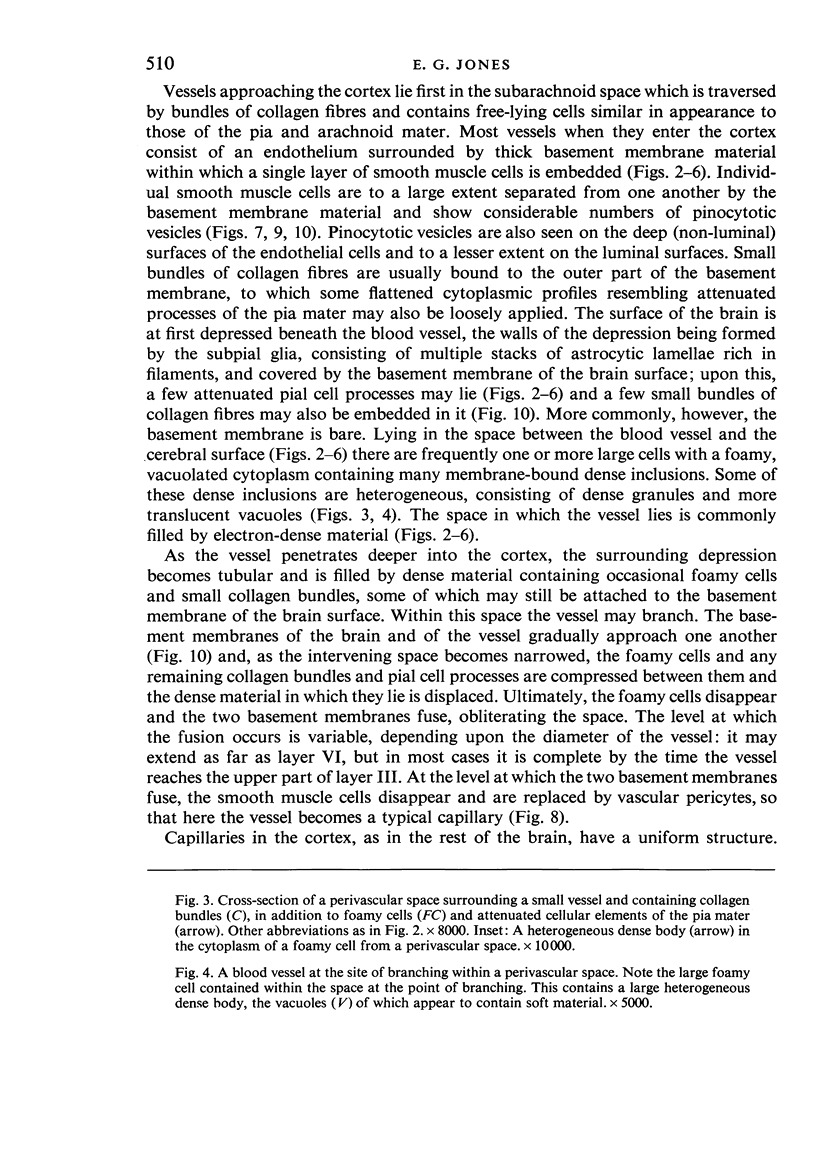
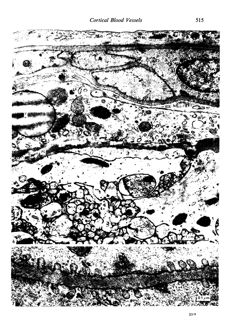
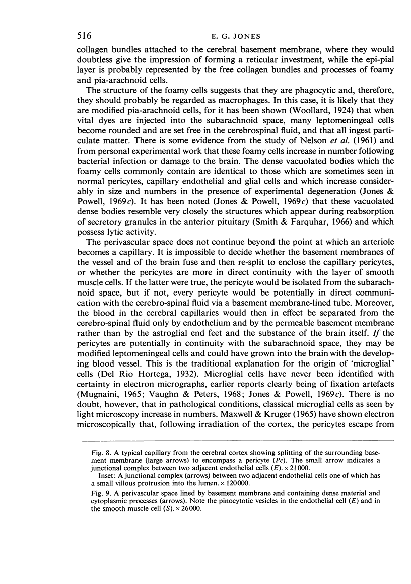
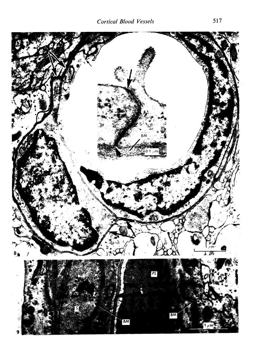
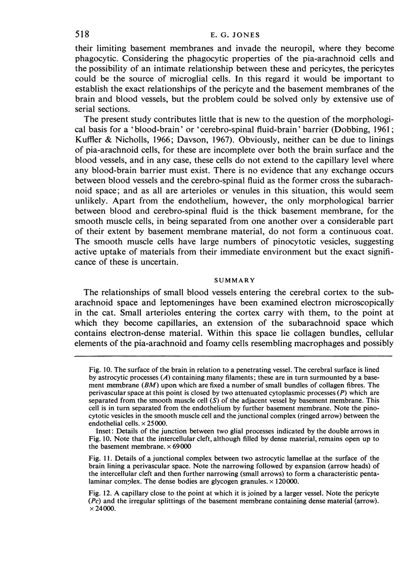
Full text
PDF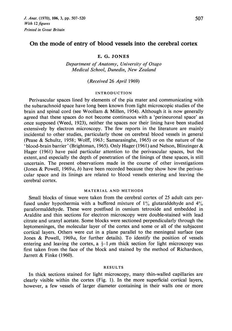

Images in this article
Selected References
These references are in PubMed. This may not be the complete list of references from this article.
- Brightman M. W. The distribution within the brain of ferritin injected into cerebrospinal fluid compartments. II. Parenchymal distribution. Am J Anat. 1965 Sep;117(2):193–219. doi: 10.1002/aja.1001170204. [DOI] [PubMed] [Google Scholar]
- DOBBING J. The blood-brain barrier. Physiol Rev. 1961 Jan;41:130–188. doi: 10.1152/physrev.1961.41.1.130. [DOI] [PubMed] [Google Scholar]
- HAGER H. [Electron microscopic studies on the fine structure of the blood vessels and perivascular spaces in the mammalian brain. A contribution to studies on the morphological principles of the so-called blood-brain barrier]. Acta Neuropathol. 1961 May 30;1:9–33. doi: 10.1007/BF00690476. [DOI] [PubMed] [Google Scholar]
- Jones E. G., Powell T. P. An electron microscopic study of terminal degeneration in the neocortex of the cat. Philos Trans R Soc Lond B Biol Sci. 1970 Jan 29;257(812):29–43. doi: 10.1098/rstb.1970.0006. [DOI] [PubMed] [Google Scholar]
- Jones E. G., Powell T. P. Electron microscopy of the somatic sensory cortex of the cat. I. Cell types and synaptic organization. Philos Trans R Soc Lond B Biol Sci. 1970 Jan 29;257(812):1–11. doi: 10.1098/rstb.1970.0003. [DOI] [PubMed] [Google Scholar]
- Jones E. G., Powell T. P. Electron microscopy of the somatic sensory cortex of the cat. II. The fine structure of layers I and II. Philos Trans R Soc Lond B Biol Sci. 1970 Jan 29;257(812):13–21. doi: 10.1098/rstb.1970.0004. [DOI] [PubMed] [Google Scholar]
- Kuffler S. W., Nicholls J. G. The physiology of neuroglial cells. Ergeb Physiol. 1966;57:1–90. [PubMed] [Google Scholar]
- MAXWELL D. S., KRUGER L. SMALL BLOOD VESSELS AND THE ORIGIN OF PHAGOCYTES IN THE RAT CEREBRAL CORTEX FOLLOWING HEAVY PARTICLE IRRADIATION. Exp Neurol. 1965 May;12:33–54. doi: 10.1016/0014-4886(65)90097-x. [DOI] [PubMed] [Google Scholar]
- MILLEN J. W., WOOLLAM D. H. On the nature of the pia mater. Brain. 1961 Sep;84:514–520. doi: 10.1093/brain/84.3.514. [DOI] [PubMed] [Google Scholar]
- NELSON E., BLINZINGER K., HAGER H. Electron microscopic observations on subarachnoid and perivascular spaces of the Syrian hamster brain. Neurology. 1961 Apr;11(4):285–295. doi: 10.1212/wnl.11.4.285. [DOI] [PubMed] [Google Scholar]
- PEASE D. C., SCHULTZ R. L. Electron microscopy of rat cranial meninges. Am J Anat. 1958 Mar;102(2):301–321. doi: 10.1002/aja.1001020207. [DOI] [PubMed] [Google Scholar]
- RICHARDSON K. C., JARETT L., FINKE E. H. Embedding in epoxy resins for ultrathin sectioning in electron microscopy. Stain Technol. 1960 Nov;35:313–323. doi: 10.3109/10520296009114754. [DOI] [PubMed] [Google Scholar]
- Ramsey H. J. Fine structure of the surface of the cerebral cortex of human brain. J Cell Biol. 1965 Aug;26(2):323–334. doi: 10.1083/jcb.26.2.323. [DOI] [PMC free article] [PubMed] [Google Scholar]
- Samarasinghe D. D. The innervation of the cerebral arteries in the rat: an electron microscope study. J Anat. 1965 Oct;99(Pt 4):815–828. [PMC free article] [PubMed] [Google Scholar]
- Vaughn J. E., Peters A. A third neuroglial cell type. An electron microscopic study. J Comp Neurol. 1968 Jun;133(2):269–288. doi: 10.1002/cne.901330207. [DOI] [PubMed] [Google Scholar]
- WOOLLAM D. H., MILLEN J. W. The perivascular spaces of the mammalian central nervous system and their relation to the perineuronal and subarachnoid spaces. J Anat. 1955 Apr;89(2):193–200. [PMC free article] [PubMed] [Google Scholar]
- Woollard H. H. Vital Staining of the Leptomeninges. J Anat. 1924 Jan;58(Pt 2):89–100. [PMC free article] [PubMed] [Google Scholar]