

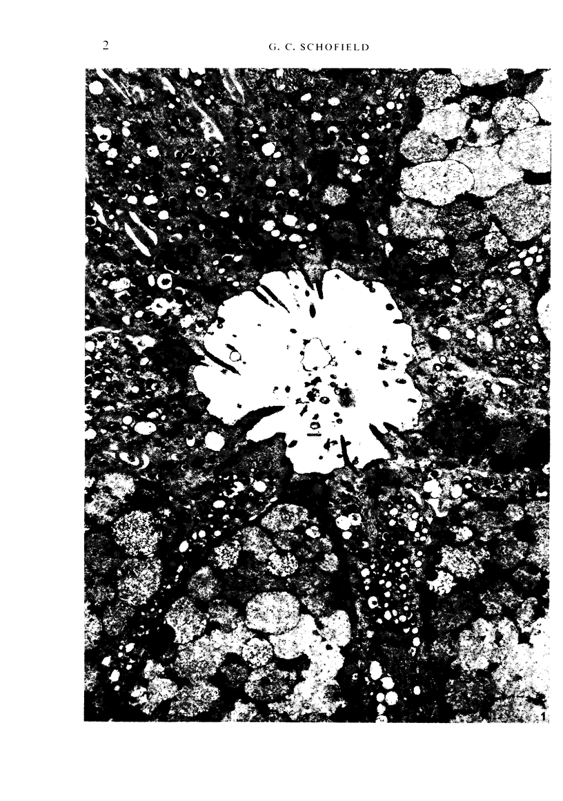
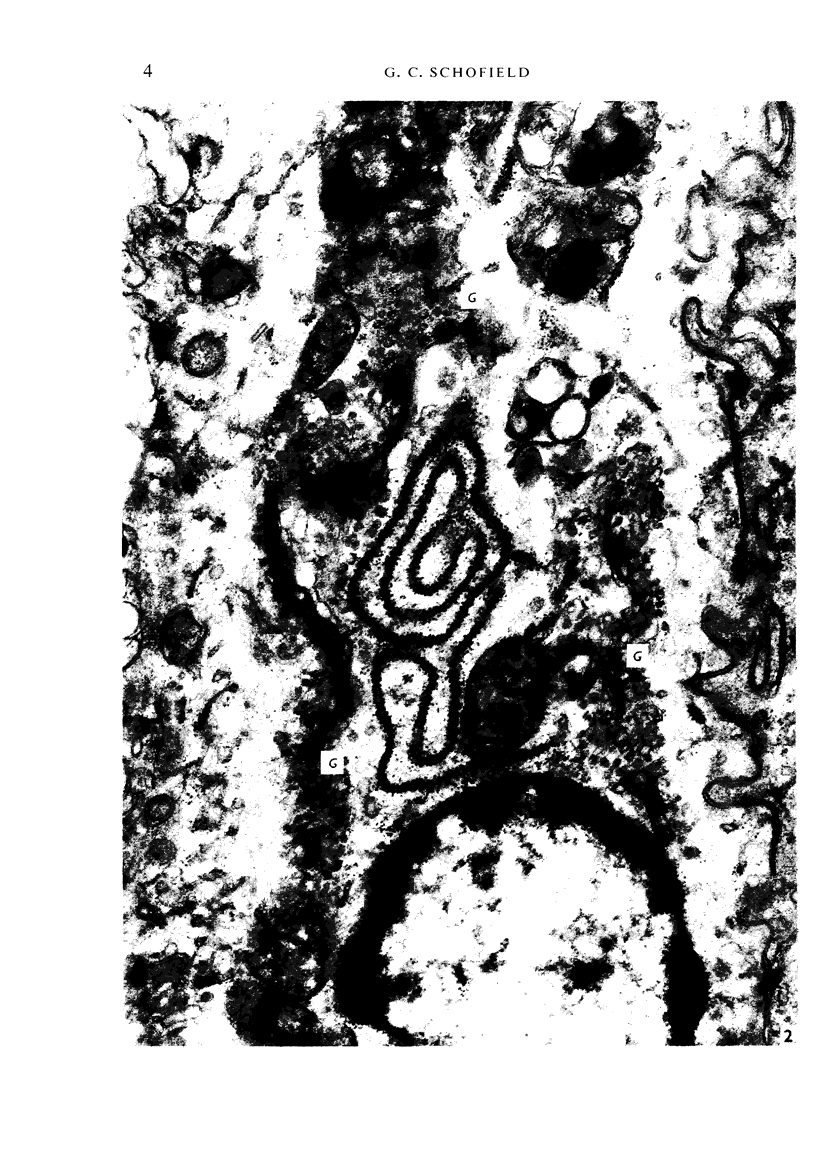
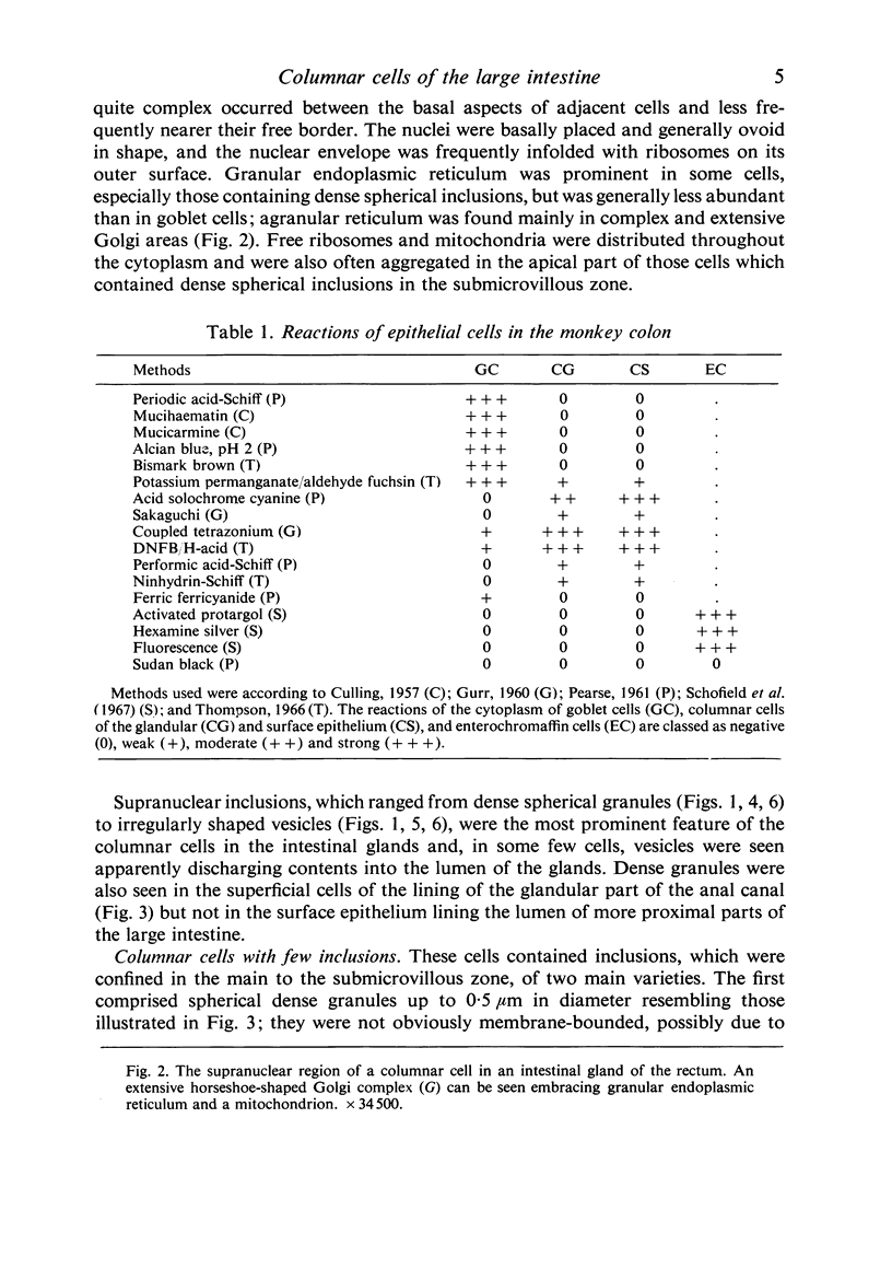
Full text
PDF
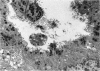
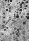
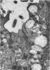
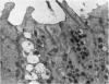
Images in this article
Selected References
These references are in PubMed. This may not be the complete list of references from this article.
- BARKA T. ELECTRON HISTOCHEMICAL LOCALIZATION OF ACID PHOSPHATASE ACTIVITY IN THE SMALL INTESTINE OF MOUSE. J Histochem Cytochem. 1964 Apr;12:229–238. doi: 10.1177/12.4.229. [DOI] [PubMed] [Google Scholar]
- Carr K. E., Whur P. Ultrastructure of globule leucocyte inclusions in the rat and mouse. Z Zellforsch Mikrosk Anat. 1968;86(2):153–162. doi: 10.1007/BF00348521. [DOI] [PubMed] [Google Scholar]
- Crabbé P. A., Heremans J. F. The distribution of immunoglobulin-containing cells along the human gastrointestinal tract. Gastroenterology. 1966 Sep;51(3):305–316. [PubMed] [Google Scholar]
- FLOREY H. W. Electron microscopic observations on goblet cells of the rat's colon. Q J Exp Physiol Cogn Med Sci. 1960 Oct;45:329–336. doi: 10.1113/expphysiol.1960.sp001487. [DOI] [PubMed] [Google Scholar]
- Freeman J. A. Goblet cell fine structure. Anat Rec. 1966 Jan;154(1):121–147. doi: 10.1002/ar.1091540111. [DOI] [PubMed] [Google Scholar]
- Gelzayd E. A., Kraft S. C., Kirsner J. B. Distribution of immunoglobulins in human rectal mucosa. I. Normal control subjects. Gastroenterology. 1968 Mar;54(3):334–340. [PubMed] [Google Scholar]
- Hollmann K. H. Uber den Feinbau des Rectumepithels. Z Zellforsch Mikrosk Anat. 1965 Nov 15;68(4):502–542. [PubMed] [Google Scholar]
- Johnson F. R., Young B. A. Undifferentiated cells in gastric mucosa. J Anat. 1968 Mar;102(Pt 3):541–551. [PMC free article] [PubMed] [Google Scholar]
- Lorenzsonn V., Trier J. S. The fine structure of human rectal mycosa. The epithelial lining of the base of the crypt. Gastroenterology. 1968 Jul;55(1):88–101. [PubMed] [Google Scholar]
- REYNOLDS E. S. The use of lead citrate at high pH as an electron-opaque stain in electron microscopy. J Cell Biol. 1963 Apr;17:208–212. doi: 10.1083/jcb.17.1.208. [DOI] [PMC free article] [PubMed] [Google Scholar]
- Rossen R. D., Morgan C., Hsu K. C., Butler W. T., Rose H. M. Localization of 11 S external secretory IgA by immunofluorescence in tissues lining the oral and respiratory passages in man. J Immunol. 1968 Apr;100(4):706–717. [PubMed] [Google Scholar]
- Schofield G. C., Ho A. K., Southwell J. M. Enterochromaffin cells and 5-hydroxytryptamine content of the colon of mice. J Anat. 1967 Sep;101(Pt 4):711–721. [PMC free article] [PubMed] [Google Scholar]
- Schofield G. C., Silva D. G. The fine structure of enterochromaffin cells in the mouse colon. J Anat. 1968 Jun;103(Pt 1):1–13. [PMC free article] [PubMed] [Google Scholar]
- Silva D. G. The fine structure of multivesicular cells with large microvilli in the epithelium of the mouse colon. J Ultrastruct Res. 1966 Dec;16(5):693–705. doi: 10.1016/s0022-5320(66)80015-1. [DOI] [PubMed] [Google Scholar]
- South M. A., Warwick W. J., Wolheim F. A., Good R. A. The IgA system. 3. IgA levels in the serum and saliva of pediatric patients--evidence for a local immunological system. J Pediatr. 1967 Nov;71(5):645–653. doi: 10.1016/s0022-3476(67)80199-9. [DOI] [PubMed] [Google Scholar]
- WATSON M. L. Staining of tissue sections for electron microscopy with heavy metals. J Biophys Biochem Cytol. 1958 Jul 25;4(4):475–478. doi: 10.1083/jcb.4.4.475. [DOI] [PMC free article] [PubMed] [Google Scholar]
- Wetzel M. G., Wetzel B. K., Spicer S. S. Ultrastructural localization of acid mucosubstances in the mouse colon with iron-containing stains. J Cell Biol. 1966 Aug;30(2):299–315. doi: 10.1083/jcb.30.2.299. [DOI] [PMC free article] [PubMed] [Google Scholar]