

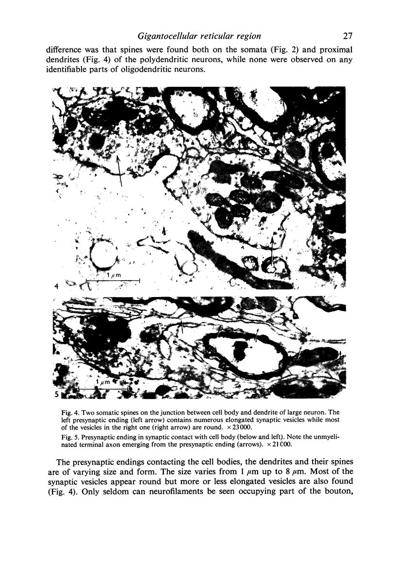
Full text
PDF


Images in this article


Selected References
These references are in PubMed. This may not be the complete list of references from this article.
- Alksne J. F., Blackstad T. W., Walberg F., White L. E., Jr Electron microscopy of axon degeneration: a valuable tool in experimental neuroanatomy. Ergeb Anat Entwicklungsgesch. 1966;39(1):3–32. doi: 10.1007/978-3-662-30450-1. [DOI] [PubMed] [Google Scholar]
- Bowsher D. Etude comparée des projections thalamiques de deux zones localisées des formations réticulées bulbaires et mésencéphaliques. C R Acad Sci Hebd Seances Acad Sci D. 1967 Jul 24;265(4):340–342. [PubMed] [Google Scholar]
- Bowsher D., Mallart A., Petit D., Albe-Fessard D. A bulbar relay to the centre median. J Neurophysiol. 1968 Mar;31(2):288–300. doi: 10.1152/jn.1968.31.2.288. [DOI] [PubMed] [Google Scholar]
- COLONNIER M. EXPERIMENTAL DEGENERATION IN THE CEREBRAL CORTEX. J Anat. 1964 Jan;98:47–53. [PMC free article] [PubMed] [Google Scholar]
- Conradi S. Ultrastructural specialization of the initial axon segment of cat lumbar motoneurons. Preliminary observations. Acta Soc Med Ups. 1966;71(5):281–284. [PubMed] [Google Scholar]
- Gray E. G., Hamlyn L. H. Electron microscopy of experimental degeneration in the avian optic tectum. J Anat. 1962 Jul;96(Pt 3):309–316.5. [PMC free article] [PubMed] [Google Scholar]
- Holländer H., Brodal P., Walberg F. Electronmicroscopic observations on the structure of the pontine nuclei and the mode of termination of the corticopontine fibres. An experimental study in the cat. Exp Brain Res. 1969;7(2):95–110. doi: 10.1007/BF00235436. [DOI] [PubMed] [Google Scholar]
- Johnstone G., Bowsher D. A new method for the selective impregnation of degenerating axon terminals. Brain Res. 1969 Jan;12(1):47–53. doi: 10.1016/0006-8993(69)90054-7. [DOI] [PubMed] [Google Scholar]
- Mannen H. Contribution to the morphological study of dendritic arborization in the brain stem. Prog Brain Res. 1966;21:131–162. doi: 10.1016/s0079-6123(08)62975-1. [DOI] [PubMed] [Google Scholar]
- Mugnaini E., Walberg F. An experimental electron microscopical study on the mode of termination of cerebellar corticovestibular fibres in the cat lateral vestibular nucleus (Deiters' nucleus). Exp Brain Res. 1967;4(3):212–236. doi: 10.1007/BF00248023. [DOI] [PubMed] [Google Scholar]
- Palay S. L., Sotelo C., Peters A., Orkand P. M. The axon hillock and the initial segment. J Cell Biol. 1968 Jul;38(1):193–201. doi: 10.1083/jcb.38.1.193. [DOI] [PMC free article] [PubMed] [Google Scholar]
- REYNOLDS E. S. The use of lead citrate at high pH as an electron-opaque stain in electron microscopy. J Cell Biol. 1963 Apr;17:208–212. doi: 10.1083/jcb.17.1.208. [DOI] [PMC free article] [PubMed] [Google Scholar]
- ROSSI G. F., BRODAL A. Terminal distribution of spinoreticular fibers in the cat. AMA Arch Neurol Psychiatry. 1957 Nov;78(5):439–453. doi: 10.1001/archneurpsyc.1957.02330410003001. [DOI] [PubMed] [Google Scholar]
- Ramón-Moliner E., Nauta W. J. The isodendritic core of the brain stem. J Comp Neurol. 1966 Mar;126(3):311–335. doi: 10.1002/cne.901260301. [DOI] [PubMed] [Google Scholar]
- SCHEIBEL M., SCHEIBEL A., MOLLICA A., MORUZZI G. Convergence and interaction of afferent impulses on single units of reticular formation. J Neurophysiol. 1955 Jul;18(4):309–331. doi: 10.1152/jn.1955.18.4.309. [DOI] [PubMed] [Google Scholar]
- Segundo J. P., Takenaka T., Encabo H. Somatic sensory properties of bulbar reticular neurons. J Neurophysiol. 1967 Sep;30(5):1221–1238. doi: 10.1152/jn.1967.30.5.1221. [DOI] [PubMed] [Google Scholar]
- Szentágothai J., Hámori J., Tömböl T. Degeneration and electron microscope analysis of the synaptic glomeruli in the lateral geniculate body. Exp Brain Res. 1966;2(4):283–301. doi: 10.1007/BF00234775. [DOI] [PubMed] [Google Scholar]
- TABER E. The cytoarchitecture of the brain stem of the cat. I. Brain stem nuclei of cat. J Comp Neurol. 1961 Feb;116:27–69. doi: 10.1002/cne.901160104. [DOI] [PubMed] [Google Scholar]
- WATSON M. L. Staining of tissue sections for electron microscopy with heavy metals. J Biophys Biochem Cytol. 1958 Jul 25;4(4):475–478. doi: 10.1083/jcb.4.4.475. [DOI] [PMC free article] [PubMed] [Google Scholar]
- Westman J. The lateral cervical nucleus in the cat. II. An electron microscopical study of the normal structure. Brain Res. 1968 Oct;11(1):107–123. doi: 10.1016/0006-8993(68)90076-0. [DOI] [PubMed] [Google Scholar]