Abstract
Gamma phage specifically lyses vegetative cells of Bacillus anthracis and serves as part of the basis for identification of isolates from agar cultures. We report our study to standardize gamma phage production and preparation and to validate the assay for routine use. Unstable phage preparations were largely reduced through propagation of phage on blood agar cultures of the avirulent B. anthracis strain CDC684 and were adequately stable for extended storage beyond 1 to 2 years at 4°C, provided that the preparation initially gave rise to clearly discernible plaques (macroplaques, 5 to 10 mm in diameter) on dilution at 1:8 or greater during potency testing with the Sterne strain or its equivalent. The primary intent of the assay was to test nonhemolytic, ground-glass-appearing bacterial B. anthracis-like colonies arising from culture of clinical or nonclinical samples on 5% sheep blood agar. Specifically, the assay was designed to show clear or primarily clear circular zones of lysis on bacterial lawns at the site of gamma phage inoculation after incubation at 35 °C ± 2°C for 20 h. When tested with 51 B. anthracis strains and 49 similar non-B. anthracis Bacillus species, the analytical specificity was >95%, a value that is intentionally low because our study design included two rare nonsusceptible B. anthracis strains as well as a rare susceptible non-B. anthracis strain, B. cereus ATCC 4342. Repeatability, day-to-day precision, and analyst-to-analyst precision were superior. The assay was rugged to variations among phage lots, phage concentration, amounts of bacterial inoculum, and incubation times as short as 6 to 8 h. System suitability evaluation showed improved robustness when bacterial lawns were tested with high- and low-density inoculum using the first and second quadrants of a serial four-quadrant streak on 5% sheep blood agar plates.
Bacteriophages that are active on B. anthracis but not specific were first described in 1931 by Cowles (7). In 1951, McCloy isolated a bacteriophage called “phage W” from what she described as an atypical Bacillus cereus strain, designated strain W, ATCC 11950 (9). The phage was highly specific for 171 strains of B. anthracis and failed to react with 19 other Bacillus species, except for two unusual strains of B. cereus, strains NCTC 1651 and NCTC 6222 (ATCC 4342). In a subsequent study, McCloy (10) isolated two types of phage from aging cultures of Bacillus strain W, referred to as “phage α” (rare mutant) and “phage β” (dominant form), which specifically produced plaques on agar cultures of B. anthracis. The two phages differed only in that phage α attacked strains of B. anthracis and Bacillus strain W. Phage β attacked all B. anthracis strains tested by McCloy but not strain W; however, it was not able to attack smooth or encapsulated forms of B. anthracis. In 1955, Brown and Cherry (4) isolated a variant of the original phage W, designated gamma phage. This phage differed from phage α and phage β in that it lysed encapsulated smooth forms of B. anthracis, failed to lyse any of the strains of B. cereus tested, and could lyse and be propagated on the Bacillus W strain. Use of gamma phage as a diagnostic tool for identification of B. anthracis has been cited in a number of articles (2-5, 12). Other sensitive Bacillus species included a soil-derived B. megaterium strain at Kansas State University (6) and four nonrhizoid strains of B. cereus var. mycoides (5). Buck et al. (6) reported these two strains to be negative in the “string-of-pearls” assay, which is typically positive for B. anthracis strains. In contrast, several purported B. anthracis isolates have been reported as not susceptible to gamma phage through either the inability of phage to bind or the specific lytic action of the PlyG lysin enzyme (11) isolated from gamma phage.
The goals of this study were to document and validate the performance characteristics of the gamma phage lysis assay for B. anthracis identification, standardize gamma phage production methods, and determine stability. The validation of the assay considered the following analytical performance parameters as listed and defined in the United States Pharmacopeia, section 1225, as appropriate: precision, accuracy, selectivity/specificity, quantification limit, detection limit, linearity, and range (13). To establish a series of system suitability parameters, elements of precision and robustness were considered. Under criteria in United States Pharmacopeia, section 1225 (10th Supplement), this procedure falls within the category of an identification test (category IV). Thus, specificity is the most important performance characteristic. Elements of accuracy, quantification limit, detection limit, linearity, and range were not considered because this is a qualitative test.
MATERIALS AND METHODS
Gamma phage and Bacillus strains.
Gamma phage was originally obtained from the Centers for Disease Control and Prevention (CDC), Atlanta, Ga., courtesy of William Cherry, as a liquid suspension during the early 1980s. Bacillus species used in this study (listed in Table 1) were from the U.S. Army Medical Research Institute for Infectious Diseases (USAMRIID) culture collection and were comprised of various species and strains originally obtained from the CDC (designated CDC or SK), the American Type Culture Collection (ATCC), and the National Collection of Type Cultures (NCTC) (8). One of the non-B. anthracis strains, originally designated by the CDC as B. mycoides strain CDC680 in the early 1980s, was later identified as a B. anthracis strain lacking both plasmids (as discussed below). The identities of all other B. anthracis strains were verified by PCR and shown to possess one or both of the anthrax virulence plasmids (pXO1 and pXO2). Additionally, strains were determined to be nonhemolytic on 5% sheep blood agar (SBA; Remel, Lenexa, Kans.) with a rough, ground-glass appearance and to present fatty acid profiles for B. anthracis when assayed for fatty acid methyl esters when subjected to MIDI analysis (MIDI, Inc., Newark, Del.). MIDI analysis was also used to verify the identification of non-B. anthracis strains in conjunction with culture characteristics, spore location, and Gram stain characteristics. The positive control used throughout this study was a suspension of B. anthracis Pasteur strain spores (2 × 10⁸ spores/ml) (plasmid profile, pXO1 negative [pXo1−]/pXO2 positive [pXO2+] [ATCC 4229]), whereas B. cereus spores (2 × 10⁸ spores/ml) (NCTC 2599) served as the negative control. Fresh SBA cultures (18- to 20-h incubation) were prepared for each day of gamma phage testing for both test and control strains.
TABLE 1.
List of test organisms for specificity determination
| Organism and strain (origin) | Organism and strain (origin) | |
|---|---|---|
| B. anthracis strains | ||
| B. anthracis (resistant) | ||
| B. anthracis C-4880C | ||
| B. anthracis 57 | ||
| B. anthracis Albia (Iowa) | ||
| B. anthracis New Hampshire | ||
| B. anthracis Pen. Res. 22-5-85 | ||
| B. anthracis ATCC 4728 | ||
| B. anthracis Buffalo | ||
| B. anthracis Nebraska | ||
| B. anthracis Ames | ||
| B. anthracis SK-61 | ||
| B. anthracis M | ||
| B. anthracis Colorado | ||
| B. anthracis Vollum 1B | ||
| B. anthracis SK-102 | ||
| B. anthracis LA-1 | ||
| B. anthracis Scotland | ||
| B. anthracis 1928 (cow, Iowa) | ||
| B. anthracis (resistant) | ||
| B. anthracis EB1, 87-3-21, R3 | ||
| B. anthracis delta-Ames | ||
| B. anthracis CDC673 (England) | ||
| B. anthracis ST1 | ||
| B. anthracis CDC572 (Argentina) | ||
| B. anthracis Okonedka | ||
| B. anthracis G-28 | ||
| B. anthracis FLA-V770 | ||
| B. anthracis CDC608 (Canada) | ||
| B. anthracis SK-128 | ||
| B. anthracis Sterne | ||
| B. anthracis LA-2 (phage sensitive) | ||
| B. anthracis CDC477 (Pakistan) | ||
| B. anthracis V770-NP1-R | ||
| B. anthracis V770 | ||
| B. anthracis SK-465 | ||
| B. anthracis SK-31 (South Africa) | ||
| B. anthracis SK-162 (Haiti) | ||
| B. anthracis Pen. Res. 9-12-83 | ||
| B. anthracis Z-6 28-5-85 | ||
| B. anthracis VH | ||
| B. anthracis CDC476 (Pakistan) | ||
| B. anthracis Arkansas | ||
| B. anthracis delta-ANR | ||
| B. anthracis ZIM 89 | ||
| B. anthracis Minnesota | ||
| B. anthracis 002BSI | ||
| B. anthracis LA2 isolate 2 | ||
| B. anthracis LA2 isolate 3 | ||
| B. anthracis Weybridge Sterne A | ||
| B. anthracis CDC607 | ||
| Non-Bacillus anthracis strains | ||
| B. mycoides ATCC 10206 | ||
| B. cereus NRS 820 | ||
| B. mycoides ATCC 6462 | ||
| B. licheniformis ATCC 12759 | ||
| B. mycoides ATCC 31101 | ||
| B. licheniformis ATCC 14580 | ||
| B. subtilus PA2 | ||
| B. subtilus PY143 | ||
| B. polymyxa ATCC 842 | ||
| B. subtilus BST-1 | ||
| B. subtilus 1S53 | ||
| B. licheniformis ATCC 6634 | ||
| B. subtilus BGSC1A2 | ||
| B. sphaericus | ||
| B. thuringiensis ATCC 10792 | ||
| B. amyloliquifaciens | ||
| B. mycoides (B. anthracis) CDC680a | ||
| B. subtilus BGSC 3A1 | ||
| B. pumilus BGSC 8A1 | ||
| B. licheniformis ATCC 12713 | ||
| B. subtilus BS-PA1 | ||
| B. cereus NCTC 8035 | ||
| Bacillus Bti-Los Alamos | ||
| B. cereus ATCC 19637 | ||
| B. licheniformis ATCC 9954A | ||
| B. cereus ATCC 9620 | ||
| B. cereus ATCC 7064 | ||
| B. megaterium BGCS 7A2 | ||
| B. megaterium BGSC 7A1 | ||
| B. megaterium ATCC 14581 | ||
| B. subtilus CDC678 | ||
| B. subtilus CDC693/NRS 744 | ||
| B. subtilus BGSC | ||
| B. licheniformis ATCC 9859 | ||
| B. medusa Delaporte ATCC 25621 | ||
| B. subtilus W-23 ATCC 23059 | ||
| B. cereus ATCC 4342 | ||
| B. cereus ATCC 11950 | ||
| B. cereus ATCC 9634 | ||
| B. subtilus BST-2 | ||
| B. subtilus niger (in house) | ||
| B. mycoides ATCC 21929 | ||
| B. mycoides ATCC 23258 | ||
| B. subtilus ATCC 6633 | ||
| B. licheniformis ATCC 12759 | ||
| B. cereus ATCC 15817 | ||
| B. thuringiensis ATCC 4040 | ||
| B. thuringiensis ATCC 4041 | ||
| B. thuringiensis ATCC 4045 | ||
| B. subtilus Niger (Dugway) |
B. mycoides CDC680 was subsequently identified by Beverly Mangold (Tetracore, Inc.) and Paul Jackson (Lawrence Livermore National Laboratories) as a plasmid-deficient Bacillus anthracis strain.
Gamma phage production.
All gamma phage production used B. anthracis strain CDC684 as the host propagation strain. This strain was originally designated in the early 1980s as B. megaterium CDC684 by the CDC but was later shown by the USAMRIID and Paul Jackson, Los Alamos National Laboratory, Los Alamos, N.M., to be a B. anthracis strain that possesses both virulence plasmids and yet lacks the galactose/N-acetylglucosamine polysaccharide (8) and is avirulent. Stock spore preparations (10 μl [approximately 105 to 106 CFU/ml]) were streaked for isolation on SBA plates in duplicate and incubated for 21 ± 3 h at 35 ± 2°C. Cultures were inspected for purity, where colonies appeared nonhemolytic, slightly raised, round with irregular edges, and gray-white with a ground-glass appearance. Bacterial inoculum for gamma phage propagation was prepared by transferring five to six isolated colonies from the SBA culture grown overnight to 5 ml of sterile 10 mM phosphate-buffered saline (PBS), pH 7.2, and mixing with a vortex mixer for at least 15 s at high speed to generate a smooth, uniform suspension. Ten SBA plates were each inoculated with 100 μl of the bacterial suspension and spread over the agar surface using a disposable plastic spreader until absorbed. Using an eight-channel multimicropipette, 5 μl of the gamma phage suspension was delivered per inoculation site in five rows (40 sites). After fluid was allowed to absorb into the agar, the plates were inverted and incubated for 21 ± 3 h at 35 ± 2°C. The cultures were inspected for plaque formation against a lawn of growth. Batch acceptance criterion was no fewer than 35 circular zones (5 to 10 mm in diameter) of distinct lysis per plate at the sites of phage application.
Harvesting and preparation of gamma phage.
Bacteriophage was harvested from the aforementioned cultures by adding 5 ml of sterile nutrient broth (Difco, Detroit, Mich.) to each plate showing the accepted number of plaques and suspending the area of growth in and around the zones of lysis by scraping the agar surface with a disposable plastic spreader. The bacterium/phage suspension was removed by slightly tilting the plate and drawing the suspension into a 10-ml serological pipette. The suspension was transferred to a centrifuge bottle held on ice, and the step was repeated with an additional 5 ml of nutrient broth. The process was repeated until all plates were harvested and combined. The number of cultures prepared was dependent on the desired quantity of end product required (empirically determined). Immediately after phage and cells were harvested from all plates, the culture material was resuspended via vortex mixing, and the majority of cells and debris were then removed via centrifugation for 10 min at 10,000 × g at 4 ± 2°C. The remaining vegetative cells, spores, and debris were further removed by filter sterilization through a 50- to 100-ml, 0.22-μm low-protein-binding filter unit. The gamma phage-containing filtrate was tested to be free of viable B. anthracis by aseptically transferring 100 μl of filtrate onto an SBA plate in triplicate and spreading the filtrate with a disposable spreader. Suspensions were deemed sterile for bacteria if no growth appeared after incubation for 72 h at 35 ± 2°C (time required by USAMRIID safety regulations), which would not be expected, as any B. anthracis growth would likely be susceptible to gamma phage in the filtrate. Fresh batches were tested for activity against a panel of known B. anthracis strains in comparison studies with previously produced batches. Acceptable batches of phage were aseptically dispensed in 0.5-ml or 1-ml aliquots into appropriately labeled 2-ml polypropylene screw-top tubes fitted with O rings and stored at 4 ± 2°C. Labels indicated contents, lot number, and an expiration date, usually set at 1 year, at which time the phage should either replaced or tested for potency.
Potency determination.
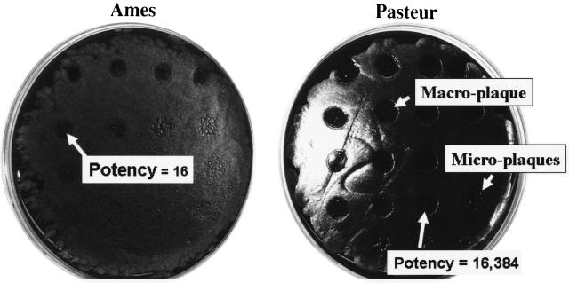
Potency is defined as the highest dilution of a gamma phage preparation which produces a distinct macroplaque (5 to 10 mm in diameter). It was determined by spotting 5 μl of twofold serial dilutions of a gamma phage test lot onto a lawn of a B. anthracis host strain lacking the pXO1 plasmid (i.e., Pasteur strain) and determining the PFU per milliliter using the dilution factor and the number of microplaques in the highest dilution showing microplaques (Fig. 1, arrow). In practice, phage lots were serially diluted twofold by transferring 50 μl of phage preparation in dilution tubes containing 50 μl of nutrient broth for 20 dilutions. Once prepared, the phage dilutions were held on ice while the bacterial cell inoculum was prepared in PBS from an overnight (18- to 20-h) culture growth of the B. anthracis Pasteur strain and either the Sterne or Ames strain to yield a turbidity approximately equal to a 0.5 McFarland standard. A volume (100 μl) of adjusted cell suspension was added to 5% SBA plates and spread over the surface of the agar using a glass or disposable spreader to create a bacterial lawn, and the fluid was allowed to absorb. The process was repeated with either the Sterne or Ames strain (Fig. 1). The dilutions of the gamma phage were added (5 μl) to premarked plates with up to 20 spots per plate. This process was repeated for the reference phage lots. The potency was determined using the last dilution of gamma phage which gave rise to a distinct macroplaque (5 to 10 mm in diameter), determined to be 1:16 for the Ames strain and 1:16,384 for the Pasteur strain (Fig. 1). The small microplaques (1 to 2 μm) within the site of application of higher dilutions permitted enumeration. As seen in Fig. 1 (bottom, right), for the Pasteur strain (see arrow), microplaques were counted, and the dilution factor was used to calculate the PFU per milliliter. For example, in Fig. 1, the gamma phage underwent 19 twofold dilutions for a final dilution of 1:262,144. In that dilution, 5 μl (1/200 of a milliliter) was spotted and four microplaques were counted in the last area of application (Fig. 1, arrow), and the PFU per milliliter was determined by an algorithm (4 PFU × 200 ml−1 × 262,144) to be 2.1 × 10⁸ PFU/ml.
FIG. 1.
Potency determination of B. anthracis strain Ames compared to that of the Pasteur strain. Nineteen twofold dilutions of gamma phage preparation were spotted identically on 5% sheep blood agar inoculated with B. anthracis strain Ames (left) versus strain Pasteur (right) and then incubated at 35°C for 18 h. Arrows identify a macroplaque and an area containing microplaques used for calculating PFU per milliliter.
Determination of the concentration by pour plate.
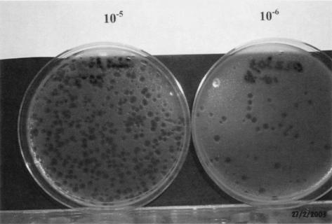
The PFU per milliliter were alternatively determined by serially diluting phage 10-fold and incubating phage in the presence of B. anthracis in semisolid LB top agar (1). Growth of B. anthracis Pasteur vaccine strain cultures, incubated 18 to 20 h at 35°C on SBA, was used to prepare a suspension comparable to a McFarland standard of 0.5 at 600 nm in PBS using a spectrophotometer. Aliquots (1.35 ml) of this suspension were prepared for each 10-fold dilution of phage (10−1 to 10−6) diluted in nutrient broth. Starting with the phage dilution of 10−3, 0.15 ml was transferred to each tube in a triplicate set of 1.35-ml cell suspensions, gently subjected to vortex mixing, and then allowed to react for 10 min at 35 ± 2°C. A total of 0.4 ml of phage/bacterial suspension was added to 9.6 ml of melted overlay agar (held at 53°C), gently mixed by inverting the tube several times, immediately poured into a sterile petri dish, covered, and allowed to solidify at room temperature. The process was repeated twice for each phage dilution to be tested. Control plates consisting of B. anthracis Pasteur strain devoid of phage were prepared. Cultures were then inverted and incubated at 35 ± 2°C. For each dilution, the presence of distinct phage microplaques was determined, and the total number of plaques was counted in the agar using a dilution plate that yielded 30 to 300 plaques (Fig. 2). The total PFU were calculated in the phage preparation.
FIG. 2.
Pour-plate plaque counts using B. anthracis strain Pasteur. The example shows 10−5 and 10−6 dilutions in pour plates.
Gamma phage lysis assay.
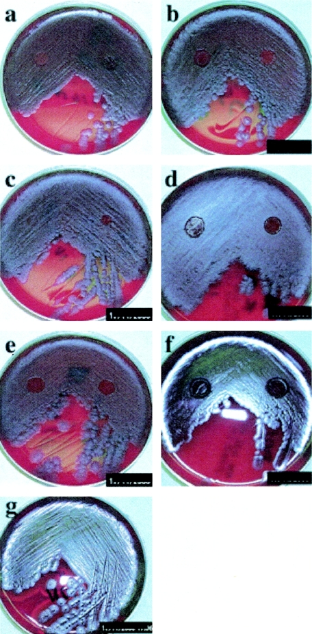
Culture isolates to be tested for gamma phage sensitivity were pure cultures or had well-defined single colonies in a mixed bacterial population. If culture integrity with respect to age or purity was in doubt, the culture was subcultured to produce isolated colonies on 5% SBA. Suspect colonies selected for testing were nonhemolytic, opaque, slightly raised, irregular (although round colonies can form) with serrated edges, and gray-white with a ground-glass appearance. Suspect colonies typically showed tenacity when the colony was probed with an inoculation loop or needle and “whipped up.” Although the assay was only validated here using vegetative culture growth from SBA cultures, spore suspensions with adequate concentration to yield confluent lawns could also be tested directly and backed up with the validated process (unpublished observation). Positive and negative control cultures were tested concomitantly. Inoculation of test samples and controls was standardized using a 1-μl loop, with which sufficient culture growth was removed to make an approximate 1-mm bead of cells, preferably from an individual colony. The growth was transferred to a fresh SBA plate by streaking a vertical line from the edge towards the center (approximately 1 in. in length) in the first quadrant. The inoculum was spread in close horizontal streaks across the vertical streak site in the first quadrant. Use of the flat side of a 10-μl disposable loop facilitated creation of a uniform inoculation. Using the same loop, several light sweeps were made across one end or edge of the first quadrant, and the close horizontal spreading was repeated in the second quadrant of the plate. This was continued as a streak for isolation in the remaining quadrants as a test of culture purity. Gamma phage suspension (5 μl) was placed on the agar surface in centers of both the first and second quadrants for each test and control plate (as shown in Fig. 3) using a clean micropipettor tip for each application. After replacing the plate lid, circles were drawn on the lid above the sites where phage was applied. The sides of the plate lid and bottom were marked to allow for realignment of the top and bottom before the plates were read postincubation. Agar cultures were incubated at 35°C ± 2°C for 20 ± 4 h. Acceptance criteria for positive assay results were that there must be a clear zone (macroplaque approximately 5 to 10 mm in diameter) of no growth where phage was applied to the positive control in either the first or second quadrant. (Note that it is possible for a few colonies to emerge within the clear zone on the positive control.) A lawn of confluent growth must be present in the first or second quadrants on all culture plates (controls and test unknowns), and quadrants 3 and 4 were inspected to confirm purity. A positive test yielded plaque formation (usually 5 to 10 mm in diameter) at the point of gamma phage application after 20 ± 4 h incubation. In practice, plaques are often seen in 4 to 8 h against the agar surface dulled by early bacterial growth around the site of gamma phage application.
FIG. 3.
Variations in gamma phage susceptibility among Bacillus anthracis strains. Shown is gamma phage macroplaque formation with various B. anthracis strains. (a) Sterne (pX01+, pX02−); (b) Colorado (pX01+, pX02+); (c) Ames (pX01+, pX02+); (d) Vollum 1B (pX01+, pX02+); (e) delta-Ames (pX01+, pX02−); (f) Pasteur (pX01+, pX02−) (positive control); (g) B. cereus (negative control).
Protocol, procedure documentation, and training.
The study was described prospectively by USAMRIID validation protocol (VP012). The test method was defined by a study-specific procedure (AN-D9-01) that was used throughout the study. Four analysts participated in the study and are identified as analysts A through D. Results were recorded on standard study data forms. Personnel were trained in the test method, were familiar with good laboratory practices, and were certified for biosafety level 3 laboratory operations.
Culture observations and gamma phage plaque formation were recorded on all plates by digital photography with a Kodak DC290 digital camera. All plates were photographed within 36 h of the end of the test. Pixel resolution was set to medium (1,140 × 960 = 1.382 megapixels), and picture format was uncompressed (TIFF). The camera was set to record date/time on the image and watermark text indicating the protocol number. Images were numbered with Kodak's absolute numbering method. Memory card number, image name, camera settings, and comments were recorded on data sheets. Image data were uploaded from compact flash cards through a personal computer and directly downloaded to a read-only compact disk. Data files were also backed up to an Iomega Zip Drive in picture albums (folders) as captured to the card.
Equipment and reference materials.
The bacteriological incubator, biological safety cabinets, and micropipettes were calibrated and maintained by the USAMRIID Medical Maintenance Branch and monitored by users. Digital camera and compact flash cards were used and monitored by the study director (J.E.B.).
Archiving.
Gamma phage and positive and negative control strains along with Bacillus species used for specificity testing and testing of precision parameters are maintained as part of the USAMRIID Diagnostic Systems Division bacterial reference library collection.
RESULTS
Selection of the gamma phage method as an identification method for B. anthracis required documentation of the performance characteristics of the assay. The method was assessed for sensitivity and specificity, precision parameters, and various elements of robustness.
Variation in gamma phage susceptibility.
A key finding of this study was that various B. anthracis strains may differ in their response to gamma phage and their sensitivity to bacterial inoculum level. Representative results are shown in Fig. 3. Susceptibility, as assessed by plaque formation, can vary as a function of the strain and plasmid profile. Fully virulent strains possessing both virulence plasmids tended to produce more clearly defined plaques in the second quadrant than in the first as shown in Fig. 3b to d in that they appeared to be more sensitive to inoculum overload. However, avirulent strains lacking the pX01 toxin plasmid (i.e., Pasteur strain) and delta-Ames routinely produced large, clearly defined plaques in both quadrants as shown in Fig. 3e and f. The Sterne strain, which lacks the pXO2 capsule-encoding plasmid but possesses the toxin-encoding pXO1 plasmid, formed more clearly defined plaques in the first quadrant. As previously stated, B. anthracis Pasteur (Fig. 3f) and B. cereus NCTC 2599 (Fig. 3g) served as positive and negative controls throughout the study.
Potency and determination of PFU per milliliter of gamma phage preparations.
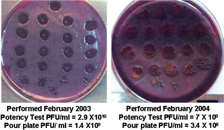
The previously mentioned variations in gamma phage susceptibility of Bacillus strains led to variations in determining the PFU per milliliter and potency of phage preparations. B. anthracis strains lacking the pXO1 plasmid (i.e., Pasteur and delta-Ames [Fig. 3e and f]) were more susceptible to the gamma phage than those possessing the plasmid (i.e., Sterne, Ames, etc. [Fig. 3a to d]). As a result, only strains lacking the pXO1 plasmid could be used for determining PFU per milliliter. When higher dilutions of phage were plated on the Pasteur strain, individual microplaques were readily detected as small 1- to 2-mm clear zones against confluent bacterial growth (Fig. 1, arrows); however, such plaques were not well formed with the Ames or Sterne strain. The use of the Pasteur strain allowed counting of the PFU per milliliter in preparations held over long periods of storage at 4 ± 2°C. The concentration of phage lot TA101201 was prepared in October 2001 and was tested in February 2002, 2003, and 2004 and found to contain 7.5 × 10⁸, 3.0 × 10⁸, and 3.23 × 10⁸ PFU/ml, respectively, by the pour-plate technique. Likewise, phage lot TA022303, prepared in February 2003, had a concentration of 1.4 × 10⁹ PFU/ml, and in February 2004, the concentration dropped to only 3.8 × 10⁸ PFU/ml. As shown in Fig. 4, the PFU per milliliter determined using the potency assay was 2.9 × 10¹⁰ PFU/ml on February 2003 and dropped to 7 × 10⁹ PFU/ml after a year. It is clear that there was less than a log10 loss in activity over 1 year of storage. As for potency determination, in which serial dilutions of phage were placed on a confluent inoculum of B. anthracis Sterne or Ames, a much higher concentration of gamma phage was required to generate macroplaques than that of Pasteur, which formed macroplaques and, ultimately, microplaques at much higher dilutions (Fig. 2 and 4). Using the potency assay, the counts for microplaques were typically higher than those determined using the pour-plate technique, a phenomenon which is not understood. In practice, the gamma phage potency of production lots was assessed using either the Ames or Sterne strain in combination with the Pasteur strain of B. anthracis. For laboratories acquiring gamma phage from outside sources, the potency test may be implemented as part of the laboratory quality control for monitoring gamma phage during long-term storage at 4°C. At a minimum, the Sterne strain or its equivalent should be used as a positive control since it more readily reflects the characteristics of fully virulent strains such as Ames.
FIG. 4.
Potency assay performed after 12 months of storage on phage lot TA022303 tested after its preparation in February 2003 and later in February 2004, with results for both the potency and pour plate presented for direct comparison.
Demonstration of precision parameters.
Repeatability was examined by testing multiple (n = 5) replicate tests of five independent B. anthracis strains (Table 2, analyst A, test 1). In the second quadrant, the test was 100% positive for all five test strains, whereas results were variable in the first quadrant where bacterial lawns were heaviest. Two strains, delta-Ames and Vollum 1B, tested consistently positive, but the Sterne and Ames strains tested consistently false negative in the first quadrant. Overall, first-quadrant results were 56% (14/25) positive for this test of intra-assay variability.
TABLE 2.
Precision within assay, between days, and between analysts for identification of B. anthracis with gamma phage lysis assay
| Assay set (B. anthracis strain) | No. of positives expected | No. of observed positives
|
% Positivea | |||||||||
|---|---|---|---|---|---|---|---|---|---|---|---|---|
| Quadrant 1 by analyst:
|
Quadrant 2 by analyst:
|
|||||||||||
| A
|
Bb | Cb | A
|
Bb | Cb | |||||||
| Test 1 | Test 2 | Test 3 | Test 1 | Test 2 | Test 3 | |||||||
| Sterne | 5 | 0 | 0 | 0 | 5 | 3 | 5 | 5 | 5 | 5 | 5 | 100 |
| Ames | 5 | 0 | 0 | 0 | 1 | 2 | 5 | 5 | 5 | 5 | 5 | 100 |
| delta-Ames | 5 | 5 | 5 | 5 | 5 | 5 | 5 | 5 | 5 | 5 | 5 | 100 |
| Vollum 1B | 5 | 5 | 5 | 5 | 5 | 5 | 5 | 5 | 5 | 5 | 5 | 100 |
| Colorado | 5 | 4 | 5 | 2 | 1 | 4 | 5 | 5 | 5 | 5 | 5 | 100 |
| Total | 25 | 14 | 15 | 12 | 16 | 19 | 25 | 25 | 25 | 25 | 25 | 100 |
Defined as positive in either the first or second quadrant.
Test 1.
Several components of intermediate precision were tested, such as day-to-day variability, analyst-to-analyst variability, and phage lot-to-lot variability. For day-to-day variability, tests performed by an analyst were 100% positive in the second quadrant over 3 days (Table 2, analyst A, tests 1, 2, and 3). In the first quadrant, results for tests 2 and 3 were similar to those seen in test 1, with 60% and 48% of plates positive. Over time, delta-Ames and Vollum 1B strains were consistently most sensitive in this quadrant, whereas Sterne and Ames strains were consistently among the least sensitive. These strains appeared to have heavy growth in the first quadrant. Comparison of results among three analysts showed similar patterns (Table 2, test 1 for analysts A, B, and C). All tests were 100% positive in the second quadrant, whereas first-quadrant results were 56%, 64%, and 56%, respectively, for the three analysts. Between analysts, the Sterne strain tested inconsistently in the first quadrant. Table 3 shows results of phage lot-to-lot variability using the Sterne strain as a test strain. In the second quadrant, the test was 100% positive for the three lots tested, and no lot provided acceptable results for the first quadrant (23% positive).
TABLE 3.
Effect of phage lot on intermediate precision for identification of B. anthracis by gamma phage lysis
| Assay set (lot) | No. of expected positives | No. of observed positives
|
% Positivea | |
|---|---|---|---|---|
| 1st quadrant | 2nd quadrant | |||
| 1 | 7 | 0 | 7 | 100 |
| 2 | 7 | 2 | 7 | 100 |
| 3 | 7 | 3 | 7 | 100 |
Defined as positive in either the first or second quadrant.
Demonstration of robustness parameters.
Three parameters of assay robustness were evaluated: phage concentration, inoculum size, and incubation time, using the Ames strain as the test strain for all tests. The effect of phage concentration was evaluated as a measure of phage activity or potency by comparing undiluted phage preparations to preparations diluted to 90%, 80%, 70%, and 60% of the original concentration (Table 4). All tests were acceptable at up to a 40% dilution (i.e., 60% of the original concentration) for second-quadrant observations. Again, first-quadrant results were 20 to 40% positive, with no observable trend. This is consistent with the findings that gamma phage preparations can be highly diluted and still form macroplaques (Fig. 1 and 4). When different bacterial inoculum quantities were compared, the data demonstrated that quantity, within the range tested, did not matter (Table 5). Inoculum volumes were approximately 1-, 2-, and 3-mm-diameter beads of inoculum. At all three levels, the assay was 100% positive in the second quadrant but 28% (6/21) positive under heavier bacterial lawn conditions in the first quadrant. The variation in macroplaque formation was shown in Fig. 3. Finally, the method was assessed after incubation for 8 to 24 h. The assay was acceptable after incubation periods as short as 8 h (Table 6). At 16 h, four of five replicates were positive; however, the one negative result at 16 h for the second quadrant was positive in the plate's first quadrant, nevertheless, giving an overall positive observation.
TABLE 4.
Effect of phage concentration on robustness of identification of B. anthracis by gamma phage lysis
| Assay set (% phage) | No. of expected positives | No. of observed positives
|
% Positivea | |
|---|---|---|---|---|
| 1st quadrant | 2nd quadrant | |||
| 100 | 5 | 1 | 5 | 100 |
| 90 | 5 | 1 | 4 | 80 |
| 80 | 5 | 2 | 5 | 100 |
| 70 | 5 | 2 | 5 | 100 |
| 60 | 5 | 1 | 5 | 100 |
Defined as positive in either the first or second quadrant.
TABLE 5.
Effect of inoculum size on robustness of identification of B. anthracis by gamma phage lysis
| Size (load) (mm) | No. of expected positives | No. of observed positives
|
% Positivea | |
|---|---|---|---|---|
| 1st quadrant | 2nd quadrant | |||
| 1 | 7 | 3 | 7 | 100 |
| 2 | 7 | 3 | 7 | 100 |
| 3 | 7 | 0 | 7 | 100 |
Defined as positive in either the first or second quadrant.
TABLE 6.
Effect of incubation time on robustness of identification of B. anthracis by gamma phage lysis
| Time (h) | No. of expected positives | No. of observed positives
|
% Positivea | |
|---|---|---|---|---|
| 1st quadrant | 2nd quadrant | |||
| 8 | 5 | 3 | 5 | 100 |
| 16 | 5 | 2 | 4 | 100 |
| 20 | 5 | 1 | 5 | 100 |
| 24 | 5 | 0 | 5 | 100 |
Defined as positive in either the first or second quadrant.
Demonstration of specificity.
Species sensitivity and specificity were considered the most important performance parameters of this assay. The assay was used to screen 50 independent, known strains of B. anthracis and 50 strains of non-B. anthracis Bacillus species. We intentionally included two nonsusceptible strains in the B. anthracis panel and two sensitive non-B. anthracis Bacillus strains in the non-B. anthracis panel (Table 1). Results initially appeared to correspond exactly with expected outcomes. However, we report 96% positive specificity for B. anthracis reflecting that two known nonsusceptible strains were not positive (Table 7). First-quadrant results were 72% positive. For the non-B. anthracis Bacillus strains, there were 48 of 50 strains that tested negative, demonstrating a specificity of at least 96%. However, in subsequent analysis, one of the two positive strains previously catalogued as a B. mycoides strain (CDC680) was found by molecular methods after this study to be a B. anthracis isolate devoid of both plasmids. Further analysis of strain CDC680 by Tetracore, Inc., Gaithersburg, Md., using their Red Line assay, revealed that the strain tested positive for B. anthracis. This identity was confirmed by Paul Jackson, Los Alamos National Laboratories, Los Alamos, N.M., and tested positive by PCR at USAMRIID for B. anthracis-specific chromosomal markers but was negative for both virulence plasmids pXO1 and PXO2 (personal communication with D. Norwood and M. Frye, USAMRIID). B. cereus ATCC 4342 was negative for chromosomal, pX01, and pX02 PCR targets, whereas the two B. anthracis strains that were resistant to gamma phage were positive for the B. anthracis-specific chromosomal target sequence and for pXO1. Additionally, one of the isolates was positive for pXO2, and the other was negative. For this strain, there was no observed lysis in the second quadrant, whereas it was positive in the first quadrant. Recalculated results indicate 98% (48/49) negative specificity for non-B. anthracis strains. Similar results were obtained when plates were read independently by a second reader (analyst D) on the same day (Table 7). Identities of the resistant B. anthracis strains are withheld from open publication. Information on the identity of these nonsusceptible strains may be obtained by authorized persons upon request (Commander, USAMRIID, 1425 Porter Street, Fort Detrick, MD 21702-5011).
TABLE 7.
Sensitivity and specificity of identification of B. anthracis by gamma phage lysis
| Analysts | Isolate (no.) | No. of expected positives | No. of observed positivesd
|
% Positivea | |
|---|---|---|---|---|---|
| 1st quadrant | 2nd quadrant | ||||
| A and D | B. anthracis (50) | 50 | 36/47 | 48/50b | 96 |
| A and D | Non-B. anthracis (50) | 0 | 2/2c | 1/1c | 2 |
Defined as positive in either the first or second quadrant.
Two of 50 B. anthracis isolates were known to be nonsusceptible and were included in this study for completeness.
Two of 50 non-B. anthracis Bacillus isolates were known to be sensitive and were included for completeness. One of the strains, Bacillus mycoides CDC680, was identified by Beverly Mangold (Tetracore, Inc.) and Paul Jackson (Lawrence Livermore National Laboratories) as a plasmid-deficient Bacillus anthracis strain.
Values are observed results of analyst A/observed results of analyst D.
Systems suitability considerations.
The data demonstrate a tendency for analysts to streak too heavily when spreading a bacterial inoculum. This behavior is noticeable when one compares the difference in response rates between the first and second quadrants. In many circumstances, first-quadrant lawns were too heavily streaked to observe phage lysis. On average, first-quadrant results were not acceptable when considered alone at any time point. The assay reliability depended on observation of second-quadrant results. Analysts should ensure that bacterial lawns are not streaked too heavily in the first quadrant. In contrast, some strains of B. anthracis may not grow to a confluent lawn in the second quadrant under assay conditions. For these isolates, testing phage sensitivity in the first quadrant may provide a satisfactory alternative.
B. anthracis Pasteur proved to be an excellent positive control strain. It was 100% positive in both the first and second quadrants of the assay throughout the study. B. cereus was 100% negative for sensitivity to phage over the course of the study and was therefore an excellent negative control strain. Although the Pasteur strain performed well in this study, it may not serve as well for day-to-day use of the assay as discussed below.
DISCUSSION
The classical gamma phage identification assay for B. anthracis was standardized and evaluated for routine test laboratory use. The acceptance criteria set forth in validation protocol USAMRIID VP012 required at least an 80% expected response for precision and robustness characteristics. For sensitivity and specificity, acceptance criteria were 90% of the expected response, a response set high to reflect the importance of correctly identifying tested isolates. The data in Tables 2 to 7 show that the acceptance criteria were met or exceeded. The assay exhibited a high level of precision for identical strains and between strains on the same day of testing. It displayed high reproducibility for day-to-day and analyst-to-analyst comparisons (Table 2) and produced high precision between the three lots of phage tested (Table 3). The robustness portion of the protocol tested for variations in phage activity (concentration), phage inoculum size, and assay incubation time (Tables 4 to 6). The assay was unaffected by these parameters within the limits tested. Sensitivity and specificity were tested by screening 51 independent B. anthracis and 49 other Bacillus species and strains (Table 7). Two sensitive non-B. anthracis strains, B. cereus ATCC 4342 and B. mycoides CDC680, along with two nonsusceptible strains of B. anthracis were included in this study (Table 1). As mentioned above, B. mycoides CDC680 was subsequently found to be a B. anthracis strain lacking both plasmids, thereby altering the original design of the study from 50 B. anthracis and 50 non-B. anthracis strains to 51 and 49 strains, respectively. Overall, the assay passed all validation acceptance criteria and was determined to be fit for its intended use and for screening of putative culture isolates to identify B. anthracis.
As stated, the choice of B. anthracis strain for use in measuring potency has been controversial due to gamma phage sensitivity variations among strains and bacterial inoculum size. As shown in Fig. 3, strains respond differently to heavy and light bacterial inocula. Those strains possessing the pXO1 plasmid give more distinct clear plaques in the second quadrant, whereas those strains lacking the pXO1 plasmid produce distinct plaques in both quadrants. These and other observations of variation in phage susceptibility provided the basis for application of phage to both the first and second quadrants as a standard operating procedure. McCloy (9) made similar observations in which she reported that her “phage preparation had an appreciable action only if the bacterial inoculum was large,” whereas “if the inoculum was smaller, the phage produced no detectable effect after 18 h.” She goes on to state that “there were sometimes minor differences in the degree to which it (bacterial strain) was attacked; these differences might perhaps have been due the fact that the bacterial inoculum was not standardized.”
It is obvious from these studies that an accurate assessment of phage preparation activity and quality control requires the use of both pXO1-positive and pXO1-negative strains. The negative strains (such as the Pasteur strain) allow accurate plaque counts, whereas the pXO1-positive strains assess the potency of the preparation to identify fully virulent strains such as the Ames strain. The basis of the increased sensitivity of strains lacking the pXO1 plasmid compared to that of positive strains is not understood. As the pXO2 plasmid appears to have no effect on the response to the gamma phage, avirulent strains such as the Sterne strain can be used with the avirulent Pasteur strain to determine potency and to perform quality control assessments. Recent data, not reported here, suggest that the effect of the pXO1 plasmid may be more indirect. It has been our observation and that of others that strains possessing the pXO1 plasmid sporulate more readily than those lacking pXO1. As was seen in this study (data not shown), Gram-stained 18- to 20-h growth from pXO1-positive strain cultures on SBA showed high numbers of spores, whereas those from Pasteur-like strains lacking pXO1 typically showed very little, if any, spores. The net result is that growth material used from primary culture of a pXO1-positive strain to test for gamma phage sensitivity would contain larger amounts of spores, cell wall debris, and dead cells containing endospores than material from a pXO1-negative strain. In theory, when gamma phage is added to the inoculated surface, much of the phage may be binding to receptors in this debris and once bound are incapable of infecting bacilli later emerging from the germinating spores. However, when inoculated onto Pasteur-like strains, there is minimal competing debris, and phage binds to and infects viable bacilli. The net result is that fewer phages are required to generate a plaque. This is supported by our observation that when washed Sterne spores were used as the inoculum rather than growth from primary culture, clearly defined macroplaques were generated at much higher dilutions, similar to those of the Pasteur strain.
To date, gamma phage prepared using the host strain, B. anthracis CDC684, and filter sterilized using low-protein-binding 0.22-μm filters to remove bacteria has shown remarkable stability over 2 years while being refrigerated at 4°C. This is contrary to the previous findings of others (6), who found it necessary to frequently prepare fresh batches of gamma phage. The stability of gamma phage preparations produced as described here allows individual gamma phage preparations to be employed for long-term use and dispersal to multiple laboratory locations with minimal loss in activity. At this juncture, several preparations have undergone analysis for potency and pour-plate PFU per milliliter determinations. Phage lot TA101201, prepared in October 2001, showed less than a log10 drop in PFU concentration from 2001 to 2004 and retained its activity towards fully virulent B. anthracis strains when tested for potency. Likewise, other lots such as TA022303 also showed less than a log10 decrease from February 2003 to February 2004, whether measured using the potency or the pour-plate assay. Since there is less than a log10 decrease in potency, it is reasonable to assign a preparation an expiration date of at least a year if it produces distinct macroplaques when diluted 1:8 to 1:10 or more in nutrient broth and applied to the first and second quadrants of a test strain possessing the pXO1 plasmid, such as Sterne. In general, our preparations are sufficiently stable for 18 months to 2 years. According to an unpublished method dated January 1971, originally provided by W. Cherry, Analytical Bacteriology Unit, CDC, acceptable phage preparations can be used either undiluted or diluted 1:10, and phage preparations having concentrations less than 107 PFU/ml should not be used. Our observations were consistent with those statements.
The findings show the reliability of this simple biological assay for identifying B. anthracis. However, we continue to stress that confirmatory tests should be used along with this gamma phage assay for positive identification. Validation studies showed lytic behavior to be as expected for all isolates, giving an assay specificity of at least 98% for B. anthracis. Gamma phage application in the two quadrants was an essential element of robustness and tolerated variation in inoculum load. Finally, B. anthracis strain CDC684 appears to be an excellent host strain for production of batches of high-titer, stable gamma phage when produced in the manner described here.
International events have focused attention on methods for reliable and affordable methods for detecting microorganisms of biowarfare or bioterrorism concern. In 2002, the Food and Drug Administration Division of Clinical Laboratory Devices recognized that assay methods for B. anthracis and Yersinia pestis were eligible for classification as preamendment in vitro diagnostic products. Recently, AOAC International has recognized the method using gamma phage lysis and direct fluorescence assay as satisfactory for first action as an AOAC Official Method of Analysis (Scott Coates, personal communication).
Acknowledgments
We thank Stephanie Redus and Renee Aguirre for acting as additional analysts during demonstrations of intra-assay precision. We also thank USAMRIID editor Katheryn Kenyon for her editorial comments. We thank Beverly Mangold, Paul Jackson, Melissa Frye, and David Norwood for experimental assistance.
Opinions, interpretations, conclusions, and recommendations are those of the authors and are not necessarily endorsed by the U.S. Army in accordance with (IAW) AR 70-31.
REFERENCES
- 1.Atlas, R. M. 1997. LB medium, p. 748. In L. C. Parks (ed.), Handbook of microbiological media, 2nd ed. CRC Press, Inc., Boca Raton, Fla.
- 2.Baron, S. (ed.). 1991. Medical microbiology, 3rd ed., p. 259. Churchill Livingston, New York, N.Y.2030501
- 3.Boyd, R. F., and B. G. Hoerl. 1991. Basic medical microbiology, 4th ed., p. 442. Little, Brown and Co., Boston, Mass.
- 4.Brown, E. R., and W. B. Cherry. 1955. Specific identification of Bacillus anthracis by means of a variant bacteriophage. J. Infect. Dis. 96:34-39. [DOI] [PubMed] [Google Scholar]
- 5.Brown, E. R., M. D. Moody, E. L. Treece, and C. W. Smith. 1958. Differential diagnosis of Bacillus cereus, Bacillus anthracis, and Bacillus cereus var. mycoides. J. Bacteriol. 75:499-509. [DOI] [PMC free article] [PubMed] [Google Scholar]
- 6.Buck, C. A., R. L. Anacker, F. S. Newman, and A. Eisenstark. 1963. Phage isolated from lysogenic Bacillus anthracis. J. Bacteriol. 85:1423-1430. [DOI] [PMC free article] [PubMed] [Google Scholar]
- 7.Cowles, P. B. 1931. A bacteriophage for B. anthracis. J. Bacteriol. 21:161-169. [DOI] [PMC free article] [PubMed] [Google Scholar]
- 8.Ezzell, J. W., T. G. Abshire, S. F. Little, B. C. Lidgerding, and C. Brown. 1990. Identification of Bacillus anthracis by using monoclonal antibody to cell wall galactose-N-acetylglucosamine polysaccharide. J. Clin. Microbiol. 28:223-231. [DOI] [PMC free article] [PubMed] [Google Scholar]
- 9.McCloy, E. W. 1951. Studies on a lysogenic Bacillus strain. I. A bacteriophage specific for Bacillus anthracis. J. Hyg. 49:114-125. [DOI] [PMC free article] [PubMed] [Google Scholar]
- 10.McCloy, E. W. 1951. Unusual behavior of a lysogenic Bacillus strain. J. Gen. Microbiol. 51:xiv-xvi. [PubMed] [Google Scholar]
- 11.Schuch, R., D. Nelson, and V. A. Fischetti. 2002. A bacteriolytic agent that detects and kills Bacillus anthracis. Nature 418:825-826. [DOI] [PubMed] [Google Scholar]
- 12.Sneath, P. H. A. 1986. Endospore-forming gram-positive rods and cocci, p. 1131. In P. H. A. Sneath, N. S. Mair, M. E. Sharpe, and J. G. Holt (ed.), Bergey's manual of systematic bacteriology, vol. 2. Williams & Wilkins, Baltimore, Md. [Google Scholar]
- 13.United States Pharmacopeial Convention, Inc. 1999. Chapter 1225. Validation of compendial methods, United States Pharmacopeia, 10th Supplement, p. 5059-5062. United States Pharmacopeial Convention, Inc., Rockville, Md.


