Abstract
Diagnostic systems based on reverse transcription (RT)-PCR are widely used for the detection of viral genomes in different human specimens. The application of internal controls (IC) to monitor each step of nucleic acid amplification is necessary to prevent false-negative results due to inhibition or human error. In this study, we designed various real-time RT-PCRs utilizing the coliphage MS2 replicase gene, which differ in detection format, amplicon size, and efficiency of amplification. These noncompetitive IC assays, using TaqMan, hybridization probe, or duplex scorpion probe techniques, were tested on the LightCycler and Rotorgene systems. In our approach, clinical specimens were spiked with the control virus to monitor the efficiency of extraction, reverse transcription, and amplification steps. The MS2 RT-PCR assays were applied for internal control when using a second target hepatitis C virus RNA in duplex PCR in blood donor screening. The 95% detection limit was calculated by probit analysis to 44.9 copies per PCR (range, 38.4 to 73.4). As demonstrated routinely, application of MS2 IC assays exhibits low variability and can be applied in various RT-PCR assays. MS2 phage lysates were obtained under standard laboratory conditions. The quantification of phage and template RNA was performed by plating assays to determine PFU or via real-time RT-PCR. High stability of the MS2 phage preparations stored at −20°C, 4°C, and room temperature was demonstrated.
Reverse transcription (RT)-PCR is used widely as a diagnostic tool to detect, quantify, or differentiate viral RNA (11). An inherent problem in diagnostic PCR is the presence of amplification inhibitors which may cause false-negative results. Therefore, the addition of an amplifiable nucleic acid in the PCR assay serves as an internal control (IC). The use of an IC is an important quality control and has already been described for early PCR experiments (for reviews, see references 8 and 11). An IC for diagnostic RT-PCR assays should be easy to produce and to standardize. Additionally, ICs should be stable, noninfectious, absent from clinical samples, and suitable for different assays.
An endogenous IC is a template that occurs naturally within the specimen being analyzed. In gene expression analysis and virus screenings, housekeeping genes are often used as ICs and references for transcript quantification (7, 16), but they have to be proven for each experiment and target. Exogenous ICs are added before nucleic acid isolation (extraction control) or amplification (amplification control), where coamplification is performed within the same reaction. Ideally, these ICs hybridize to the same primers, have identical amplification efficiencies, and contain discriminating features, such as length or sequence variations, targeted by hybridization probes. However, these competitive ICs can lower the amplification efficiency, which results in a lower detection limit (8). Therefore, noncompetitive IC templates are used, where the target and IC are amplified with different primer sets. The disadvantage is that amplification of the IC may not accurately reflect amplification of the target.
Currently, most diagnostic assays in which viral RNA is detected or quantified rely on RNA standards (19). Some assays use RNA standards synthesized by in vitro transcription, which are very susceptible to degradation by RNases. Therefore, the armored RNA technology produces RNase-resistant RNA controls and standards by assembling specific RNA sequences and viral coat proteins to pseudoviral particles (6, 15). This IC RNA contains the same primer binding sites as the target RNA but has a different probe region (3, 6).
For viral nucleic acid amplification tests (NAT), the detection of model viruses has been described. In these approaches, clinical specimens were spiked with a known amount of an animal virus (4, 13, 23) to monitor the efficiency of extraction, reverse transcription, and amplification. The advantage of such model viruses is the stability of RNA and the control of decapsulation of the viral RNA during the extraction procedure. The production of these animal pathogenic viruses may raise issues of safety, and virus cultivation needs substantial technical skill. Therefore, it should be demanded that the preparation of the control viruses be performed under standard laboratory conditions. Regarding this requirement, it is simple to establish the cultivation of Escherichia coli bacteriophages, such as Q-beta or MS2, in every laboratory. The F+-specific coliphage MS2 has been widely used as a surrogate for human enteric viruses in many studies on virus transport, disinfection, and fate (1, 5, 17, 20, 21). Here we present an MS2 NAT for different real-time RT-PCR approaches, which was used to monitor RT-PCRs for the detection of human RNA viruses.
MATERIALS AND METHODS
Phage isolation.
Phage MS2 DSM13767 was purchased from DSMZ (Braunschweig, Germany). MS2 was grown in F plasmid-harboring E. coli strain XL10-Gold (Stratagene, La Jolla, CA) by the double-agar-layer plaque technique (2). The top agar layer showing confluent lysis of the host cells was harvested by being scraped into a small amount of suspension medium (SM) (0.1 M NaCl, 8 mM MgSO4, 0.05 M Tris-HCl, pH 7.5, 0.01% [wt/vol] solid gelatin). The virus particles were extracted with an equal volume of chloroform, and the supernatant was recovered by low-speed centrifugation (4,000 × g) for 30 min at 4°C. Finally, the virus particles were diluted in SM buffer and stored at −20°C for further analysis.
The PFU were determined by plating assays. The MS2 RNA was quantified (copies per ml) with a genomic MS2 RNA purchased from Roche Diagnostics (Mannheim, Germany). RNA quantitation was carried out with a sensitive fluorescence-based solution assay for RNA, using RiboGreen RNA quantitation reagent (Molecular Probes, Leiden, The Netherlands) as described by the manufacturer. The MS2 stock solution contained at least 6 × 10¹⁰ PFU per ml and was quantified to 6 × 10¹² copies per ml. As an internal control of RT-PCR, MS2 phage dilutions were spiked into plasma pools.
Nucleic acid isolation.
RNA was extracted from 140 μl plasma with a QIAamp viral RNA kit (QIAGEN, Hilden, Germany) according to the manufacturer's protocol. The RNA was eluted with 60 μl AVE buffer (QIAGEN). For blood donor screening NAT, RNA was prepared from EDTA plasma of volunteer blood donors spiked with MS2 phage by using a QIAamp UltraSens virus kit (QIAGEN). For this purpose, the working lysis solution was prepared with 5.6 μl carrier RNA, 50 μl MS2 phage lysate containing 6 × 10⁴ PFU per ml (final concentration of 3,000 PFU MS2 per ml of plasma), and 800 μl AC buffer (QIAGEN). Total nucleic acid from up to 1 ml of plasma was eluted in 60 μl AVE buffer, of which 15 μl was applied to the RT-PCR. Each extraction run included a negative control plasma and two low-copy positive run controls for the corresponding RT-PCR assay.
Primer and probe design.
The oligonucleotides were designed by utilizing OLIGO 5.0 primer analysis software (National Biosciences, Plymouth, Minn.), Primer Express software (Applied Biosystems, Darmstadt, Germany), and LightCycler Probe Design Software, v. 1.0 (Roche Diagnostics, Germany). The degree of nucleotide sequence homology was checked by using the BLAST algorithm (www.ncbi.nlm.nih.gov/BLAST), which searches the EMBL, GenBank, and DDBJ databases.
Real-time RT-PCR using hybridization probes.
A Superscript II one-step RT-PCR with Platinum Taq kit (Invitrogen, Karlsruhe, Germany) was used as the basis for the reaction mixture in the LightCycler (LC) RT-PCR assay. A volume of 20 μl was used in each reaction capillary. An aliquot of 5 μl of the RNA was added to 15 μl of the reaction mixture containing 1× Reaction Mix (Invitrogen), 4.5 mM MgSO4 (Invitrogen), 500 ng per μl nonacetylated bovine serum albumin (BSA) (Sigma-Aldrich, Taufkirchen, Germany), 600 nM of forward primer KY-78s (5′-CAA GCA CCC TAT CAG GCA GT), 600 nM of reverse primer KY-80s (5′-AGC GTC TAG CCA TGG CGT), 250 nM of donor probe HCV-3FL (GCA GCC TCC AGG ACC CCC C-FAM [6-carboxyfluorescein]), 250 nM of acceptor probe HCV-5LC (5′-LC Red 640 [LightCycler-Red 705-phosphoramidite]-CCC GGG AGA GCC ATA GTG GTC TG-Ph [3′-phosphate]) (18), 300 nM of each MS2 primer (MS2-2717F and MS2-3031R), 50 nM of each MS2 probe (MS2-FL and MS2-LC) (Table 1), and 0.6 μl RT-Platinum Taq mix (Invitrogen). In addition to the positive run control, each test run included one no-target control containing 15 μl of the reaction mixture and 5 μl PCR-grade water. The reaction capillaries were capped, centrifuged, and placed into the carousel of the LightCycler instrument (Roche Diagnostics).
TABLE 1.
Primers and probes used for RT-PCR
| Primer or probe | Type | PCR systema | Sequenceb | Positionc |
|---|---|---|---|---|
| MS2-2717F | Sense | LC | CTGGGCAATAGTCAAA | 2717-2732 |
| MS2-3031R | Antisense | LC | CGTGGATCTGACATAC | 3031-3016 |
| MS2-LC-FL | Donor probe | LC | GACAATCTCTTCGCCCTGATGC-[FL] | 2955-2976 |
| MS2-LC-LC | Acceptor probe | LC | [Red 705]-ATATTAAATCGGCTACGGGGTTGG-[Ph] | 2979-3002 |
| MS2-TM2-F | Sense | TM2 RG | TGCTCGCGGATACCCG | 3169-3184 |
| MS2-TM2-R | Antisense | TM2 RG | AACTTGCGTTCTCGAGCGAT | 3229-3210 |
| MS2-TM3-F | Sense | TM3 LC/RG | GGCTGCTCGCGGATACCC | 3166-3183 |
| MS2-TM3-R | Antisense | TM3 RG | TGAGGGAATGTGGGAACCG | 3367-3349 |
| MS2-TM2JOEd | TaqMan probe | TM2/3 RG | [JOE]-ACCTCGGGTTTCCGTCTTGCTCGT-[BHQ1] | 3186-3209 |
| Sc-MS2-3R | Biprobe scorpion | LC/RG | [ROX]-CGTTA[X]ACGCACTTCGGAGTGGCTA-[HEG]- TGAGGGAATGTGGGAACCG | |
| QSc-MS2-3R | Quencher scorpion | LC/RG | ACTCCGAAGTGCGT[Z]TAACG-[Q] |
LC, LightCycler; RG, Rotorgene; TM2 and TM3, TaqMan RT-PCR assays 2 and 3, respectively.
[FL], fluorescein; [Red 705], LightCycler-Red 705-phosphoramidite; [Ph], 3′-phosphate; [JOE], 6-carboxy-4′,5′-dichloro-2′,7′-dimethoxyfluorescein; [ROX], carboxy-X-rhodamine; [BHQ1], black hole quencher 1; [Q], dR methyl red; [HEG], hexaethylene glycol; [X], FAM Cap Prop dU; and [Z], methyl red Prop deaza dA.
Positions according to bacteriophage MS2 (accession number V00642).
Reporter dye HEX was used for detection on the LightCycler platform.
The LC RT-PCR protocol included the following parameters: an initial 1-min incubation at 50°C, followed by 10 min at 48°C (reverse transcription), 2 min at 95°C (Taq DNA polymerase activation), subsequent 40 cycles at 95°C for 2 s, annealing and fluorescence detection at 50°C for 12 s, and extension at 72°C for 10 s. The data were obtained during the annealing period in the “single” mode with the channel settings F2/F1 (hepatitis C virus [HCV] specific) and F3 (internal control MS2), respectively.
Real-time RT-PCR using scorpion primers.
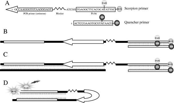
The specific fluorescence resonance energy transfer scorpion primer for MS2 is shown in Fig. 1. The oligonucleotide consists of a probe region with a 5′-ROX (carboxy-X-rhodamine) dye, an internal fluorescein (FAM), a PCR blocker (HEG [hexaethylene glycol]), and a 3′-PCR primer sequence. The scorpion was quenched by the second primer reverse complementary to the probe region of the scorpion. The primer QSc-MS2-3R was labeled with the dark quencher methyl red twice, once at its 3′ end and once internally. Each reaction mixture contained 1× Reaction Mix (Invitrogen), 4.5 mM MgSO4, 500 ng per μl nonacetylated BSA (Sigma-Aldrich), 300 nM of scorpion primer Sc-MS2-3R, 900 nM of quencher primer QSc-MS2-3R, 300 nM of forward primer MS2-TM3-F (Table 1), and 0.6 μl per 20 μl RT-Platinum Taq mix (Invitrogen). The PCRs were carried out on the LightCycler instrument. The cycling conditions were as follows: reverse transcription at 50°C for 10 min, denaturation at 95°C for 2 min, 40 cycles at 95°C for 0 s, annealing at 50°C for 3 s, monitoring at 60°C for 4 s (channel F2), and elongation at 72°C for 5 s. A single fluorescence measurement was made in each cycle during the monitoring step.
FIG. 1.
Duplex scorpion structure. The elements of the MS2 duplex scorpion showing the probe sequence (box), primer (arrow), PCR blocker, and fluorophores FAM and ROX (A). Excitation of the ROX dye is mediated by the emission of FAM (fluorescence resonance energy transfer scorpion). The quencher oligonucleotide is reverse complementary to the probe sequence and labeled internally and at the 3′ end with the dark quencher methyl red (MR) (B). After amplification, the scorpion primer is incorporated into the amplicon, while the cDNA strand is terminated by the PCR blocker that prevents separation of the scorpion quencher primer complex (C). During the next cycle, the probe region of the scorpion hybridizes intramolecularly to the newly synthesized target sequence (D).
TaqMan real-time RT-PCR.
For the LightCycler RT-PCR assay, an aliquot of 5 μl RNA was added to 15 μl of the reaction mixture containing 1× Reaction Mix (Invitrogen), 4.5 mM MgSO4, 500 ng per μl nonacetylated BSA (Sigma-Aldrich), 500 nM of forward primer HCV-C53-F (5′-AYCACTCCCCTGTGAGGAACT), 600 nM of reverse primer HCV-C33-R (5′-GGKCCTGGAGGYTGYACG), 200 nM of probe HCV-CTPR (5′-FAM-TGTCTTCACGCRGAAAGCGTCTAGCCAT-BHQ1 [black hole quencher 1]) (12), 300 nM of each MS2 primer (MS2-TM3-F and MS2-TM3-R), and 100 nM of dual-labeled probe MS2-TM2 (HEX and TAMRA [6-carboxytetramethylrhodamine]) (Table 1). Reaction capillaries were loaded, centrifuged, and placed in the carousel of the LightCycler 2.0 instrument. The RT-PCR protocol was as follows: reverse transcription for 1 min at 50°C and 10 min at 42°C and denaturation for 2 min at 95°C were followed by 40 cycles of 2 s for denaturation at 95°C, 20 s for annealing at 55°C, and 30 s for extension at 65°C. Temperature transitions were set to 20°/s. The fluorescence was measured once per cycle in the 530-nm channel (FAM) and the 555-nm channel (HEX) channel at 55°C to generate amplification plots.
The Rotorgene RT-PCR assay reactions were performed in a volume of 50 μl including 15 μl nucleic acid extract. The reaction mixture was done as described above, but without BSA, and the MS2 probe was labeled with the reporter dye JOE (6-carboxy-4′,5′-dichloro-2′,7′-dimethoxyfluorescein). Cycling conditions were 50°C for 10 min and 95°C for 2 min, followed by 45 cycles at 95°C for 10 s and 60°C for 45 s. Amplification, detection, and data analysis were performed with the Rotorgene 3000 cycler system (Corbett Research, Sydney, Australia). This HCV/MS2 RT-PCR assay was validated and compared with the RealArt HCV RG RT-PCR reagents on the Rotorgene 3000 (Artus GmbH, Hamburg, Germany). This assay is validated for HCV RNA screening of blood donations according to the criteria released by the Paul Ehrlich Institute, the federal licensing agency of Germany, for routine NAT.
Stability of the MS2 phage.
Purified MS2 phage with 1.7 × 10⁵ PFU per ml (1.7 × 10⁷ copies per ml) SM buffer was aliquoted in single-time-point samples of 0.2 ml. Samples were stored at the assigned temperatures until they were assayed. In this study, three different storage temperatures (−20, 4, and 22°C) were examined and samples were assayed in quadruplicate at every time point from day 1 to day 7. RNA extraction of 140-μl samples was performed with a QIAamp viral RNA kit (QIAGEN). RNA samples were stored at −80°C. All of the samples were assayed in duplicate with real-time RT-PCR in a single run on the Rotorgene platform.
Probit analysis on experimental data.
Probit analysis to determine the lower detection limit of NAT assays was performed using SPSS 10.0 software (SPSS GmbH Software, Munich, Germany).
RESULTS
General strategy for controlling real-time RT-PCR.
In order to establish an internal control system for clinically relevant RT-PCR assays, we designed four different RT-PCRs targeting the replicase gene of MS2. The primer and probe systems differ in kind of detection, amplicon size, and amplification efficiency. In order to obtain accurate and reproducible results, reactions should have an efficiency as close to 100% as possible. At this efficiency, the template doubles after each cycle during exponential amplification. For determination of efficiencies of MS2 RT-PCR assays, the slopes of standard curves were used (Fig. 2).
FIG. 2.
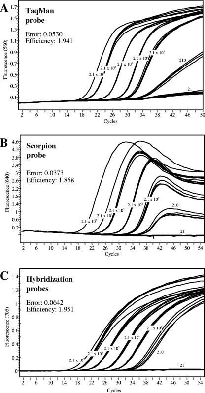
MS2 RT-PCRs with different probe formats. RT-PCR was performed on the LightCycler 2.0 with TaqMan (A), duplex scorpion (B), or hybridization (C) probes, respectively. MS2 RNA (Roche Diagnostics) was spiked into the RT-PCRs in a range of 21 to 2.1 × 10⁷ copies per reaction. Samples were assayed in quadruplicate at every concentration, and amplification efficiencies were determined by the slopes of standard curves. CT values were determined by the second derivative maximum method.
Two TaqMan RT-PCRs were tested on the Rotorgene 3000 platform with the same TaqMan probe, although the amplicons differed in size. While the amplicon of the MS2-TM2 system is 80 bp in size, the length of the product of the MS2-TM3 RT-PCR is 203 bp. In real-time RT-PCR with these MS2 TaqMan systems, the cycle threshold (CT) values vary by five to six cycles. When using RNA preparations spiked with the same amount of MS2 phage, the mean value ± standard deviation of the MS2-TM3 RT-PCR is 36.2 ± 0.4, compared to 30.5 ± 0.3 of the MS2-TM2 RT-PCR (n = 8). With HEX as the reporter dye, both RT-PCR systems were used on the LightCycler 2.0 platform, and multiplexing with a second FAM-labeled TaqMan probe was possible. In addition, an MS2 hybridization probe system was constructed and used as an IC on the LightCycler. Fluorescence detection was performed in channel F3, whereas the second target was detected in channel F2 by using LC Red640 as the acceptor fluorescence dye. Additional self-probing amplicons were generated with a duplex scorpion, where the quencher was on a different oligonucleotide to the scorpion primer (Fig. 1).
The comparison of the three probe formats with MS2 RNA demonstrated very good reaction efficiencies in a range of 1.868 to 1.951 (Fig. 2), which is near the optimal PCR efficiency. The MS2 RT-PCR assays were suited for internal control when using a second target (e.g., HCV RNA) in duplex PCR (data not shown).
Sensitivity of the MS2 RT-PCR assay.
To determine the analytical sensitivity of the MS2 assay, we used human plasma spiked with different MS2 titers from 1.5 × 10³ to 1.5 × 10⁵ copies per ml plasma, corresponding to 21 to 2,097 copies of MS2 per RT-PCR. Eight plasma pools of each concentration were processed through all steps of nucleic acid isolation and RT-PCR. The 95% detection limit was calculated by probit analysis to 44.9 copies per PCR (range, 38.4 to 73.4) when using the MS2 TaqMan assay on the Rotorgene 3000.
Stabilities of the MS2 phage.
We investigated the stabilities of the MS2 phage preparations stored at −20°C, 4°C, and room temperature for 7 days. The MS2 phage aliquots were used for RNA isolation and assayed for MS2 RNA with RT-PCR. Starting from 1.9 × 10⁵ PFU per ml, the copy number was determined to 1.9 × 10⁴ copies per RT-PCR. The CT values were compared to the starting values (Table 2). Probes stored at the three different temperatures showed no significant loss in copy number over the time period analyzed. Long-time storage (over 6 months) of MS2 phage in SM buffer at −20°C showed no decline of copy number compared to the original input (data not shown).
TABLE 2.
Stabilities of MS2 phage at different incubation temperaturesa
| Incubation time (days) |
CT mean ± SD at:
|
||
|---|---|---|---|
| −20°C | +4°C | +22°C | |
| 0 | 29.98 ± 0.06 | 29.98 ± 0.06 | 29.98 ± 0.06 |
| 1 | 29.65 ± 0.07 | 30.09 ± 0.12 | 30.35 ± 0.04 |
| 2 | 29.87 ± 0.16 | 30.08 ± 0.33 | 29.74 ± 0.17 |
| 3 | 29.82 ± 0.24 | 30.11 ± 0.06 | 30.05 ± 0.27 |
| 4 | 30.17 ± 0.28 | 30.35 ± 0.10 | 30.15 ± 0.03 |
| 5 | 29.85 ± 0.17 | 29.99 ± 0.31 | 30.09 ± 0.17 |
| 6 | 30.16 ± 0.41 | 30.00 ± 0.15 | 29.72 ± 0.28 |
| 7 | 30.24 ± 0.23 | 29.45 ± 0.28 | 29.98 ± 0.14 |
MS2 phage (1.7 × 10⁵ PFU per ml [1.7 × 10⁷ copies per ml]) was incubated in SM buffer for 7 days at different temperatures. The determination of stability was performed by the MS2 TaqMan assay on the Rotorgene platform. Each sample was assayed in quadruplicate, and standard deviations (SD) were determined.
Implementation of the MS2 phage internal control in clinical assays.
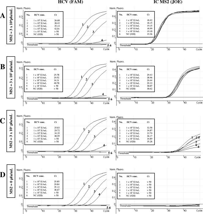
For implementation of an internal control reaction, which would be useful for continuous monitoring of the sample preparation process, we used an HCV RT-PCR assay as described previously (14). To ensure that the MS2 RNA internal control sequence does not suppress amplification of HCV RNA in routine screening tests, we spiked pooled plasma samples containing 10 to 10,000 IU HCV per ml with increasing amounts of MS2 phage (Fig. 3). Nucleic acid extracts from 1 ml plasma were amplified using the PCR protocol given above. The aim was accurate detection and quantification of low target concentrations without affecting the sensitivity limit of the assay. As a result, the sensitivity for the HCV/MS2 duplex assay was not significantly reduced compared to that of the HCV RT-PCR without IC. However, near the lower detection limit of HCV, an increase in CT values and a decrease in fluorescence were observed.
FIG. 3.
Determination of MS2 IC concentrations (conc.) in HCV RT-PCRs. Four sets of RT-PCR samples were prepared, each with an identical dilution series of HCV (0 to 105 IU/ml). Each set of HCV sample was spiked with a different amount of MS2 phage: (A) 3 × 10⁴ PFU/ml; (B) 3 × 10² PFU/ml; (C) 3 × 101 PFU/ml; and (D) 0 PFU/ml. RNA was prepared from 1 ml EDTA plasma by using a QIAamp UltraSens virus kit (QIAGEN). RT-PCR was performed on the Rotorgene 3000 platform with the MS2-TM3 RT-PCR system and HCV primer and probes as described previously (12). Norm. Fluoro., normalized fluorescence; NC, negative control (H2O).
On the basis of these results, 3,000 PFU (3 × 10⁵ copies) per ml of plasma was used as an internal control for routine screening of plasma from blood donors on the Rotorgene 3000 with TaqMan probes. The results of routine RT-PCR runs for HCV screening of blood donations indicated that the presence of the internal control did not compromise the detection limits of the HCV RT-PCR. The two HCV run controls were detected in each experiment with a mean CT value of 32.76 ± 0.79 and a coefficient of variation (CV) of CT of <2.4% (Table 3).
TABLE 3.
Precision testing of the HCV/MS2 RT-PCRa
| No. of expt | No. of samples | Result for:
|
||||
|---|---|---|---|---|---|---|
| MS2 intra-assay variation
|
HCV run control
|
|||||
| CT mean | SD | CV | A CT | B CT | ||
| 1 | 9 | 22.41 | 0.296 | 1.33 | 31.61 | 32.26 |
| 2 | 10 | 22.09 | 0.335 | 1.52 | 32.59 | 32.42 |
| 3 | 10 | 22.46 | 0.523 | 2.33 | 32.49 | 32.50 |
| 4 | 9 | 21.53 | 0.381 | 1.77 | 33.23 | 33.31 |
| 5 | 11 | 22.36 | 0.344 | 1.54 | 31.94 | 31.52 |
| 6 | 10 | 22.31 | 0.446 | 2.00 | 33.36 | 31.83 |
| 7 | 9 | 22.77 | 0.218 | 1.00 | 33.40 | 33.20 |
| 8 | 11 | 23.48 | 0.724 | 1.33 | 32.61 | 31.93 |
| 9 | 11 | 23.67 | 0.603 | 1.52 | 32.99 | 32.35 |
| 10 | 14 | 22.53 | 0.408 | 2.33 | 33.84 | 33.62 |
| 11 | 10 | 22.41 | 0.646 | 1.77 | 34.32 | 33.91 |
MS2 phage was spiked into 1 ml plasma pool (3,000 PFU per ml [3 × 10⁵ copies per ml]). Nucleic acid was extracted with a QIAamp Ultrasens virus kit (Qiagen, Hilden, Germany) and analyzed by RT-PCR on the Rotorgene platform with coamplification of HCV-RNA, as described in Materials and Methods. Two HCV RNA-positive control pools with 208 IU HCV per ml (run controls A and B) were included in each experiment. SD, standard deviation.
Additionally, HCV RT-PCR (21) was performed on the LightCycler instrument with the implementation of MS2 IC. The coamplification of MS2 RNA did not interfere with detection of the HCV RNA. All of the samples tested were positive for the IC.
Precision testing.
The results of the experiments for the screening of blood donation pools for HCV RNA were used to calculate the intra-assay variation and the total variation of the assay. Inter- and intra-assay variations were calculated for CT values. The assay was used in parallel with our established blood donor screening PCR to screen pools of plasma in a 2-week test period. The reproducibility of the method was demonstrated by intra-assay analysis (Table 3). The CVof CT was <2.6% for all 114 plasma pools tested. The interassay variability was calculated from 11 independent RT-PCR runs with CVs ranging from 1.33 to 2.33% and a mean CT of 22.54 ± 0.63 (Table 3).
DISCUSSION
In this study, the MS2 phage was successfully used as an internal control for routine clinical diagnostic RT-PCRs. We developed four MS2 specific real-time RT-PCRs that are based on different probe formats and serve as noncompetitive ICs. For example, we demonstrated the coamplification of MS2 with HCV RNA on different PCR platforms. Additionally, we used these RT-PCR assays to control assays for other viral pathogens, e.g., norovirus genogroups I and II, hepatitis A virus, and human immunodeficiency virus (data not shown). The incorporation of an internal control into each RT-PCR tube is important to identify inhibitors and to eliminate false-negative results, even when problematic specimens such as stool or bronchial lavage fluids are used (9, 10, 22). Commercially produced diagnostic kits are available for relatively few pathogens. Furthermore, armored RNAs for various RT-PCR assays are commercially available, but their cost and lack of versatility have so far prevented the widespread adoption of these ICs. Hence, many in-house assays using both gel-based and real-time detection systems have been developed for the detection of additional targets. Such assays are economical, but they generally lack reaction-specific internal controls to monitor the extraction, reverse transcription, amplification, and detection steps (8).
The use of nucleic acid-based assays for the diagnosis and monitoring of viral RNA is widespread. Most of these assays depend on the use of synthetic RNAs such as in vitro transcripts or armored RNA as the positive control, internal control, or external standards (3, 6, 14, 15). For that, the control RNA has to be placed in long-term storage without degradation or loss of copy numbers. Naked RNA molecules are often affected by hydrolysis due to an insufficient storage environment or minor contamination with RNase. Therefore, the development of armored RNA technology overcomes the problem of instability of control RNA, as demonstrated previously (6, 15). The disadvantage of these pseudoviral particles is that they are difficult to synthesize and represent only a minor part of the viral genome. For highly conserved targets, such as the 5′ untranslated region of the HCV genome, the construction of a “universal” armored RNA control is significant. Viruses with divergent subtypes, like human immunodeficiency virus, are problematic because commercial and in-house NATs use different targets and primers.
As an alternative, the use of intact viruses for external standards in absolute quantification assays or positive control is preferred (4, 13, 23), but the risk of infection for laboratory workers has to be considered when human or animal pathogenic viruses are used. Our approach avoids these disadvantages by using E. coli phage MS2 as a target for the IC. A resistance to RNase degradation, even at high storage temperatures, was demonstrated. The precision of the MS2 RT-PCR was high. We observed no failure of the internal control in the 2-week test period, and the coamplification of MS2 RNA did not prohibit day-to-day application of the assay. The analytical sensitivity of the MS2 NAT was determined by probit regression analysis with different input titers of MS2 phage. The MS2 RNA was sufficiently stable for routine use and did not decrease the detection limit of the multiplex RT-PCRs in which it was used.
For routine clinical applications, the laboratory can maximize the test sensitivity by using an IC to monitor amplification in every specimen. We used the MS2 RT-PCR assays to monitor amplification by spiking MS2 RNA into the RT-PCR master mixture and also MS2 phage for controlling the nucleic acid extraction and all subsequent steps of the procedure. The control phage was simple to produce in high yields, and standardization was possible by plating assays to determine PFU. The MS2 genome sequence was absent from the human specimens, cell cultures, and veterinary samples. Therefore, this approach should be used for different diagnostic NATs, as demonstrated in several RT-PCR assays.
In conclusion, the use of MS2 phage to control clinical diagnostic NAT was demonstrated. The present study supplies evidence that noncompetitive ICs are suitable for many different assays and combine most of the features that are required for valid ICs.
Acknowledgments
We thank Michael Schmidt for his critical reading of the manuscript and Sarah Kirkby for her linguistic advice.
REFERENCES
- 1.Allwood, P. B., Y. S. Malik, C. W. Hedberg, and S. M. Goyal. 2003. Survival of F-specific RNA coliphage, feline calicivirus, and Escherichia coli in water: a comparative study. Appl. Environ. Microbiol. 69:5707-5710. [DOI] [PMC free article] [PubMed] [Google Scholar]
- 2.Ausubel, F. M., R. Brent, R. E. Kingston, D. D. Moore, J. G. Seidman, J. A. Smith, and K. Struhl (ed.). 1999. Short protocols in molecular biology, 4th edition. John Wiley & Sons, New York, N.Y.
- 3.Beld, M., R. Minnaar, J. Weel, C. Sol, M. Damen, H. van der Avoort, P. Wertheim-van Dillen, A. van Breda, and R. Boom. 2004. Highly sensitive assay for detection of enterovirus in clinical specimens by reverse transcription-PCR with an armored RNA internal control. J. Clin. Microbiol. 42:3059-3064. [DOI] [PMC free article] [PubMed] [Google Scholar]
- 4.Cleland, A., P. Nettleton, L. Jarvis, and P. Simmonds. 1999. Use of bovine viral diarrhoea virus as an internal control for amplification of hepatitis C virus. Vox Sang. 76:170-174. [DOI] [PubMed] [Google Scholar]
- 5.Croci, L., D. De Medici, C. Scalfaro, A. Fiore, M. Divizia, D. Donia, A. M. Cosentino, P. Moretti, and G. Costantini. 2000. Determination of enteroviruses, hepatitis A virus, bacteriophages and Escherichia coli in Adriatic Sea mussels. J. Appl. Microbiol. 88:293-298. [DOI] [PubMed] [Google Scholar]
- 6.Drosten, C., E. Seifried, and W. K. Roth. 2001. TaqMan 5′-nuclease human immunodeficiency virus type 1 PCR assay with phage-packaged competitive internal control for high-throughput blood donor screening. J. Clin. Microbiol. 39:4302-4308. [DOI] [PMC free article] [PubMed] [Google Scholar]
- 7.Hennig, H., J. Luhm, D. Hartwig, H. Kluter, and H. Kirchner. 2001. A novel RT-PCR for reliable and rapid HCV RNA screening of blood donations. Transfusion 41:1100-1106. [DOI] [PubMed] [Google Scholar]
- 8.Hoorfar, J., B. Malorny, A. Abdulmawjood, N. Cook, M. Wagner, and P. Fach. 2004. Practical considerations in design of internal amplification controls for diagnostic PCR assays. J. Clin. Microbiol. 42:1863-1868. [DOI] [PMC free article] [PubMed] [Google Scholar]
- 9.Kim, K., J. Park, Y. Chung, D. Cheon, I. B. Lee, S. Lee, J. Yoon, H. Cho, C. Song, and K. H. Lee. 2002. Use of internal standard RNA molecules for the RT-PCR amplification of the faeces-borne RNA viruses. J. Virol. Methods 104:107-115. [DOI] [PubMed] [Google Scholar]
- 10.Lachnik, J., B. Ackermann, A. Bohrssen, S. Maass, C. Diephaus, A. Puncken, M. Stermann, and F. C. Bange. 2002. Rapid-cycle PCR and fluorimetry for detection of mycobacteria. J. Clin. Microbiol. 40:3364-3373. [DOI] [PMC free article] [PubMed] [Google Scholar]
- 11.Mackay, I. M. 2004. Real-time PCR in the microbiology laboratory. Clin. Microbiol. Infect. 10:190-212. [DOI] [PubMed] [Google Scholar]
- 12.Mitsunaga, S., K. Fujimura, C. Matsumoto, R. Shiozawa, S. Hirakawa, K. Nakajima, K. Tadokoro, and T. Juji. 2002. High-throughput HBV DNA and HCV RNA detection system using a nucleic acid purification robot and real-time detection PCR: its application to analysis of posttransfusion hepatitis. Transfusion 42:100-106. [DOI] [PubMed] [Google Scholar]
- 13.Neisters, H. G. M. 2002. Clinical virology in real time. J. Clin. Virol. 25:S3-S12. [DOI] [PubMed] [Google Scholar]
- 14.Panning, M., M. Asper, S. Kramme, H. Schmitz, and C. Drosten. 2004. Rapid detection and differentiation of human pathogenic orthopox viruses by a fluorescence resonance energy transfer real-time PCR assay. Clin. Chem. 50:702-708. [DOI] [PubMed] [Google Scholar]
- 15.Pasloske, B. L., C. R. Walkerpeach, R. D. Obermoeller, M. Winkler, and D. B. DuBois. 1998. Armored RNA technology for production ribonuclease-resistant viral RNA controls and standards. J. Clin. Microbiol. 36:3590-3594. [DOI] [PMC free article] [PubMed] [Google Scholar]
- 16.Poon, L. L., B. W. Wong, K. H. Chan, C. S. Leung, K. Y. Yuen, Y. Guan, and J. S. Peiris. 2004. A one step quantitative RT-PCR for detection of SARS coronavirus with an internal control for PCR inhibitors. J. Clin. Virol. 30:214-217. [DOI] [PMC free article] [PubMed] [Google Scholar]
- 17.Pyra, H., J. Boni, and J. Schupbach. 1994. Ultrasensitive retrovirus detection by a reverse transcriptase assay based on product enhancement. Proc. Natl. Acad. Sci. USA 91:1544-1548. [DOI] [PMC free article] [PubMed] [Google Scholar]
- 18.Ratge, D., B. Scheiblhuber, M. Nitsche, and C. Knabbe. 2000. High-speed detection of blood-borne hepatitis C virus RNA by single-tube real-time fluorescence reverse transcription-PCR with the LightCycler. Clin. Chem. 46:1987-1989. [PubMed] [Google Scholar]
- 19.Rosenstraus, M., Z. Wang, S. Y. Chang, D. DeBonville, and J. P. Spadoro. 1998. An internal control for routine diagnostic PCR: design, properties, and effect on clinical performance. J. Clin. Microbiol. 36:191-197. [DOI] [PMC free article] [PubMed] [Google Scholar]
- 20.Shin, G. A., and M. D. Sobsey. 2003. Reduction of Norwalk virus, poliovirus 1, and bacteriophage MS2 by ozone disinfection of water. Appl. Environ. Microbiol. 69:3975-3978. [DOI] [PMC free article] [PubMed] [Google Scholar]
- 21.Thompson, S. S., and M. V. Yates. 1999. Bacteriophage inactivation at the air-water-solid interface in dynamic batch systems. Appl. Environ. Microbiol. 65:1186-1190. [DOI] [PMC free article] [PubMed] [Google Scholar]
- 22.Ursi, D., K. Dirven, K. Loens, M. Ieven, and H. Goossens. 2003. Detection of Mycoplasma pneumoniae in respiratory samples by real-time PCR using an inhibition control. J. Microbiol. Methods 55:149-153. [DOI] [PubMed] [Google Scholar]
- 23.van Doornum, G. J. J., J. Guldemeester, A. D. M. E. Osterhaus, and H. G. M. Niesters. 2003. Diagnosing herpesvirus infections by real-time amplification and rapid culture. J. Clin. Microbiol. 41:576-580. [DOI] [PMC free article] [PubMed] [Google Scholar]


