



ABSTRACT
Bioengineered tubular bile ducts are an emerging strategy in liver regenerative medicine and extrahepatic bile duct reconstruction, offering potential solutions to address biliary diseases and improve outcomes in liver transplantation. Disorders of the biliary system, such as biliary atresia, primary sclerosing cholangitis, and bile duct injury, often result in irreversible damage and cannot be adequately treated with conventional therapies. This review explores the latest advancements in the bioengineering of both intrahepatic and extrahepatic bile ducts, focusing on approaches that leverage cholangiocyte biology, stem cell technologies, and biomaterial innovations. Key cell sources, including primary cholangiocytes, induced pluripotent stem cells (iPSCs), liver progenitor cells (LPCs), and transdifferentiated hepatocytes, are discussed for their potential to generate functional bile duct epithelial cells capable of restoring biliary function. The role of biomaterials in providing structural support and promoting cellular growth and differentiation is critically examined, emphasizing synthetic and natural scaffolds, such as poly(lactic acid) (PLA) and collagen‐based materials, for creating viable tubular structures. This review provides a comprehensive overview of the current progress and challenges in bioengineered bile duct tissue, focusing on the goals of enhancing liver regeneration and enabling extrahepatic bile duct reconstruction for clinical applications. Integrating cellular, biomaterial, and bioengineered strategies offers promising avenues for advancing treatments for biliary diseases and improving liver transplantation outcomes.
Keywords: bioartificial, bioengineered bile duct, biomaterials, cholangiopathies, liver regeneration
1. Introduction
The biliary system is a complex structural and functional network within the human body, comprising the liver, bile ducts, and gallbladder. The bile duct system is composed of intra‐ and extrahepatic ducts. The formation of intrahepatic bile ducts refers to the process during embryonic development in which cholangiocytes surround the portal vein to form ductal plates, which then develop into small luminal spaces and mature into the intrahepatic bile duct system [1]. This process, known as “ductal hollowing,” leads to the formation of bile ducts [1, 2]. The organization of the biliary system integrates the apical‐basal polarity of hepatocytes and cholangiocytes. In contrast, the extrahepatic biliary system originates from the foregut and follows a distinct developmental pathway [3]. The main function of the bile duct system is the transportation of bile, which is produced by hepatocytes and secreted into the bile canaliculi, subsequently flowing through the bile ducts. The inner lining of the bile ducts is covered by biliary epithelial cells, also known as cholangiocytes, which are responsible for regulating the final composition of bile [4]. The functions crucial for tissue homeostasis of cholangiocytes were reviewed in detail and include production, transportation, modification of bile, and liver's regenerative response following injury or disease [5, 6].
The Intrahepatic and extrahepatic bile duct disorders result in significant morbidity and mortality. Notably, biliary atresia (BA) that results from an idiopathic progressive fibrosclerosing obliteration of extrahepatic bile ducts is a leading cause of pediatric liver transplantation [7, 8], accounting for approximately 70% of such procedures [8], while primary sclerosing cholangitis (PSC) alone accounts for 5% of liver transplantation [9]. Additionally, biliary complications are the leading cause of graft failure after deceased donor liver transplantation [10, 11, 12]. However, the therapeutic approaches remain limited due to the insufficient availability of healthy cell and tissue sources to reconstruct or replace the primarily diseased bile ducts [13]. For primary intrahepatic cholangiopathies, there are no effective treatment options, and the disease progression can only be delayed with limited pharmacological interventions, ultimately necessitating liver transplantation [14]. However, chronic rejection reactions may also lead to chronic cholangiopathies, which can impair graft function, potentially requiring retransplantation [15, 16]. The lack of sufficient donor livers poses a significant health burden, which highlights the importance of maintaining cholangiocytes and generating engineered bile duct tissue in vitro for extended periods, crucial for drug testing and tissue bioengineered applications.
Recent studies have increasingly shown that the use of bile duct organoids for intrahepatic transplantation can effectively delay disease progression and repair damaged bile duct structures [17, 18]. Also, for extrahepatic bile duct damage or deficiency, advancements in stem cell research and biomedical bioengineered artificial bile duct (ABD) have led to significant progress in the development of replacement therapies [19, 20]. Bioengineered bile ducts, comprising biomaterials and cholangiocytes or liver progenitor cells (LPCs), are emerging as multifunctional platforms in both research and clinical contexts. Beyond their use in drug screening, they hold great promise in clinical therapies, disease modeling, transplantation, and regenerative medicine. In clinical therapies, bioengineered ducts are being developed as grafts to treat bile duct injuries and congenital defects such as BA and choledochal cysts [21]. Studies have shown that three‐dimensional (3D)‐printed, cell‐laden scaffolds (e.g., PLGA/PCL‐based) can successfully replace damaged extrahepatic bile ducts in animal models, restoring bile flow and preserving liver function [22]. Additionally, transplantation of cholangiocyte organoids (COs) into human donor livers ex vivo has demonstrated the potential for repairing biliary injury [21]. For disease modeling, bile duct organoids and engineered duct‐like structures enable the study of cholangiopathies such as PSC, BA, and cholangiocarcinoma in controlled in vitro systems [14]. These 3D models, including bile duct‐on‐chip platforms, replicate duct architecture, ciliary function, and bile acid transport more accurately than 2D cultures, allowing for high‐fidelity disease modeling and therapeutic testing [14, 17]. In transplantation, preclinical models have demonstrated that bioengineered bile ducts can be surgically implanted to replace resected ducts in mice, rabbits, and pigs [23]. These grafts integrate with host tissue and maintain patency and function, highlighting their potential as alternative treatments to conventional liver transplantation or biliary reconstruction procedures [24, 25, 26]. In regenerative medicine, bile duct engineering supports liver repair by delivering cholangiocyte‐like cells capable of restoring damaged biliary epithelium. Organoid transplantation into damaged bile ducts or liver grafts has shown successful engraftment, epithelial regeneration, and improved bile secretion [21, 27]. Moreover, hepatobiliary organoids—containing both hepatocyte‐ and cholangiocyte‐like cells—have been developed to mimic functional liver‐bile duct units, supporting future applications in liver tissue engineering and regeneration [17, 28, 29].
Overall, bioengineered bile ducts represent a versatile and rapidly advancing field with wide‐ranging translational applications. Continued research in material optimization, cell sourcing, and long‐term function will be key to clinical implementation. In this review, we describe the methods for the engineered production of cholangiocytes for research purposes, COs, cholangiopathy models, and bioengineered bile duct tissues that may be applicable for clinical treatment.
2. Cell Sources for Bile Duct Engineering
2.1. Primary Cholangiocytes, Immortalized Cholangiocytes, and Hepatocytes‐Cholangiocyte Transdifferentiation
Cholangiocytes are a heterogeneous, highly dynamic population of epithelial cells that line a 3D network of biliary trees, including the intra‐ and extrahepatic ducts [5]. Hepatocytes and cholangiocytes originate from a common precursor, the hepatoblasts, which derive from the endoderm during embryonic development [30, 31]. In the early stages of embryogenesis, hepatoblasts differentiate from the anterior portion of the endoderm, which has the bipotential capacity of differentiation into both hepatocytes and cholangiocytes [32]. The differentiation of hepatocytes is regulated by multiple signaling pathways, including the WNT/β‐catenin pathway, while the Notch signaling pathway plays a key role in the differentiation of cholangiocytes [32]. In contrast, the development of extrahepatic bile ducts is more closely related to the development of the duodenum and pancreas. The bile duct epithelium of the extrahepatic bile ducts and gallbladder originates from the caudal part of the hepatic diverticulum, with these cells, along with pancreatic tissue, originating from the same region and differentiating from pancreatic and duodenal homeobox 1 (Pdx1+) cells [33]. The preferred cell source for bile duct tissue engineering is intrahepatic and extrahepatic cholangiocytes due to their functional properties, regenerative potential, and compatibility with the liver microenvironment [34]. Fotios et al. have reported that they used a new method for the isolation and propagation of human cholangiocytes from the extrahepatic biliary tree in the form of extrahepatic COs and bioengineered artificial ducts for the reconstruction of the biliary tree [20, 35]. The potential applications of cholangiocytes have been demonstrated [36, 37]; however, isolated primary cholangiocytes, limited in availability, experience a decline in functional characteristics and restricted expansion during long‐term 2D culture, which is due to the absence of the extracellular matrix and microenvironmental factors critical for cholangiocyte growth [38]. While 3D culture systems can promote expansion and delay the loss of functionality [20, 39, 40, 41] the availability of primary cholangiocytes is limited by poor access to primary biliary tissue, difficulties in culturing primary cholangiocytes in vitro, and inadequate animal disease models [35, 42]. Long‐term studies and applications involving cholangiocytes can also utilize immortalized cholangiocyte cell lines, such as the MMNK‐1 line, which is established through Simian Virus 40 T antigen (SV40T) and hTERT transduction [43]. The H69 cell line is an SV40‐immortalized human intrahepatic biliary epithelial cell line derived from normal human liver tissue. The HepaRG cell line is a bipotent progenitor cell line established from a liver tumor associated with chronic hepatitis C, exhibiting both hepatocyte‐like and biliary epithelial‐like phenotypes [44, 45, 46, 47]. But due to safety concerns, immortalized or tumor‐derived cell lines may carry a risk of tumorigenicity, raising safety concerns for clinical applications.
Hepatocytes and cholangiocytes maintain a low turnover rate to compensate for occasional cell loss, such as apoptosis and senescence, and have a remarkable ability to self‐renew, particularly in response to liver damage or loss [48, 49]. Hepatocytes exhibit heterogeneity, with different subpopulations having specific proliferative and differentiative capacities under various physiological and pathological conditions [50]. Following hepatic injury or surgical resection, the liver can restore its volume through cellular hypertrophy and compensatory proliferation, and various signaling pathways are activated to initiate liver regeneration. These pathways include Wnt/β‐catenin, NF‐κB, and MAPK, which regulate cellular growth, proliferation, and survival [50, 51, 52]. The plasticity of hepatocytes is not only reflected in their intrinsic proliferation and differentiation but also manifests in their capacity for transdifferentiation observed in cholangiopathies [53, 54]. Schaub et al. demonstrated that TGFβ‐mediated transdifferentiation of hepatocytes in the mouse liver could build a structure that failed to form in development—the biliary system in a mouse model that mimics the hepatic phenotype of human Alagille syndrome (ALGS) [54]. Sekiya et al. also indicate that albumin‐positive hepatocytes residing in the portal areas give rise to the majority of the biliary lineage cells that form primitive ductules in DDC‐induced chronically injured liver through Notch‐mediated cell lineage conversion [55]. A study employing Tg(fabp10a:CFP‐NTR) zebrafish also demonstrated that hepatocyte‐to‐cholangiocyte conversion occurs through transdifferentiation independently of proliferation, and Notch and Yap signaling control the process in parallel with a mutually positive interaction [56]. This process is mediated by specific signaling pathways, including the Notch and Wnt pathways, which are known to play roles in biliary development and regeneration. For instance, activation of the Notch signaling pathway has been correlated with the upregulation of cholangiocyte markers, suggesting a lineage conversion [57]. Such transdifferentiation can be observed in livers affected by injury or congenital bile duct defects [58]. However, the large‐scale expansion of cholangiocytes for the engineering of bile ducts in vitro appears to be impractical, as hepatocytes exhibit limited proliferative capacity ex vivo [59]. The limited expansion of hepatocytes is partly intrinsic—adult hepatocytes have short telomeres, rapidly enter senescence, and proliferate poorly outside the body [60]. Even with optimized 3D culture, only a tiny fraction of adult hepatocytes form expanding organoids (on the order of 0.5%–1.0%), and growth invariably plateaus after just a few months [59, 60]. In contrast, cholangiocyte‐derived organoids can be maintained for ≥ 6 months [60]. Bile duct engineering research has focused on bona fide biliary cells since these can be expanded and form ducts readily. For example, human COs have been cultured long‐term and successfully used to repair biliary injuries. A study showed that scaffolds seeded with human extrahepatic cholangiocyte organoids repaired damaged bile ducts in mice, and human gallbladder‐derived COs engrafted and rebuilt intrahepatic ducts in perfused human livers [61]. Hepatocyte‐cholangiocyte conversion only occurs under forced or chronic injury conditions (e.g., with specific signaling cues in severe cholangiopathies) and is typically very limited [62]. Although it is possible to construct engineered bile ducts by transdifferentiating hepatocytes from limited sources, this approach is neither efficient nor meets the functional requirements and normal physiological conditions. Consequently, the potential for utilizing liver cells to undergo transdifferentiation into cholangiocytes for engineering applications has not garnered significant attention, and they are not considered a primary cell source.
2.2. Stem Cells
Recent studies have focused on the culture and application of cholangiocytes from stem cells, including induced pluripotent stem cells (iPSCs), chemically induced LPCs, human embryonic stem cells (hESCs), and others. iPSCs can self‐renew and differentiate into various cell types [63]. Functional cholangiocytes were generated from human differentiating PSCs, including human iPSCs and hESCs, under the induction of growth factors, EGF, IL‐6, and sodium taurocholate [45]. Induction of functional, mature cholangiocytes from iPSCs typically involves a staged process dependent on specific growth factors that mimic embryonic development. Initially, iPSCs are stepwise differentiated into hepatoblast‐like cells. Subsequently, through 3D culture models, a combination of growth factors and small molecules further induces the formation of cholangiocyte‐like cells or bile duct‐like organoids [41, 64, 65, 66, 67]. As the direct developmental source of cholangiocytes, LPCs can proliferate and expand in vitro. Cholangiocytes have been generated from LPCs in vitro [68, 69, 70, 71]. Unfortunately, the low number (0.5%–2.5%) and lack of specific surface markers of LPCs limit their isolation and purification and hamper research on progenitor cell‐derived cholangiocytes [72, 73]. In recent years, small molecules for somatic cell dedifferentiation have garnered attention. Studies have shown that a combination of small molecules, such as Y‐27632, A‐83‐01, CHIR99021, and HGF, successfully induces LPCs, named CLiPs, from isolated mature hepatocytes through chemical reprogramming [74, 75, 76, 77]. These LPCs, with bidirectional differentiation potential, can rapidly expand in vitro, enrich the source of cholangiocytes, and provide a cell source for constructing tubular bile ducts in vitro. Using this method, we successfully generated various cholangiocyte‐like structures and bile duct‐like organoids from CLiPs generated from mature hepatocytes [77, 78, 79, 80, 81, 82]. Mature hepatocytes are abundant compared to LPCs and primary cholangiocytes. After expansion, CLiPs can serve as a cell bank, offering potential clinical applications. Compared to iPSCs, gene‐editing‐free CLiPs demonstrate higher clinical safety and practicality in liver regeneration (Figure 1).
FIGURE 1.
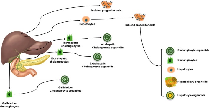
Cholangiocyte organoids could be generated from patients' tissue‐derived cell sources, including primary cholangiocytes, mature hepatocyte‐derived chemically indued progenitor cells and isolated progenitor cells.
3. Materials for Bioengineering Tubular Grafts for Artificial Extrahepatic Bile Ducts
Generating bioengineered bile ducts requires incorporating the aforementioned cell types into suitable materials to form tubular structures. In tissue engineering, various synthetic and biological materials can be used to create scaffolds that support the maintenance and growth of cell populations, either in vitro or in vivo [83]. These scaffolds provide the necessary structural framework, biochemical cues, and mechanical properties to promote cell adhesion, proliferation, and differentiation, ultimately facilitating the creation of functional bile duct‐like structures. The selection of materials for bioengineered tubular bile ducts not only needs to support the growth of the relevant cells but must balance characteristics such as tissue compatibility, ease of surgical manipulation, and functionality [84]. The primary function of the bioengineered bile ducts is to maintain an unobstructed lumen for bile drainage, which requires an appropriate size and effective anastomosis with host tissue. Biological tissue compatibility requires that the grafts induce minimal or no inflammatory and immune rejection responses after implantation [85]. These materials should promote cell adhesion, proliferation, and differentiation while ensuring biocompatibility to minimize immune rejection. Combining scaffold materials, cell seeding substrates, and bioactive factors is regarded as the three key elements of tubular bile duct engineering [86].
Scaffold materials could be further divided into non‐degradable and degradable materials. Nondegradable materials of synthetic polymers, including expanded poly(tetrafluoroethylene) (PTFE) and poly(urethane) (PU), are easily processed into 3D structures, which have been used for vascular grafts [87, 88, 89]. Although these materials can provide long‐term mechanical support, they may inhibit the attachment and proliferation of cholangiocytes due to low hydrophilicity. Therefore, carefully considering their impact on biocompatibility and functionality is essential when selecting non‐degradable materials [90]. Therefore, it is generally no longer common to use non‐degradable polymer materials alone for engineered bile ducts; instead, a combination of biodegradable and bioresorbable materials is often required [91].
Synthetic biodegradable materials, poly(glycolic) acid (PGA), poly(ε‐caprolactone) (PCL), poly(lactic) acid (PLA), and biodegradable poly(urethane) (BPU) have been used for bile duct repair [19, 92, 93, 94, 95]. Jiang et al. reported that an ABD was fabricated for extrahepatic bile duct regeneration based on biodegradable poly(urethane) (BPU) and ureter acellular matrix (UAM) to endow it with favorable biocompatibility and eliminate bile leakage during in vivo bile duct regeneration [93]. Later observation revealed continuous biliary epithelial layer formation, demonstrating good biocompatibility and bile drainage. In a porcine model, a tubular prosthesis fabricated by electrospinning poly(d,l‐lactide‐co‐glycolide) (PLGA) and poly(caprolactone) (PCL) blended with gelatin (Gel) successfully replaced the extrahepatic bile duct—supporting cell adhesion, migration, and proliferation, preserving duct patency, and maintaining normal liver function tests [86]. This composite of synthetic biodegradable materials and biological extracellular matrix provides both sufficient strength and a favorable extracellular matrix environment for cell growth, indicating it may be a promising direction [20, 22, 95, 96, 97].
The natural material scaffolds, including decellularized tissues and naturally derived materials of collagen and polysaccharides, have also been used for bioengineered bile ducts with the advantages of biocompatibility and bioabsorption, but the disadvantages of insufficient strength to resist tensile forces and stenosis propensity [23, 98, 99, 100]. In addition, Eguchi et al. used 3D printing to create scaffold‐free extrahepatic bile ducts from allogeneic pig fibroblasts and successfully implanted them to replace the original bile ducts [101]. The fibroblast‐derived grafts remained stable in vivo and effectively drained bile, maintaining stable serum liver and bile duct enzyme levels [101]. This bioengineered bile duct is an extracellular matrix scaffold‐free but cell‐formed scaffold. Sampaziotis et al. generated tubular scaffolds of densified collagen that were populated with extrahepatic COs maintaining expression of biliary markers such as KRT19, KRT7, HNF‐1β, SOX9, and CFTR and exhibiting GGT and ALP enzymatic activity, which further confirmed the unique capacity of extrahepatic COs in generating bile‐resistant bioengineered bile ducts [20]. Yan et al. created an induced‐mesenchymal stem cell (MSC)‐laden gelatin methacrylate (GelMA)/poly(ethylene glycol) diacrylate (PEGDA)/poly(lactic‐co‐glycolic acid) (PLGA) dual‐layer tubular scaffold to replace the common bile duct with end‐to‐end anastomosis in Bama pig models, which could support biliary regeneration and ultimately help in the reconstruction of the injured part of the bile duct [102]. These recent studies have confirmed that the engineered bile ducts composed of different cell types combined with bioabsorbable scaffolds exhibit enhanced biocompatibility, supporting the regeneration, integration, and functional maintenance of biliary cells [103, 104, 105, 106, 107]. Finally, the fabrication techniques of bioengineered bile duct include rolling of a polymeric sheet, molding, 3D printing techniques, electrospinning, freeze‐drying, and cellular self‐assembly approaches [22, 24, 101, 103, 106, 108, 109]. These technologies are applied in bioengineering to construct new ABDs, holding significant potential for medical applications.
4. Bioengineered Bile Duct for Modeling Cholangiopathies and Liver Regeneration
Dysfunction of cholangiocytes leads to cholangiopathies, including ALGS [110], PSC [111], autoimmune cholangitis [112], BA, cystic fibrosis‐associated liver disease [113], and cholangiocarcinoma [114], can affect both the intrahepatic and extrahepatic biliary trees [115]. These cholangiopathies, caused by genetic, viral, and inflammatory stimuli, lead to damage of the biliary epithelial cells, resulting in cholestasis, bile duct fibrosis, and hepatic parenchymal injury, ultimately progressing to end‐stage liver disease requiring liver transplantation. The pathogenesis of these primary or secondary cholangiopathies requires further in vitro and in vivo studies to develop genuinely effective therapeutic drugs and strategies. Bioengineered bile duct models, particularly hepatobiliary organoids, serve as excellent in vitro platforms and have even been employed in in vivo therapeutic applications [21, 81]. We have established hepatobiliary organoids or hepatobiliary‐like tissues derived from human or animal sources using CLiPs [78, 80, 82]. This methodology is beneficial for studying the mechanisms of bile secretion and hepatobiliary diseases [116]. Using patients' tissue‐derived cells to establish hepatobiliary organoids will significantly advance individualized diagnosis and drug screening [81].
As mentioned above, in vitro COs, constructed using cells from various sources, mimic the characteristics and progression of cholangiopathies [33, 41, 65, 81, 117]. PSC is a cholestatic liver disease characterized by ductular reaction, biliary inflammation, and fibrosis and caused by the interaction of predisposing genetic factors and environmental exposures in local biological processes [118]. PSC's pathogenesis still involves many unresolved questions and remains a scientific and clinical challenge. Bioengineered bile duct organoids provide new tools for studying the mechanisms and treatments of PSC. COs derived from bile ducts of PSC patients who developed dysplasia indicate that COs may retain certain disease‐relevant features despite their adaption toward the in vitro environment and suggest a future potential application of COs as a diagnostic tool for cholangiocarcinoma [119]. COs sourced from PSC and non‐PSC patients could raise the understanding of PSC, such as the contribution of IL‐17 stimulation in the pathogenesis of PSC [120]. Moreover, Zhang et al. generated scaffold‐free multicellular 3D COs from primary liver cells isolated from non‐PSC and PSC patients to study the pathophysiological mechanisms driving PSC progression. The COs produced many of the hallmark pathophysiological features of PSC, such as biliary inflammation, fibrosis, angiogenesis, and neuroendocrine phenotypes [121]. These models have facilitated a better understanding of PSC and provided a platform to advance therapeutic strategies for treating PSC. Yao et al. explored the therapeutic potential of human placental mesenchymal stem cells in PSC through the Takeda G protein‐coupled receptor 5 receptor pathway [122].
BA is a cholangiopathy affecting infants exclusively, characterized by biliary inflammation and liver fibrosis, and the exact cause of BA remains unclear [123]. BA‐related organoids provide new insights into the disease mechanisms and therapeutic strategies [124]. The rotavirus infection has been suggested as a contributing factor in some patients, but concrete proof remains lacking. Chen et al. consolidated this evidence in human biliary organoids that the organoids are highly susceptible to rotavirus infection, supporting its full life cycle and triggering host defense and injury responses. The findings provide evidence linking rotavirus infection to the development of BA and reveal the efficacy of mycophenolic acid, interferon alpha, and a VP7‐targeting neutralizing antibody in inhibiting infection [27]. In addition, Chung et al. also explored the creation of BA‐like organoids by treating non‐BA human liver organoids with poly(inosinic): poly(cytidylic) acid (poly I: C) [125]. Treated organoids developed aberrant morphologies resembling BA organoids and showed transcriptomic similarities, including the expression of inflammatory and immune‐mediated signaling genes. These findings suggest that poly I: C‐treated normal liver organoids could be valuable models for studying immune‐mediated inflammation in BA [125]. A study utilizing a toxic model of BA induced by biliatresone revealed that microtubule instability, along with disruptions in Wnt and Hippo signaling pathways, plays significant roles in the disease's pathogenesis [126]. Results from these in vitro bioengineered models may provide opportunities to explore human‐relevant molecules bearing structural similarities and help gain insights into the mechanisms of BA. Recently, a discovery based on COs could lead to better treatment for BA. The researchers generated biliary organoids from liver biopsies of infants with BA and controls, observing that organoids from BA exhibited structural and functional abnormalities, including misoriented cilia and increased permeability [127]. Treatment with EGF and FGF2 improved differentiation marker expressions of cytokeratin 7 and hepatocyte nuclear factor 1 homeobox B, restored cell polarity with improved localization of F‐actin, β‐catenin, and tight junction protein zonula occludens‐1, increased CFTR function, and decreased uptake of rodamine‐123 [127]. Based on this finding, activating EGF and FGF2 in bile ducts might be the potential treatment for BA.
In addition to primary diseases, liver ductal organoids can serve as disease models and drug screening platforms for secondary cholangiopathies [128]. Ischemia of the bile duct is a common feature in liver disease and transplantation [129]. Shi et al. created COs to screen the novel cholangio‐protective agents for bile duct ischemia and reoxygenation injury, and they found that alpha1‐antitrypsin reduced apoptosis and necroptosis at both hypoxia and reoxygenation phases [130]. Chusilp et al. investigated the potential therapeutic effects of human amniotic fluid stem cells (hAFSCs) on cholangiocyte apoptosis in a bile duct injury model using liver ductal organoids [131]. This study demonstrated that hAFSCs can attenuate cholangiocyte apoptosis, promoting cell survival and recovery in bile duct injury. These findings suggested that hAFSCs may have regenerative properties that could be leveraged for treating bile duct‐related diseases, such as BA and other forms of liver injury, by protecting cholangiocytes from apoptosis and aiding in tissue repair [131]. In addition to being used as a research tool, COs can also be used as a therapeutic tool to treat bile duct injury. Sampaziotis et al. used organoid technology to develop a cell‐based therapy using cholangiocytes from different regions of intrahepatic and common bile ducts from patients [21]. COs were transplanted into the intrahepatic ducts of deceased human donor livers undergoing ex vivo normothermic machine perfusion. The livers could be maintained for up to 100 h, and the transplanted organoids engrafted, exhibited function, and could repair bile ducts [21]. Engineered bile duct organoids or tissues, as research tools or therapeutic agents, are an imaginative and promising tool for studying liver and biliary diseases, and a potential cell therapy approach.
5. Biofabrication Technology for Tubular Bioengineered Bile Duct
Biofabrication technology for bioengineered bile ducts involves the integration of advanced biomaterials, 3D bioprinting, and tissue engineering strategies to create functional substitutes or models for studying biliary diseases (Table 1). Recent biofabrication techniques enable the creation of 3D tubular scaffolds combined with cells and biologically active molecules to develop functional bile duct structures [19, 24, 101, 133].
TABLE 1.
Represent studies on the replacement of the bile duct with engineered tissue.
| Refs. | Scaffold materials | Cells | Synthetic/biological | Fabrication | Animals | Replacement | Character. | Follow time |
|---|---|---|---|---|---|---|---|---|
| [19] | Poly(caprolactone) (PCL), poly(lactic) acid (PLA) and poly(glycolic) acid (PGA) | Bone marrow cells | Hybrid | Freeze‐drying | Pigs | Common bile duct (CBD) | 6 and 10 weeks: incomplete epithelialization; 6 months: Morphology of native CBD | 6 weeks, 10 weeks or 6 months |
| [102] | Poly (lactic‐co‐glycolic) acid (PLGA) and gelatin methacrylate (gelma)/poly (ethylene glycol) diacrylate (PEGDA) | Mesenchymal stem cells | Hybrid | Modified immersion precipitation and solution casting | Pigs | CBD | Promoted bile duct repairing; enhanced more intact biliary epithelium regenerating | 12 weeks |
| [26] | Poly(ε‐caprolactone) methacrylate (PCLMA), Biocompatible methacrylated recombinant type I collagen(rcolma) and ε‐poly(l‐lysine)‐methacrylamide (EPLMA) | Bone marrow mesenchymal stem cells | Hybrid | 3D printing | Rabbits | Gallbladder | Promoting the differentiation of BMSCs into biliary epithelial cells | 2 weeks |
| [101] | Scaffold‐free Porcine skin fibroblasts spheroids | Fibroblasts | Biological | Bio‐3D printing | Pigs | CBD | Angiogenesis in the fibroblast tube and absence of extensions of the biliary epithelium | 2 weeks |
| [95] | Poly(caprolactone) (PCL) and poly(lactic) acid (PLA) (50:50) reinforced with poly(glycolic) acid (PGA) fibers | Acellular | Synthetic | Freeze‐drying | Pigs | CBD | Grafts revealed complete freedom of stricture; neo‐bile duct form | 4 months |
| [91] | Gelatin hydrogel nonwoven fabric | Murine fibroblast L929 cells | Hybrid | Solution blow spinning | Rats | CBD | Regenerated epithelium and angiogenesis | 2, 6, and 12 weeks |
| [132] | Collagen sponge reinforced with a poly(propylene) (PP) mesh framework | Acellular | Synthetic | Freeze‐drying | Dogs | CBD | Newly formed biliary epithelium and connective tissue | 2 months |
| [100] | Decellularization of allogeneic blood vessels | Autologous cholangiocytes | Biological | Decellularization | Pig | CBD | Neoangiogenesis was observed around and into the implanted tissue | 2 weeks |
Cell seeding and self‐assembly refer to the process of distributing cells onto a scaffold to promote their attachment, growth, and proliferation, thereby spontaneously forming tissue [134]. Scaffolds used for cell seeding are typically made from natural or synthetic polymers, or decellularized tubular tissues from animal or human donors, while the cells usually include bile duct cells as mentioned above [18]. Subsequently, cells are seeded on the surface or inside the scaffold, followed by self‐assembly, resulting in the fabrication of 3D composite materials with cells, offering improved biological functionality and biocompatibility [18]. Tomofuji et al. demonstrated that liver ductal organoids can serve as a promising cell source for reconstructing bile ducts in bioengineered liver grafts using 3D decellularized scaffolds, addressing challenges in cell sourcing and distribution while advancing regenerative medicine [18]. However, this method of cell self‐assembly combined with decellularized scaffolds is only suitable for studying the mechanisms of bile duct regeneration and in vivo bile duct repair.
For engineering extrahepatic bile ducts, more robust bile duct scaffolds are required. The development of a scaffold that incorporates stem cells is the gold standard of tissue engineering [135]. Scaffolds are composed of various biocompatible materials, including animal‐derived collagen and synthetic polymers of nondegradable and biodegradable materials. Various methods with good manufacturing practice (GMP) standards, such as polymer sheet rolling, molding, 3D printing, electrospinning, freeze‐drying, and decellularization, can be used to fabricate the materials into bioengineered tubular bile ducts.
Rolling involves the rolling of a flat substrate, which is the manufacturing method that most specifically applies to tubular scaffold production [136, 137]. The flat substrate is often already seeded with cells or engineered with specific material properties. Cheng et al. have reported that they developed a multilayered vascular scaffold by electrospinning PLGA and PCL onto a poly(dimethylsiloxane) (PDMS) substrate, followed by sequential seeding of endothelial cells, smooth muscle cells, and fibroblasts [137]. Mechanical release of the pre‐stressed substrate induced controlled rolling, achieving spatially stratified cell layers that mimic the layered structure of natural blood vessels [137]. A key advantage of this approach, particularly with biological polymers, is the ability to seed cells directly into the precursor polymer solution, enabling the integration of cells within a dense scaffold [138]. The cells can be homogeneously seeded and cultured in 2D and then rolled into a 3D tube. The synthetic polymers can be molded to form a variety of structures including tubes. The molding method is also a traditional fabrication technique. Yan et al. reported a combination method in which dual‐layer tubular scaffolds were manufactured layer‐by‐layer using the two‐stage molding method that included modified immersion precipitation and solution casting [102]. This inner layer was made of PLGA polymer that provided mechanical support, while the outer layer was composed of GelMA/PEGDA hydrogel embedded with MSCs that provided a biological microenvironment [102]. These dual‐layer induced‐cell‐laden scaffolds could promote biliary regeneration in vivo. Zhang et al. also reported a similar dual‐layer multifunctional hydrogel scaffold based on recombinant collagen fabricated using advanced 3D printing technology [26]. These two fabrication methods offer valuable insights into bile duct tissue regeneration and replacement research, which could enhance implanted‐cells proliferation and their differentiation into bile duct epithelial cells, or achieve functional replacement in vivo. The 3D printing technology applied here enables rapid, multilayer, and multi‐structural printing using different bioink materials [139, 140]. Hamada et al. used scaffold‐free tubular constructs of allogeneic pig fibroblasts created by a Bio‐3D printer to implant into pigs as interposition grafts for duct‐to‐duct biliary reconstruction [101]. As a result, micro‐computed tomography showed no biliary strictures, leakages, or bile duct dilations, while immunohistochemistry revealed angiogenesis in the fibroblast tube and no biliary epithelium extension into its lumen [101]. 3D‐printed materials offer diverse sources, and the tubular structures combined with cells or growth factors significantly enhance the in vivo strength and biocompatibility of bioengineered bile ducts. As described in previous experiments, the availability of materials required for 3D printing, especially biomaterials, is a significant limitation [101]. Hamada et al. used fibroblast spheroids as printing material for bio‐3D printing; the fragile tubular fiber scaffolds had to be cultured for another > 90 days before their strength became barely sufficient, but it was still relatively weak [101]. However, with technological advancements in large‐scale production of cells and organoids, this challenge can potentially be overcome. For small‐sized or thin tubular structures, issues such as printing and mechanical strength can be addressed by integrating other techniques and incorporating fiber scaffolds.
Electrospinning for bioengineered tubular ducts refers to the fabrication of scaffolds using electrospinning techniques, which produce nanofibrous materials mimicking the extracellular matrix [141]. These nanofiber scaffolds provide an excellent environment for cell attachment, proliferation, and differentiation. Electrospun materials can be engineered to form tubular structures for applications such as bile ducts, blood vessels, or other tubular tissues [142].
Freeze‐drying is a process that freezes and dehydrates a polymer solution, resulting in a macro‐porous sponge‐like structure [19, 25, 95]. After freezing the swollen hydrogel, the ice formed within the hydrogel network sublimates through freeze‐drying. This simple method produces a porous hydrogel. Regardless of any rinsing or cleaning processes, water is evenly distributed within the hydrogel network, allowing the hydrogel to swell uniformly, while the ice acts as a template for the porous structure during freezing [143]. However, the porous fibrous scaffolds formed in this way lack sufficient strength to withstand the pressure required for bile ducts, often requiring the addition of other synthetic fibers to enhance their strength [19]. The porous structure also facilitates cell growth within it, enhancing its biocompatibility and functionality. Adding growth factors or inducers to the filler materials further promotes stem cell differentiation or the functional maturation of mature cells [95, 144].
Finally, when producing artificial duct scaffolds using related manufacturing techniques, multiple approaches can be combined to enhance their bioactivity and functionality. After fabricating the fibrous scaffold, cell seeding with stem cells or functional cells, such as bile duct epithelial cells, can be performed to promote vascular growth and vascularization. In addition, mixing appropriate materials with growth factors, such as fibroblast growth factor and vascular endothelial growth factor, can further enhance vascularization [23].
6. Outstanding Questions and Conclusion
Despite significant progress in the biomedical engineering of bile ducts and bile duct reconstruction using related technologies in recent years, challenges and controversies persist. For instance, using iPSCs, immortalized cholangiocyte cells, and other stem cells for bile duct regeneration and bioengineered bile ducts still raises safety concerns. With the development of biocompatible materials such as hydrogels and decellularized scaffolds, bile duct materials combining hydrogels and synthetic extracellular matrices have already undergone testing in vivo. Recent studies have highlighted the potential of resorbable materials and cells, supported by intraluminal stents to maintain patency during early biointegration and tissue maturation. Despite promising results from the use of bioengineered tubes in animals, all studies were performed in healthy animals. Results demonstrate their potential for further practical application and industrialization. But to date, no material has been routinely adopted as a bile duct wall substitute in clinical practice. Although further research is needed, especially in cell seeding and tissue integration, current advancements bring us closer to developing a clinically viable bile duct substitute.
However, is it still necessary to combine cellular bioengineering with bile ducts? Chemically induced LPCs might primarily solve the issue of the source of cholangiocytes, but safety and availability require more preclinical and clinical validation. Meanwhile, organoids derived from multipotent stem cells and tissue sources for intrahepatic bile duct regeneration and liver regeneration are gaining attention in both in vitro models and in vivo treatments, offering hope for improved therapeutic prospects for biliary diseases. Additionally, the long‐term functional maintenance of these implanted materials in vivo remains one of the current challenges. Cellularization and vascularization are potential strategies to improve the long‐term functionality of these materials in the body. Finally, GMP materials and cells are essential prerequisites for medical devices and biomaterials.
In conclusion, as medical materials for extrahepatic bile duct reconstruction, these bioengineered bile ducts bring new hope for treating biliary diseases. Organoid‐based cell therapy and in vitro research models provide new insights for treating intrahepatic bile duct diseases. However, hope comes with challenges, and many issues in basic research and clinical application still need to be overcome.
Conflicts of Interest
The authors declare no conflicts of interest.
Li P., Huang D., Gu W., Gao Y., and Huang Y., “Bioengineered Bile Duct for Liver Regenerative Medicine and Bile Duct Reconstruction,” JGH Open 9, no. 8 (2025): e70254, 10.1002/jgh3.70254.
Funding: This work was supported by the Science and Technology Projects in Guangzhou (2024A03J1016), the Guangzhou Science and Technology Project of Traditional Chinese Medicine and Combined Chinese and Western Medicine (20242A011001), the Guangdong Medical Science and Research Foundation (A2024088), and the Scientific Research Project of Guangdong Provincial Bureau of Traditional Chinese Medicine (20231254).
Contributor Information
Yi Gao, Email: gaoyi6146@163.com.
Yu Huang, Email: dr.hyu@outlook.com.
Data Availability Statement
Data sharing is not applicable to this article as no data sets were generated or analyzed during the current study.
References
- 1. Ober E. A. and Lemaigre F. P., “Development of the Liver: Insights Into Organ and Tissue Morphogenesis,” Journal of Hepatology 68, no. 5 (2018): 1049–1062, 10.1016/j.jhep.2018.01.005. [DOI] [PubMed] [Google Scholar]
- 2. Iruela‐Arispe M. L. and Beitel G. J., “Tubulogenesis,” Development (Cambridge, England) 140, no. 14 (2013): 2851–2855, 10.1242/dev.070680. [DOI] [PMC free article] [PubMed] [Google Scholar]
- 3. Tomita H. and Hara A., “Development of Extrahepatic Bile Ducts and Mechanisms of Tumorigenesis: Lessons From Mouse Models,” Pathology International 72, no. 12 (2022): 589–605, 10.1111/pin.13287. [DOI] [PMC free article] [PubMed] [Google Scholar]
- 4. Strazzabosco M. and Fabris L., “Functional Anatomy of Normal Bile Ducts,” Anatomical Record (Hoboken) 291, no. 6 (2008): 653–660, 10.1002/ar.20664. [DOI] [PMC free article] [PubMed] [Google Scholar]
- 5. Tabibian J. H., Masyuk A. I., Masyuk T. V., O'Hara S. P., and LaRusso N. F., “Physiology of Cholangiocytes,” Comprehensive Physiology 3, no. 1 (2013): 541–565, 10.1002/cphy.c120019. [DOI] [PMC free article] [PubMed] [Google Scholar]
- 6. Zhang H., Leung P. S. C., Gershwin M. E., and Ma X., “How the Biliary Tree Maintains Immune Tolerance?,” Biochimica et Biophysica Acta 1864, no. 4 Pt B (2018): 1367–1373, 10.1016/j.bbadis.2017.08.019. [DOI] [PubMed] [Google Scholar]
- 7. Kasahara M., Umeshita K., Sakamoto S., Fukuda A., Furukawa H., and Uemoto S., “Liver Transplantation for Biliary Atresia: A Systematic Review,” Pediatric Surgery International 33, no. 12 (2017): 1289–1295, 10.1007/s00383-017-4173-5. [DOI] [PubMed] [Google Scholar]
- 8. Perkins J. D., “Are We Reporting the Same Thing?,” Liver Transplantation 13, no. 3 (2007): 465–466. [PubMed] [Google Scholar]
- 9. Carbone M., Della Penna A., Mazzarelli C., et al., “Liver Transplantation for Primary Sclerosing Cholangitis (PSC) With or Without Inflammatory Bowel Disease (IBD)‐A European Society of Organ Transplantation (ESOT) Consensus Statement,” Transplant International 36 (2023): 11729, 10.3389/ti.2023.11729. [DOI] [PMC free article] [PubMed] [Google Scholar]
- 10. Ammori B. J., Joseph S., Attia M., Lodge J. P. A., Beger H., and Schwarz A., “Biliary Strictures Complicating Pancreaticoduodenectomy,” International Journal of Pancreatology 28 (2000): 15–22. [DOI] [PubMed] [Google Scholar]
- 11. Enestvedt C. K., Malik S., Reese P. P., et al., “Biliary Complications Adversely Affect Patient and Graft Survival After Liver Retransplantation,” Liver Transplantation 19, no. 9 (2013): 965–972, 10.1002/lt.23696. [DOI] [PubMed] [Google Scholar]
- 12. Felder S. I., Menon V. G., Nissen N. N., Margulies D. R., Lo S., and Colquhoun S. D., “Hepaticojejunostomy Using Short‐Limb Roux‐En‐Y Reconstruction,” JAMA Surgery 148, no. 3 (2013): 253–257. [DOI] [PubMed] [Google Scholar]
- 13. Gallo A. and Esquivel C. O., “Current Options for Management of Biliary Atresia,” Pediatric Transplantation 17, no. 2 (2013): 95–98. [DOI] [PubMed] [Google Scholar]
- 14. Buchanan‐Peart K. A. and Levy C., “Novel Therapies in Primary Biliary Cholangitis: What Is in the Pipeline?,” Clinics in Liver Disease 26, no. 4 (2022): 747–764, 10.1016/j.cld.2022.06.013. [DOI] [PubMed] [Google Scholar]
- 15. Halme L., Hockerstedt K., and Lautenschlager I., “Cytomegalovirus Infection and Development of Biliary Complications After Liver transplantation1,” Transplantation 75, no. 11 (2003): 1853–1858. [DOI] [PubMed] [Google Scholar]
- 16. Sugawara Y., Makuuchi M., Takayama T., et al., “Small‐for‐Size Grafts in Living‐Related Liver Transplantation,” Journal of the American College of Surgeons 192, no. 4 (2001): 510–513. [DOI] [PubMed] [Google Scholar]
- 17. Shiota J., Samuelson L. C., and Razumilava N., “Hepatobiliary Organoids and Their Applications for Studies of Liver Health and Disease: Are We There Yet?,” Hepatology 74, no. 4 (2021): 2251–2263, 10.1002/hep.31772. [DOI] [PMC free article] [PubMed] [Google Scholar]
- 18. Tomofuji K., Fukumitsu K., Kondo J., et al., “Liver Ductal Organoids Reconstruct Intrahepatic Biliary Trees in Decellularized Liver Grafts,” Biomaterials 287 (2022): 121614, 10.1016/j.biomaterials.2022.121614. [DOI] [PubMed] [Google Scholar]
- 19. Miyazawa M., Torii T., Toshimitsu Y., Okada K., Koyama I., and Ikada Y., “A Tissue‐Engineered Artificial Bile Duct Grown to Resemble the Native Bile Duct,” American Journal of Transplantation 5, no. 6 (2005): 1541–1547, 10.1111/j.1600-6143.2005.00845.x. [DOI] [PubMed] [Google Scholar]
- 20. Sampaziotis F., Justin A. W., Tysoe O. C., et al., “Reconstruction of the Mouse Extrahepatic Biliary Tree Using Primary Human Extrahepatic Cholangiocyte Organoids,” Nature Medicine 23, no. 8 (2017): 954–963, 10.1038/nm.4360. [DOI] [PubMed] [Google Scholar]
- 21. Sampaziotis F., Muraro D., Tysoe O. C., et al., “Cholangiocyte Organoids Can Repair Bile Ducts After Transplantation in the Human Liver,” Science 371, no. 6531 (2021): 839–846, 10.1126/science.aaz6964. [DOI] [PMC free article] [PubMed] [Google Scholar]
- 22. Xiang Y., Wang W., Gao Y., et al., “Production and Characterization of an Integrated Multi‐Layer 3D Printed PLGA/GelMA Scaffold Aimed for Bile Duct Restoration and Detection,” Frontiers in Bioengineering and Biotechnology 8 (2020): 971, 10.3389/fbioe.2020.00971. [DOI] [PMC free article] [PubMed] [Google Scholar]
- 23. Li Q., Tao L., Chen B., et al., “Extrahepatic Bile Duct Regeneration in Pigs Using Collagen Scaffolds Loaded With Human Collagen‐Binding bFGF,” Biomaterials 33, no. 17 (2012): 4298–4308, 10.1016/j.biomaterials.2012.03.003. [DOI] [PubMed] [Google Scholar]
- 24. Li H., Yin Y., Xiang Y., Liu H., and Guo R., “A Novel 3D Printing PCL/GelMA Scaffold Containing USPIO for MRI‐Guided Bile Duct Repair,” Biomedical Materials 15, no. 4 (2020): 045004, 10.1088/1748-605X/ab797a. [DOI] [PubMed] [Google Scholar]
- 25. Tao L., Li Q., Ren H., et al., “Repair of Extrahepatic Bile Duct Defect Using a Collagen Patch in a Swine Model,” Artificial Organs 39, no. 4 (2015): 352–360, 10.1111/aor.12388. [DOI] [PubMed] [Google Scholar]
- 26. Zhang J., Gao Y., Yang Y., et al., “3D Printing of Multifunctional Hydrogel Scaffolds Based on Recombinant Collagen for Bile Duct Repair,” APL Materials 13, no. 2 (2025): 021106, 10.1063/5.0251988. [DOI] [Google Scholar]
- 27. Chen S., Li P., Wang Y., et al., “Rotavirus Infection and Cytopathogenesis in Human Biliary Organoids Potentially Recapitulate Biliary Atresia Development,” MBio 11, no. 4 (2020), 10.1128/mbio.01968-20IF. [DOI] [PMC free article] [PubMed] [Google Scholar]
- 28. Liu S., Cheng C., Zhu L., et al., “Liver Organoids: Updates on Generation Strategies and Biomedical Applications,” Stem Cell Research & Therapy 15, no. 1 (2024): 244, 10.1186/s13287-024-03865-3. [DOI] [PMC free article] [PubMed] [Google Scholar]
- 29. Tanimizu N., Ichinohe N., Sasaki Y., et al., “Generation of Functional Liver Organoids on Combining Hepatocytes and Cholangiocytes With Hepatobiliary Connections Ex Vivo,” Nature Communications 12, no. 1 (2021): 3390, 10.1038/s41467-021-23575-1. [DOI] [PMC free article] [PubMed] [Google Scholar]
- 30. Swartley O. M., Foley J. F., D. P. Livingston, 3rd , Cullen J. M., and Elmore S. A., “Histology Atlas of the Developing Mouse Hepatobiliary Hemolymphatic Vascular System With Emphasis on Embryonic Days 11.5‐18.5 and Early Postnatal Development,” Toxicologic Pathology 44, no. 5 (2016): 705–725, 10.1177/0192623316630836. [DOI] [PMC free article] [PubMed] [Google Scholar]
- 31. Yanagida A., Mizuno N., Yamazaki Y., et al., “Investigation of Bipotent Differentiation of Hepatoblasts Using Inducible Diphtheria Toxin Receptor‐Transgenic Mice,” Hepatology Research 46, no. 8 (2016): 816–828, 10.1111/hepr.12622. [DOI] [PubMed] [Google Scholar]
- 32. Shin D. and Monga S. P., “Cellular and Molecular Basis of Liver Development,” Comprehensive Physiology 3, no. 2 (2013): 799–815, 10.1002/cphy.c120022. [DOI] [PMC free article] [PubMed] [Google Scholar]
- 33. Verstegen M. M. A., Roos F. J. M., Burka K., et al., “Human Extrahepatic and Intrahepatic Cholangiocyte Organoids Show Region‐Specific Differentiation Potential and Model Cystic Fibrosis‐Related Bile Duct Disease,” Scientific Reports 10, no. 1 (2020): 21900, 10.1038/s41598-020-79082-8. [DOI] [PMC free article] [PubMed] [Google Scholar]
- 34. Pu W., Zhu H., Zhang M., et al., “Bipotent Transitional Liver Progenitor Cells Contribute to Liver Regeneration,” Nature Genetics 55, no. 4 (2023): 651–664, 10.1038/s41588-023-01335-9. [DOI] [PMC free article] [PubMed] [Google Scholar]
- 35. Tysoe O. C., Justin A. W., Brevini T., et al., “Isolation and Propagation of Primary Human Cholangiocyte Organoids for the Generation of Bioengineered Biliary Tissue,” Nature Protocols 14, no. 6 (2019): 1884–1925, 10.1038/s41596-019-0168-0. [DOI] [PubMed] [Google Scholar]
- 36. Hallett J. M., Ferreira‐Gonzalez S., Man T. Y., et al., “Human Biliary Epithelial Cells From Discarded Donor Livers Rescue Bile Duct Structure and Function in a Mouse Model of Biliary Disease,” Cell Stem Cell 29, no. 3 (2022): 355–371.e10, 10.1016/j.stem.2022.02.006. [DOI] [PMC free article] [PubMed] [Google Scholar]
- 37. Wang Z., Faria J., van der Laan L. J. W., Penning L. C., Masereeuw R., and Spee B., “Human Cholangiocytes Form a Polarized and Functional Bile Duct on Hollow Fiber Membranes,” Frontiers in Bioengineering and Biotechnology 10 (2022): 868857, 10.3389/fbioe.2022.868857. [DOI] [PMC free article] [PubMed] [Google Scholar]
- 38. Joplin R., “Isolation and Culture of Biliary Epithelial Cells,” Gut 35, no. 7 (1994): 875–878, 10.1136/gut.35.7.875. [DOI] [PMC free article] [PubMed] [Google Scholar]
- 39. Godoy P., Hewitt N. J., Albrecht U., et al., “Recent Advances in 2D and 3D In Vitro Systems Using Primary Hepatocytes, Alternative Hepatocyte Sources and Non‐Parenchymal Liver Cells and Their Use in Investigating Mechanisms of Hepatotoxicity, Cell Signaling and ADME,” Archives of Toxicology 87, no. 8 (2013): 1315–1530, 10.1007/s00204-013-1078-5. [DOI] [PMC free article] [PubMed] [Google Scholar]
- 40. Huch M., Gehart H., van Boxtel R., et al., “Long‐Term Culture of Genome‐Stable Bipotent Stem Cells From Adult Human Liver,” Cell 160, no. 1 (2015): 299–312, 10.1016/j.cell.2014.11.050. [DOI] [PMC free article] [PubMed] [Google Scholar]
- 41. Sampaziotis F., Cardoso de Brito M., Madrigal P., et al., “Cholangiocytes Derived From Human Induced Pluripotent Stem Cells for Disease Modeling and Drug Validation,” Nature Biotechnology 33, no. 8 (2015): 845–852, 10.1038/nbt.3275. [DOI] [PMC free article] [PubMed] [Google Scholar]
- 42. Pollheimer M. J., Trauner M., and Fickert P., “Will We Ever Model PSC?—“It's Hard to Be a PSC Model!”,” Clinics and Research in Hepatology and Gastroenterology 35, no. 12 (2011): 792–804, 10.1016/j.clinre.2011.04.014. [DOI] [PubMed] [Google Scholar]
- 43. Maruyama M., Kobayashi N., Westerman K. A., et al., “Establishment of a Highly Differentiated Immortalized Human Cholangiocyte Cell Line With SV40T and hTERT,” Transplantation 77, no. 3 (2004): 446–451. [DOI] [PubMed] [Google Scholar]
- 44. Cerec V., Glaise D., Garnier D., et al., “Transdifferentiation of Hepatocyte‐Like Cells From the Human Hepatoma HepaRG Cell Line Through Bipotent Progenitor,” Hepatology 45, no. 4 (2007): 957–967, 10.1002/hep.21536. [DOI] [PubMed] [Google Scholar]
- 45. Dianat N., Dubois‐Pot‐Schneider H., Steichen C., et al., “Generation of Functional Cholangiocyte‐Like Cells From Human Pluripotent Stem Cells and HepaRG Cells,” Hepatology 60, no. 2 (2014): 700–714, 10.1002/hep.27165. [DOI] [PMC free article] [PubMed] [Google Scholar]
- 46. Grubman S. A., Perrone R. D., Lee D. W., et al., “Regulation of Intracellular pH by Immortalized Human Intrahepatic Biliary Epithelial Cell Lines,” American Journal of Physiology. Gastrointestinal and Liver Physiology 266, no. 6 (1994): G1060–G1070, 10.1152/ajpgi.1994.266.6.G1060. [DOI] [PubMed] [Google Scholar]
- 47. Ilyas S. I. and Gores G. J., “Pathogenesis, Diagnosis, and Management of Cholangiocarcinoma,” Gastroenterology 145, no. 6 (2013): 1215–1229, 10.1053/j.gastro.2013.10.013. [DOI] [PMC free article] [PubMed] [Google Scholar]
- 48. Lan T., Qian S., Tang C., and Gao J., “Role of Immune Cells in Biliary Repair,” Frontiers in Immunology 13 (2022): 866040, 10.3389/fimmu.2022.866040. [DOI] [PMC free article] [PubMed] [Google Scholar]
- 49. Stanger B. Z., “Cellular Homeostasis and Repair in the Mammalian Liver,” Annual Review of Physiology 77 (2015): 179–200, 10.1146/annurev-physiol-021113-170255. [DOI] [PMC free article] [PubMed] [Google Scholar]
- 50. Chen F., Schönberger K., and Tchorz J. S., “Distinct Hepatocyte Identities in Liver Homeostasis and Regeneration,” JHEP Reports 5, no. 8 (2023): 100779, 10.1016/j.jhepr.2023.100779. [DOI] [PMC free article] [PubMed] [Google Scholar]
- 51. Michalopoulos G. K., “Principles of Liver Regeneration and Growth Homeostasis,” Comprehensive Physiology 3, no. 1 (2013): 485–513. [DOI] [PubMed] [Google Scholar]
- 52. Pagano D., Spada M., Parikh V., et al., “Liver Regeneration After Liver Resection: Clinical Aspects and Correlation With Infective Complications,” World Journal of Gastroenterology 20, no. 22 (2014): 6953–6960, 10.3748/wjg.v20.i22.6953. [DOI] [PMC free article] [PubMed] [Google Scholar]
- 53. Michalopoulos G. K., “The Regenerative Altruism of Hepatocytes and Cholangiocytes,” Cell Stem Cell 23, no. 1 (2018): 11–12, 10.1016/j.stem.2018.06.006. [DOI] [PubMed] [Google Scholar]
- 54. Schaub J. R., Huppert K. A., Kurial S. N., et al., “De Novo Formation of the Biliary System by TGFβ‐Mediated Hepatocyte Transdifferentiation,” Nature 557, no. 7704 (2018): 247–251. [DOI] [PMC free article] [PubMed] [Google Scholar]
- 55. Sekiya S. and Suzuki A., “Hepatocytes, Rather Than Cholangiocytes, Can be the Major Source of Primitive Ductules in the Chronically Injured Mouse Liver,” American Journal of Pathology 184, no. 5 (2014): 1468–1478, 10.1016/j.ajpath.2014.01.005. [DOI] [PubMed] [Google Scholar]
- 56. Lee S.‐H., So J., and Shin D., “Hepatocyte‐to‐Cholangiocyte Conversion Occurs Through Transdifferentiation Independently of Proliferation in Zebrafish,” Hepatology 77, no. 4 (2023): 1198–1210. [DOI] [PMC free article] [PubMed] [Google Scholar]
- 57. Zong Y., Panikkar A., Xu J., et al., “Notch Signaling Controls Liver Development by Regulating Biliary Differentiation,” Development 136, no. 10 (2009): 1727–1739, 10.1242/dev.029140. [DOI] [PMC free article] [PubMed] [Google Scholar]
- 58. Lan T., Tai Y., Zhao C., et al., “Atypical Cholangiocytes Derived From Hepatocyte‐Cholangiocyte Transdifferentiation Mediated by COX‐2: A Kind of Misguided Liver Regeneration,” Inflammation and Regeneration 43, no. 1 (2023): 37, 10.1186/s41232-023-00284-4. [DOI] [PMC free article] [PubMed] [Google Scholar]
- 59. Hu H., Gehart H., Artegiani B., et al., “Long‐Term Expansion of Functional Mouse and Human Hepatocytes as 3D Organoids,” Cell 175, no. 6 (2018): 1591–1606.e19, 10.1016/j.cell.2018.11.013. [DOI] [PubMed] [Google Scholar]
- 60. Ock S. A., Kim S.‐Y., Kim Y.‐I., Lee P., and Ju W. S., “The Regenerative Capacity of Hepatocyte Organoids Following Long‐Term Cryopreservation in Republic of Korea,” Organoid 4 (2024): e9, 10.51335/organoid.2024.4.e9. [DOI] [Google Scholar]
- 61. Afonso M. B., Marques V., van Mil S. W. C., and Rodrigues C. M. P., “Human Liver Organoids: From Generation to Applications,” Hepatology 79, no. 6 (2024): 1432–1451, 10.1097/hep.0000000000000343. [DOI] [PMC free article] [PubMed] [Google Scholar]
- 62. Limaye P. B., Alarcón G., Walls A. L., et al., “Expression of Specific Hepatocyte and Cholangiocyte Transcription Factors in Human Liver Disease and Embryonic Development,” Laboratory Investigation 88, no. 8 (2008): 865–872, 10.1038/labinvest.2008.56. [DOI] [PMC free article] [PubMed] [Google Scholar]
- 63. Takahashi K., Tanabe K., Ohnuki M., et al., “Induction of Pluripotent Stem Cells From Adult Human Fibroblasts by Defined Factors,” Cell 131, no. 5 (2007): 861–872, 10.1016/j.cell.2007.11.019. [DOI] [PubMed] [Google Scholar]
- 64. Chi K. Y., Kim G., Kim H., et al., “Optimization of Culture Conditions to Generate Vascularized Multi‐Lineage Liver Organoids With Structural Complexity and Functionality,” Biomaterials 314 (2025): 122898, 10.1016/j.biomaterials.2024.122898. [DOI] [PubMed] [Google Scholar]
- 65. Ghanekar A. and Kamath B. M., “Cholangiocytes Derived From Induced Pluripotent Stem Cells for Disease Modeling,” Current Opinion in Gastroenterology 32, no. 3 (2016): 210–215, 10.1097/mog.0000000000000260. [DOI] [PubMed] [Google Scholar]
- 66. Hannan N. R. F., Segeritz C.‐P., Touboul T., and Vallier L., “Production of Hepatocyte‐Like Cells From Human Pluripotent Stem Cells,” Nature Protocols 8, no. 2 (2013): 430–437, 10.1038/nprot.2012.153. [DOI] [PMC free article] [PubMed] [Google Scholar]
- 67. Rashid S. T., Corbineau S., Hannan N., et al., “Modeling Inherited Metabolic Disorders of the Liver Using Human Induced Pluripotent Stem Cells,” Journal of Clinical Investigation 120, no. 9 (2010): 3127–3136, 10.1172/JCI43122. [DOI] [PMC free article] [PubMed] [Google Scholar]
- 68. Cardinale V., Wang Y., Carpino G., et al., “Multipotent Stem/Progenitor Cells in Human Biliary Tree Give Rise to Hepatocytes, Cholangiocytes, and Pancreatic Islets,” Hepatology 54, no. 6 (2011): 2159–2172. [DOI] [PubMed] [Google Scholar]
- 69. Lu W.‐Y., Bird T. G., Boulter L., et al., “Hepatic Progenitor Cells of Biliary Origin With Liver Repopulation Capacity,” Nature Cell Biology 17, no. 8 (2015): 971–983, 10.1038/ncb3203. [DOI] [PMC free article] [PubMed] [Google Scholar]
- 70. Tong Y., Ueyama‐Toba Y., and Mizuguchi H., “Biliary Epithelial Cell Differentiation of Bipotent Human Liver‐Derived Organoids by 2D and 3D Culture,” Biochemistry and Biophysics Reports 33 (2023): 101432, 10.1016/j.bbrep.2023.101432. [DOI] [PMC free article] [PubMed] [Google Scholar]
- 71. Vyas D., Baptista P. M., Brovold M., et al., “Self‐Assembled Liver Organoids Recapitulate Hepatobiliary Organogenesis In Vitro,” Hepatology 67, no. 2 (2018): 750–761. [DOI] [PMC free article] [PubMed] [Google Scholar]
- 72. Julich‐Haertel H., Tiwari M., Mehrfeld C., Krause E., Kornek M., and Lukacs‐Kornek V., “Isolation and Enrichment of Liver Progenitor Subsets Identified by a Novel Surface Marker Combination,” Journal of Visualized Experiments 120 (2017): e55284, 10.3791/55284. [DOI] [PMC free article] [PubMed] [Google Scholar]
- 73. Schmelzer E., Zhang L., Bruce A., et al., “Human Hepatic Stem Cells From Fetal and Postnatal Donors,” Journal of Experimental Medicine 204, no. 8 (2007): 1973–1987, 10.1084/jem.20061603. [DOI] [PMC free article] [PubMed] [Google Scholar]
- 74. Katsuda T., Kawamata M., Hagiwara K., et al., “Conversion of Terminally Committed Hepatocytes to Culturable Bipotent Progenitor Cells With Regenerative Capacity,” Cell Stem Cell 20, no. 1 (2017): 41–55, 10.1016/j.stem.2016.10.007. [DOI] [PubMed] [Google Scholar]
- 75. Katsuda T., Matsuzaki J., Yamaguchi T., et al., “Generation of Human Hepatic Progenitor Cells With Regenerative and Metabolic Capacities From Primary Hepatocytes,” eLife 8 (2019): e47313, 10.7554/eLife.47313. [DOI] [PMC free article] [PubMed] [Google Scholar]
- 76. Kim Y., Kang K., Lee S. B., et al., “Small Molecule‐Mediated Reprogramming of Human Hepatocytes Into Bipotent Progenitor Cells,” Journal of Hepatology 70, no. 1 (2019): 97–107, 10.1016/j.jhep.2018.09.007. [DOI] [PubMed] [Google Scholar]
- 77. Miyoshi T., Hidaka M., Miyamoto D., et al., “Successful Induction of Human Chemically Induced Liver Progenitors With Small Molecules From Damaged Liver,” Journal of Gastroenterology 57, no. 6 (2022): 441–452, 10.1007/s00535-022-01869-5. [DOI] [PubMed] [Google Scholar]
- 78. Fukumoto M., Miyamoto D., Soyama A., et al., “Characteristics of Chemically Induced Liver Progenitors Derived From a Pig Model of Metabolic Dysfunction‐Associated Steatotic Liver Disease,” PLoS One 19, no. 12 (2024): e0313312, 10.1371/journal.pone.0313312. [DOI] [PMC free article] [PubMed] [Google Scholar]
- 79. Huang Y., Miyamoto D., Li P. L., et al., “Chemical Conversion of Aged Hepatocytes Into Bipotent Liver Progenitor Cells,” Hepatology Research 51, no. 3 (2021): 323–335, 10.1111/hepr.13609. [DOI] [PubMed] [Google Scholar]
- 80. Huang Y., Sakai Y., Hara T., et al., “Bioengineering of a CLiP‐Derived Tubular Biliary‐Duct‐Like Structure for Bile Transport In Vitro,” Biotechnology and Bioengineering 118, no. 7 (2021): 2572–2584, 10.1002/bit.27773. [DOI] [PubMed] [Google Scholar]
- 81. Li P., Miyamoto D., Fukumoto M., et al., “Generation of Human Hepatobiliary Organoids With a Functional Bile Duct From Chemically Induced Liver Progenitor Cells,” Stem Cell Research & Therapy 15, no. 1 (2024): 269, 10.1186/s13287-024-03877-z. [DOI] [PMC free article] [PubMed] [Google Scholar]
- 82. Li P., Miyamoto D., Huang Y., et al., “Three‐Dimensional Human Bile Duct Formation From Chemically Induced Human Liver Progenitor Cells,” Frontiers in Bioengineering and Biotechnology 11 (2023): 1249769, 10.3389/fbioe.2023.1249769. [DOI] [PMC free article] [PubMed] [Google Scholar]
- 83. Hunt J. A., Chen R., van Veen T., and Bryan N., “Hydrogels for Tissue Engineering and Regenerative Medicine,” Journal of Materials Chemistry B 2, no. 33 (2014): 5319–5338, 10.1039/C4TB00775A. [DOI] [PubMed] [Google Scholar]
- 84. Xu Y., Chen C., Hellwarth P. B., and Bao X., “Biomaterials for Stem Cell Engineering and Biomanufacturing,” Bioactive Materials 4 (2019): 366–379, 10.1016/j.bioactmat.2019.11.002. [DOI] [PMC free article] [PubMed] [Google Scholar]
- 85. Chan G. and Mooney D. J., “New Materials for Tissue Engineering: Towards Greater Control Over the Biological Response,” Trends in Biotechnology 26, no. 7 (2008): 382–392, 10.1016/j.tibtech.2008.03.011. [DOI] [PubMed] [Google Scholar]
- 86. Valderrama‐Treviño A. I., Castell‐Rodríguez A. E., Hernández‐Muñoz R., et al., “Development of a Biodegradable Prosthesis Through Tissue Engineering, for the Organ‐Replacement or Substitution of the Extrahepatic Bile Duct,” Annals of Hepatology 29, no. 5 (2024): 101530, 10.1016/j.aohep.2024.101530. [DOI] [PubMed] [Google Scholar]
- 87. Ravi S. and Chaikof E. L., “Biomaterials for Vascular Tissue Engineering,” Regenerative Medicine 5, no. 1 (2010): 107–120, 10.2217/rme.09.77. [DOI] [PMC free article] [PubMed] [Google Scholar]
- 88. Ren W. and Shi D., “Experimental Study on Repair of Bile Duct Defects With Expanded Polytetrafluoroethylene,” Zhongguo Xiu Fu Chong Jian Wai Ke Za Zhi 15, no. 5 (2001): 305–307. [PubMed] [Google Scholar]
- 89. Schanaider A., Pannain V. L. N., Müller L. C. C. M., and Maya M. C. A., “Expanded Polytetrafluoroethylene in Canine Bile Duct Injury: A Critical Analysis,” Acta Cirúrgica Brasileira 26 (2011): 247–252. [DOI] [PubMed] [Google Scholar]
- 90. Aikawa M., Miyazawa M., Okamoto K., et al., “A Novel Treatment for Bile Duct Injury With a Tissue‐Engineered Bioabsorbable Polymer Patch,” Surgery 147, no. 4 (2010): 575–580, 10.1016/j.surg.2009.10.049. [DOI] [PubMed] [Google Scholar]
- 91. Uemoto Y., Taura K., Nakamura D., et al., “Bile Duct Regeneration With an Artificial Bile Duct Made of Gelatin Hydrogel Nonwoven Fabrics,” Tissue Engineering Part A 28, no. 17–18 (2022): 737–748, 10.1089/ten.tea.2021.0209. [DOI] [PubMed] [Google Scholar]
- 92. Aikawa M., Miyazawa M., Okamoto K., et al., “An Extrahepatic Bile Duct Grafting Using a Bioabsorbable Polymer Tube,” Journal of Gastrointestinal Surgery 16, no. 3 (2012): 529–534, 10.1007/s11605-011-1801-6. [DOI] [PubMed] [Google Scholar]
- 93. Jiang X., Xiong X., Lin Y., et al., “A Composite Scaffold Fabricated With an Acellular Matrix and Biodegradable Polyurethane for the In Vivo Regeneration of Pig Bile Duct Defects,” Acta Biomaterialia 150 (2022): 238–253, 10.1016/j.actbio.2022.07.032. [DOI] [PubMed] [Google Scholar]
- 94. Miyazawa M., Aikawa M., Okada K., et al., “Regeneration of Extrahepatic Bile Ducts by Tissue Engineering With a Bioabsorbable Polymer,” Journal of Artificial Organs 15, no. 1 (2012): 26–31, 10.1007/s10047-011-0590-8. [DOI] [PubMed] [Google Scholar]
- 95. Miyazawa M., Aikawa M., Okada K., et al., “An Artificial Bile Duct Made of Bioabsorbable Polymer: A Viable Substitute for Narrowed Portion of the Extrahepatic Bile Duct,” International Surgery 100, no. 11–12 (2015): 1408–1413, 10.9738/intsurg-d-15-00037.1. [DOI] [Google Scholar]
- 96. Griffith L. G. and Naughton G., “Tissue Engineering—Current Challenges and Expanding Opportunities,” Science 295, no. 5557 (2002): 1009–1014, 10.1126/science.1069210. [DOI] [PubMed] [Google Scholar]
- 97. Rosen M., Ponsky J., Petras R., Fanning A., Brody F., and Duperier F., “Small Intestinal Submucosa as a Bioscaffold for Biliary Tract Regeneration,” Surgery 132, no. 3 (2002): 480–486, 10.1067/msy.2002.126505. [DOI] [PubMed] [Google Scholar]
- 98. Pérez Alonso A. J., del Olmo Rivas C., Machado Romero I., Pérez Cabrera B., Cañizares Garcia F. J., and Torne Poyatos P., “Bile Duct Reconstruction Using 3‐Dimensional Collagen Tubes,” Cirugía Española (English Edition) 91, no. 9 (2013): 590–594, 10.1016/j.cireng.2013.12.011. [DOI] [PubMed] [Google Scholar]
- 99. Pérez Alonso A. J., Del Olmo Rivas C., Romero I. M., Cañizares Garcia F. J., and Poyatos P. T., “Tissue‐Engineering Repair of Extrahepatic Bile Ducts,” Journal of Surgical Research 179, no. 1 (2013): 18–21, 10.1016/j.jss.2012.08.035. [DOI] [PubMed] [Google Scholar]
- 100. Struecker B., Hillebrandt K.‐H., Raschzok N., et al., “Implantation of a Tissue‐Engineered Neo‐Bile Duct in Domestic Pigs,” European Surgical Research 56, no. 1–2 (2015): 61–75, 10.1159/000441720. [DOI] [PubMed] [Google Scholar]
- 101. Hamada T., Nakamura A., Soyama A., et al., “Bile Duct Reconstruction Using Scaffold‐Free Tubular Constructs Created by Bio‐3D Printer,” Regenerative Therapy 16 (2021): 81–89, 10.1016/j.reth.2021.02.001. [DOI] [PMC free article] [PubMed] [Google Scholar]
- 102. Yan J., Liu X., Liu J., et al., “A Dual‐Layer Cell‐Laden Tubular Scaffold for Bile Duct Regeneration,” Materials & Design 212 (2021): 110229, 10.1016/j.matdes.2021.110229. [DOI] [PubMed] [Google Scholar]
- 103. Geevarghese R., Sajjadi S. S., Hudecki A., et al., “Biodegradable and Non‐Biodegradable Biomaterials and Their Effect on Cell Differentiation,” International Journal of Molecular Sciences 23, no. 24 (2022): 16185. [DOI] [PMC free article] [PubMed] [Google Scholar]
- 104. Górnicki T., Lambrinow J., Golkar‐Narenji A., et al., “Biomimetic Scaffolds—A Novel Approach to Three Dimensional Cell Culture Techniques for Potential Implementation in Tissue Engineering,” Nanomaterials 14, no. 6 (2024): 531. [DOI] [PMC free article] [PubMed] [Google Scholar]
- 105. Hosseini V., Maroufi N. F., Saghati S., et al., “Current Progress in Hepatic Tissue Regeneration by Tissue Engineering,” Journal of Translational Medicine 17, no. 1 (2019): 383, 10.1186/s12967-019-02137-6. [DOI] [PMC free article] [PubMed] [Google Scholar]
- 106. Park S.‐H., Kang B.‐K., Lee J. E., et al., “Design and Fabrication of a Thin‐Walled Free‐Form Scaffold on the Basis of Medical Image Data and a 3D Printed Template: Its Potential Use in Bile Duct Regeneration,” ACS Applied Materials & Interfaces 9, no. 14 (2017): 12290–12298, 10.1021/acsami.7b00849. [DOI] [PubMed] [Google Scholar]
- 107. Sun Q., Shen Z., Liang X., et al., “Progress and Current Limitations of Materials for Artificial Bile Duct Engineering,” Materials 14, no. 23 (2021): 7468. [DOI] [PMC free article] [PubMed] [Google Scholar]
- 108. Justin A. W., Saeb‐Parsy K., Markaki A. E., Vallier L., and Sampaziotis F., “Advances in the Generation of Bioengineered Bile Ducts,” Biochimica et Biophysica Acta 1864, no. 4, Part B (2018): 1532–1538, 10.1016/j.bbadis.2017.10.034. [DOI] [PubMed] [Google Scholar]
- 109. Lee H., Won D.‐S., Park S., et al., “3D‐Printed Versatile Biliary Stents With Nanoengineered Surface for Anti‐Hyperplasia and Antibiofilm Formation,” Bioactive Materials 37 (2024): 172–190, 10.1016/j.bioactmat.2024.03.018. [DOI] [PMC free article] [PubMed] [Google Scholar]
- 110. Mitchell E., Gilbert M., and Loomes K. M., “Alagille Syndrome,” Clinics in Liver Disease 22, no. 4 (2018): 625–641, 10.1016/j.cld.2018.06.001. [DOI] [PubMed] [Google Scholar]
- 111. Karlsen T. H., Folseraas T., Thorburn D., and Vesterhus M., “Primary Sclerosing Cholangitis—A Comprehensive Review,” Journal of Hepatology 67, no. 6 (2017): 1298–1323, 10.1016/j.jhep.2017.07.022. [DOI] [PubMed] [Google Scholar]
- 112. Sarcognato S., Sacchi D., Grillo F., et al., “Autoimmune Biliary Diseases: Primary Biliary Cholangitis and Primary Sclerosing Cholangitis,” Pathologica 113, no. 3 (2021): 170–184, 10.32074/1591-951x-245. [DOI] [PMC free article] [PubMed] [Google Scholar]
- 113. Leeuwen L., Fitzgerald D. A., and Gaskin K. J., “Liver Disease in Cystic Fibrosis,” Paediatric Respiratory Reviews 15, no. 1 (2014): 69–74, 10.1016/j.prrv.2013.05.001. [DOI] [PubMed] [Google Scholar]
- 114. Razumilava N. and Gores G. J., “Cholangiocarcinoma,” Lancet 383, no. 9935 (2014): 2168–2179, 10.1016/s0140-6736(13)61903-0. [DOI] [PMC free article] [PubMed] [Google Scholar]
- 115. Tam P. K. H., Yiu R. S., Lendahl U., and Andersson E. R., “Cholangiopathies—Towards a Molecular Understanding,” eBioMedicine 35 (2018): 381–393, 10.1016/j.ebiom.2018.08.024. [DOI] [PMC free article] [PubMed] [Google Scholar]
- 116. Li P., Miyamoto D., Adachi T., et al., “Mitigation of Polystyrene Microplastic‐Induced Hepatotoxicity in Human Hepatobiliary Organoids Through Bile Extraction,” Ecotoxicology and Environmental Safety 288 (2024): 117330, 10.1016/j.ecoenv.2024.117330. [DOI] [PubMed] [Google Scholar]
- 117. Nagao M., Fukuda A., Kashima H., et al., “Cholangiocyte Organoids for Disease, Cancer, and Regenerative Medicine,” European Journal of Cell Biology 104, no. 1 (2025): 151472, 10.1016/j.ejcb.2024.151472. [DOI] [PubMed] [Google Scholar]
- 118. Lazaridis K. N. and LaRusso N. F., “Primary Sclerosing Cholangitis,” New England Journal of Medicine 375, no. 12 (2016): 1161–1170, 10.1056/NEJMra1506330. [DOI] [PMC free article] [PubMed] [Google Scholar]
- 119. Frank A. K., Chung B. K., De Novales M. L. L., et al., “Single‐Cell Transcriptomic Profiling of Cholangiocyte Organoids Derived From Bile Ducts of Primary Sclerosing Cholangitis Patients,” Digestive Diseases and Sciences 69, no. 10 (2024): 3810–3823, 10.1007/s10620-024-08570-y. [DOI] [PMC free article] [PubMed] [Google Scholar]
- 120. Garcia Moreno A. S., Guicciardi M. E., Wixom A. Q., et al., “IL‐17 Signaling in Primary Sclerosing Cholangitis Patient‐Derived Organoids,” Hepatology Communications 8, no. 6 (2024): e0454. [DOI] [PMC free article] [PubMed] [Google Scholar]
- 121. Zhang W., Kyritsi K., Isidan A., et al., “Development of Scaffold‐Free Three‐Dimensional Cholangiocyte Organoids to Study the Progression of Primary Sclerosing Cholangitis,” American Journal of Pathology 193, no. 9 (2023): 1156–1169, 10.1016/j.ajpath.2023.05.005. [DOI] [PubMed] [Google Scholar]
- 122. Yao Q., Chen W., Yu Y., et al., “Human Placental Mesenchymal Stem Cells Relieve Primary Sclerosing Cholangitis via Upregulation of TGR5 in Mdr2−/− Mice and Human Intrahepatic Cholangiocyte Organoid Models,” Research 6 (2023): 0207, 10.34133/research.0207. [DOI] [PMC free article] [PubMed] [Google Scholar]
- 123. Ortiz‐Perez A., Donnelly B., Temple H., Tiao G., Bansal R., and Mohanty S. K., “Innate Immunity and Pathogenesis of Biliary Atresia,” Frontiers in Immunology 11 (2020): 329. [DOI] [PMC free article] [PubMed] [Google Scholar]
- 124. Lendahl U., Lui V. C. H., Chung P. H. Y., and Tam P. K. H., “Biliary Atresia—Emerging Diagnostic and Therapy Opportunities,” eBioMedicine 74 (2021): 103689, 10.1016/j.ebiom.2021.103689. [DOI] [PMC free article] [PubMed] [Google Scholar]
- 125. Chung P. H.‐Y., Babu R. O., Wu Z., Wong K. K.‐Y., Tam P. K.‐H., and Lui V. C.‐H., “Developing Biliary Atresia‐Like Model by Treating Human Liver Organoids With Polyinosinic:Polycytidylic Acid (Poly (I:C)),” Current Issues in Molecular Biology 44, no. 2 (2022): 644–653. [DOI] [PMC free article] [PubMed] [Google Scholar]
- 126. Fried S., Har‐Zahav A., Hamudi Y., et al., “Biliary Atresia: Insights Into Mechanisms Using a Toxic Model of the Disease Including Wnt and Hippo Signaling Pathways and Microtubules,” Pediatric Research 97 (2024): 184–194, 10.1038/s41390-024-03335-9. [DOI] [PMC free article] [PubMed] [Google Scholar]
- 127. Amarachintha S. P., Mourya R., Ayabe H., et al., “Biliary Organoids Uncover Delayed Epithelial Development and Barrier Function in Biliary Atresia,” Hepatology 75, no. 1 (2022): 89–103, 10.1002/hep.32107. [DOI] [PMC free article] [PubMed] [Google Scholar]
- 128. Sato K., Glaser S., Kennedy L., et al., “Preclinical Insights Into Cholangiopathies: Disease Modeling and Emerging Therapeutic Targets,” Expert Opinion on Therapeutic Targets 23, no. 6 (2019): 461–472, 10.1080/14728222.2019.1608950. [DOI] [PMC free article] [PubMed] [Google Scholar]
- 129. Deltenre P. and Valla D.‐C., “Ischemic Cholangiopathy,” Journal of Hepatology 44, no. 4 (2006): 806–817, 10.1016/j.jhep.2006.01.009. [DOI] [PubMed] [Google Scholar]
- 130. Shi S., Roest H. P., van den Bosch T. P. P., et al., “Modeling Bile Duct Ischemia and Reoxygenation Injury in Human Cholangiocyte Organoids for Screening of Novel Cholangio‐Protective Agents,” eBioMedicine 88 (2023): 104431, 10.1016/j.ebiom.2022.104431. [DOI] [PMC free article] [PubMed] [Google Scholar]
- 131. Chusilp S., Lee C., Li B., et al., “Human Amniotic Fluid Stem Cells Attenuate Cholangiocyte Apoptosis in a Bile Duct Injury Model of Liver Ductal Organoids,” Journal of Pediatric Surgery 56, no. 1 (2021): 11–16, 10.1016/j.jpedsurg.2020.09.043. [DOI] [PubMed] [Google Scholar]
- 132. Nakashima S., Nakamura T., Han L.‐h., et al., “Experimental Biliary Reconstruction With an Artificial Bile Duct Using In Situ Tissue Engineering Technique,” Inflammation and Regeneration 27, no. 6 (2007): 579–585, 10.2492/inflammregen.27.579. [DOI] [Google Scholar]
- 133. Buisson E. M., Park S.‐H., Kim M., et al., “Transplantation of Patient‐Specific Bile Duct Bioengineered With Chemically Reprogrammed and Microtopographically Differentiated Cells,” Bioengineering & Translational Medicine 7, no. 1 (2022): e10252, 10.1002/btm2.10252. [DOI] [PMC free article] [PubMed] [Google Scholar]
- 134. Chan B. P. and Leong K. W., “Scaffolding in Tissue Engineering: General Approaches and Tissue‐Specific Considerations,” European Spine Journal 17, no. Suppl 4 (2008): S467–S479, 10.1007/s00586-008-0745-3. [DOI] [PMC free article] [PubMed] [Google Scholar]
- 135. O'Brien F. J., “Biomaterials & Scaffolds for Tissue Engineering,” Materials Today 14, no. 3 (2011): 88–95, 10.1016/S1369-7021(11)70058-X. [DOI] [Google Scholar]
- 136. Boys A. J., Barron S. L., Tilev D., and Owens R. M., “Building Scaffolds for Tubular Tissue Engineering,” Frontiers in Bioengineering and Biotechnology 8 (2020): 589960, 10.3389/fbioe.2020.589960. [DOI] [PMC free article] [PubMed] [Google Scholar]
- 137. Cheng S., Jin Y., Wang N., et al., “Self‐Adjusting, Polymeric Multilayered Roll That Can Keep the Shapes of the Blood Vessel Scaffolds During Biodegradation,” Advanced Materials 29, no. 28 (2017): 1700171, 10.1002/adma.201700171. [DOI] [PubMed] [Google Scholar]
- 138. Choi N. W., Cabodi M., Held B., Gleghorn J. P., Bonassar L. J., and Stroock A. D., “Microfluidic Scaffolds for Tissue Engineering,” Nature Materials 6, no. 11 (2007): 908–915, 10.1038/nmat2022. [DOI] [PubMed] [Google Scholar]
- 139. Chia H. N. and Wu B. M., “Recent Advances in 3D Printing of Biomaterials,” Journal of Biological Engineering 9 (2015): 1–14. [DOI] [PMC free article] [PubMed] [Google Scholar]
- 140. Stansbury J. W. and Idacavage M. J., “3D Printing With Polymers: Challenges Among Expanding Options and Opportunities,” Dental Materials 32, no. 1 (2016): 54–64, 10.1016/j.dental.2015.09.018. [DOI] [PubMed] [Google Scholar]
- 141. Hasan A., Memic A., Annabi N., et al., “Electrospun Scaffolds for Tissue Engineering of Vascular Grafts,” Acta Biomaterialia 10, no. 1 (2014): 11–25, 10.1016/j.actbio.2013.08.022. [DOI] [PMC free article] [PubMed] [Google Scholar]
- 142. Agarwal S., Wendorff J. H., and Greiner A., “Use of Electrospinning Technique for Biomedical Applications,” Polymer 49, no. 26 (2008): 5603–5621, 10.1016/j.polymer.2008.09.014. [DOI] [Google Scholar]
- 143. Kang H.‐W., Tabata Y., and Ikada Y., “Fabrication of Porous Gelatin Scaffolds for Tissue Engineering,” Biomaterials 20, no. 14 (1999): 1339–1344, 10.1016/S0142-9612(99)00036-8. [DOI] [PubMed] [Google Scholar]
- 144. Roh J. D., Nelson G. N., Brennan M. P., et al., “Small‐Diameter Biodegradable Scaffolds for Functional Vascular Tissue Engineering in the Mouse Model,” Biomaterials 29, no. 10 (2008): 1454–1463. [DOI] [PMC free article] [PubMed] [Google Scholar]
Associated Data
This section collects any data citations, data availability statements, or supplementary materials included in this article.
Data Availability Statement
Data sharing is not applicable to this article as no data sets were generated or analyzed during the current study.