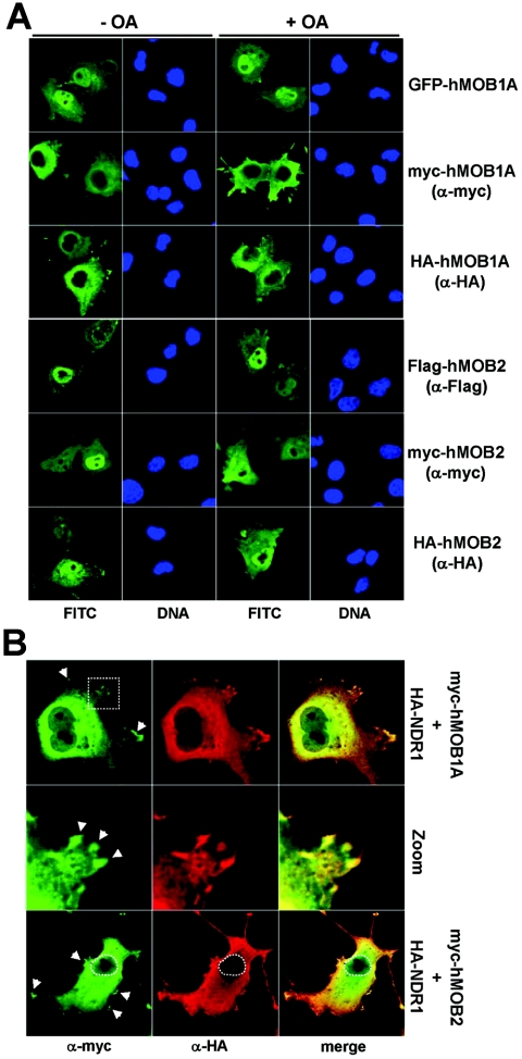
FIG. 5.
hMOB1A is mostly cytoplasmic, in contrast to hMOB2; however, both species colocalize with NDR1 in the cytoplasm/membrane. A. COS-7 cells expressing either GFP-hMOB1A (top panels), myc-hMOB1A (second top panels), HA-hMOB1A (middle top panels), Flag-hMOB2 (middle bottom panels), myc-hMOB2 (second bottom panels), or HA-hMOB2 (bottom panels) were either left untreated (− OA) or incubated with 1 μM okadaic acid (+ OA), before processing for immunofluorescence microscopy using no, anti-myc, anti-HA Y11, or anti-Flag M2 antibody. GFP/FITC signals are shown in green. Nuclei are indicated in blue. B. Cells cotransfected with myc-hMOB1A/HA-NDR1 (top panels) or myc-hMOB2/HA-NDR1 (bottom panels) were analyzed by immunofluorescence microscopy using anti-myc 9E10 (green) and anti-HA Y11 (red) antibodies. Merged images of hMOB and NDR1 staining are shown (merge). Enlargement of the region indicated by a white rectangle is shown in the middle panels. Arrowheads indicate colocalization sites at the plasma membrane.

