

Abstract
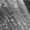
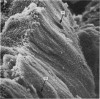
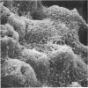
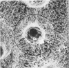
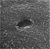
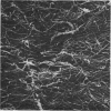
In this scanning electron microscopic study blocks of collagen fibrils were prepared from human articular cartilage, using two techinques which selectively removed either the proteoglycans alone, or both the proteoglycans and the collagen fibrils, of the non-calcified cartilage layer. Amino acid analysis of the fibrils confirmed the purity of the collagen after proteoglycan extraction. The cartilage was scanned in four different ways: (1) normal to the articular surface, (2) in superficial sections, (3) on surfaces of blocks which had been broken in planes parallel to artificial splits make by the insertion of a pin, and (4) on fracture surfaces which traversed the calcified cartilage and the subchondral bone. Five features of the organization of the collagen fibrils were specially noted: (1) Individual fibrils within the trabeculae joined to form small fibre bundles which became grouped into larger bundles at the calcified/uncalcified interface. (2) Fibrils in the deep and middle zones which, exhibiting the characteristic surface periodicity of collagen, were generally oriented towars the articular surface in large bundles approximately 55 micronm across. (3) In the superficial zone, fibrils ran parallel to the surface. (4) The surface fibrils had random orientation, even at the bases of empty lacunae vacated by chondrocytes during specimen preparation. (5) The collagen fibrils of the lacunar walls appeared to be thinner and more closely packed than thos between the lacunae. The fine collagen fibrils associated with the lacunar walls were frequently observed to pass through a large lacunar space, resulting in the formation of two or more compartments, each of which was presumably filled with a chondrocyte in the living cartilage.
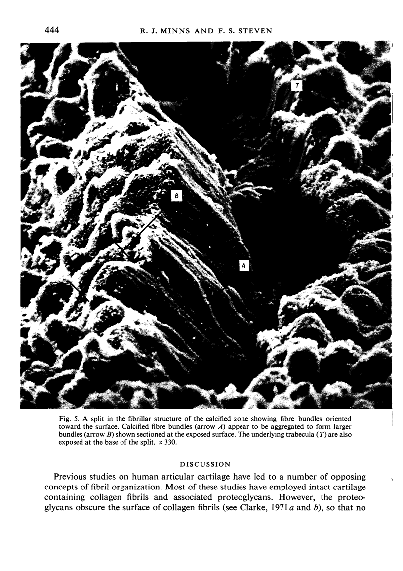
Full text
PDF


Images in this article


Selected References
These references are in PubMed. This may not be the complete list of references from this article.
- Bullough P., Goodfellow J. The significance of the fine structure of articular cartilage. J Bone Joint Surg Br. 1968 Nov;50(4):852–857. [PubMed] [Google Scholar]
- Clarke I. C. Articular cartilage: a review and scanning electron microscope study. 1. The interterritorial fibrillar architecture. J Bone Joint Surg Br. 1971 Nov;53(4):732–750. [PubMed] [Google Scholar]
- Clarke I. C. Surface characteristics of human articular cartilage--a scanning electron microscope study. J Anat. 1971 Jan;108(Pt 1):23–30. [PMC free article] [PubMed] [Google Scholar]
- Ghadially F. N., Ailsby R. L., Oryschak A. F. Scanning electron microscopy of superficial defects in articular cartilage. Ann Rheum Dis. 1974 Jul;33(4):327–332. doi: 10.1136/ard.33.4.327. [DOI] [PMC free article] [PubMed] [Google Scholar]
- Lane J. M., Weiss C. Review of articular cartilage collagen research. Arthritis Rheum. 1975 Nov-Dec;18(6):553–562. doi: 10.1002/art.1780180605. [DOI] [PubMed] [Google Scholar]
- MacCONAILL M. A. The movements of bones and joints; the mechanical structure of articulating cartilage. J Bone Joint Surg Br. 1951 May;33B(2):251–257. [PubMed] [Google Scholar]
- McCutchen C. W. A note upon tensile stresses in the collagen fibers of articular cartilage. Med Electron Biol Eng. 1965 Oct;3(4):447–448. doi: 10.1007/BF02476145. [DOI] [PubMed] [Google Scholar]
- Meachim G., Denham D., Emery I. H., Wilkinson P. H. Collagen alignments and artificial splits at the surface of human articular cartilage. J Anat. 1974 Sep;118(Pt 1):101–118. [PMC free article] [PubMed] [Google Scholar]
- Meachim G. Light microscopy of Indian ink preparations of fibrillated cartilage. Ann Rheum Dis. 1972 Nov;31(6):457–464. doi: 10.1136/ard.31.6.457. [DOI] [PMC free article] [PubMed] [Google Scholar]
- Minns R. J. Letter: Cartilage ulceration and shear fatigue failure. Lancet. 1976 Apr 24;1(7965):907–908. doi: 10.1016/s0140-6736(76)92124-3. [DOI] [PubMed] [Google Scholar]
- Mow V. C., Lai W. M. Some surface characteristics of articular cartilage. I. A scanning electron microscopy study and a theoretical model for the dynamic interaction of synovial fluid and articular cartilage. J Biomech. 1974 Sep;7(5):449–456. doi: 10.1016/0021-9290(74)90007-4. [DOI] [PubMed] [Google Scholar]
- Podrazky V., Steven F. S., Jackson D. S., Weiss J. B., Leibovich S. J. Interaction of tropocollagen with protein-polysaccharide complexes. An analysis of the ionic groups responsible for interaction. Biochim Biophys Acta. 1971 Mar 23;229(3):690–697. doi: 10.1016/0005-2795(71)90285-6. [DOI] [PubMed] [Google Scholar]
- Redler I. A scanning electron microscopic study of human normal and osteoarthritic articular cartilage. Clin Orthop Relat Res. 1974;(103):262–268. doi: 10.1097/00003086-197409000-00087. [DOI] [PubMed] [Google Scholar]
- Redler I., Mow V. C., Zimny M. L., Mansell J. The ultrastructure and biomechanical significance of the tidemark of articular cartilage. Clin Orthop Relat Res. 1975 Oct;(112):357–362. [PubMed] [Google Scholar]
- Steven F. S., Thomas H. Preparation of insoluble collagen from human cartilage. Biochem J. 1973 Sep;135(1):245–247. doi: 10.1042/bj1350245. [DOI] [PMC free article] [PubMed] [Google Scholar]
- TRUETA J., LITTLE K. The vascular contribution to osteogenesis. II. Studies with the electron microscope. J Bone Joint Surg Br. 1960 May;42-B:367–376. doi: 10.1302/0301-620X.42B2.367. [DOI] [PubMed] [Google Scholar]
- Weiss C., Rosenberg L., Helfet A. J. An ultrastructural study of normal young adult human articular cartilage. J Bone Joint Surg Am. 1968 Jun;50(4):663–674. doi: 10.2106/00004623-196850040-00002. [DOI] [PubMed] [Google Scholar]