

Abstract
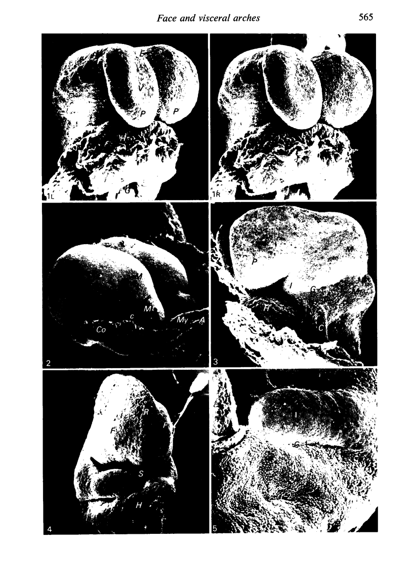
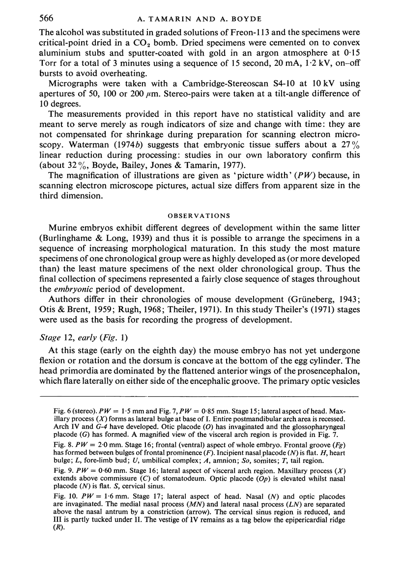
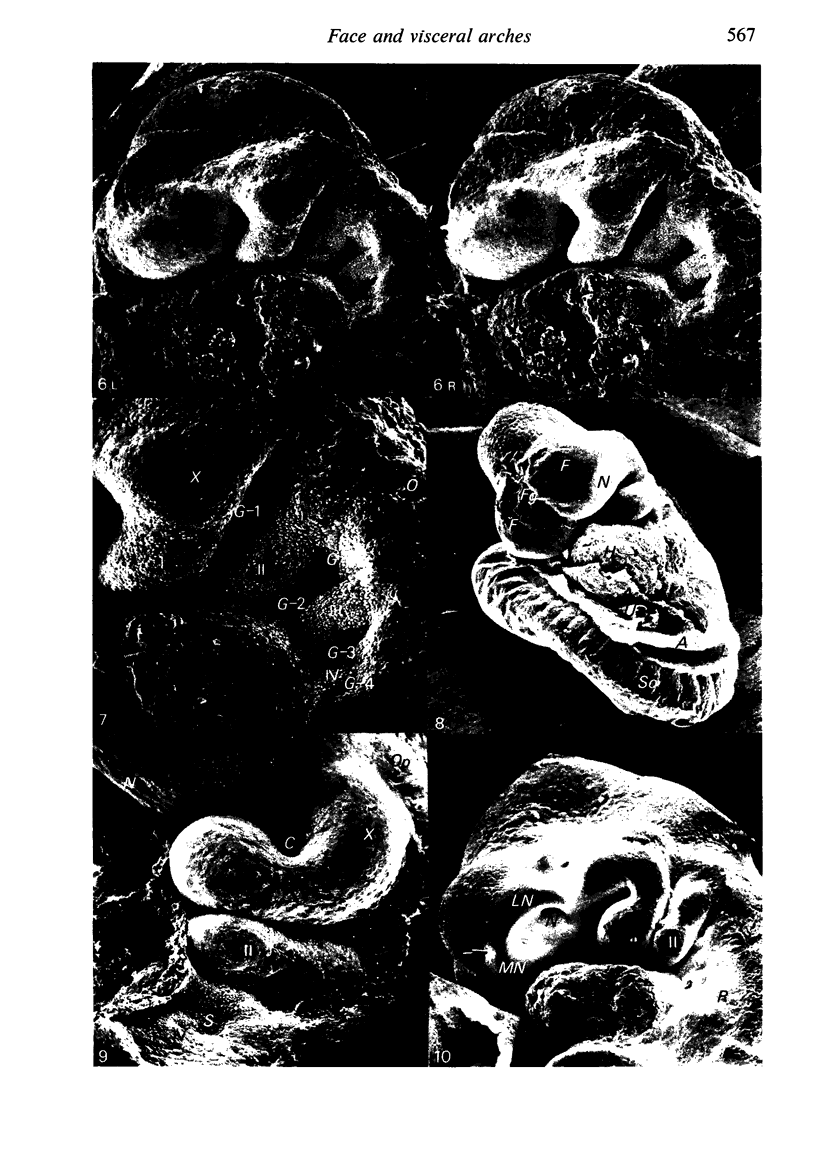
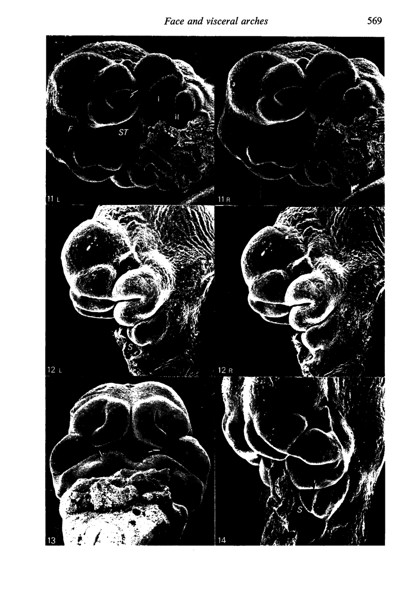
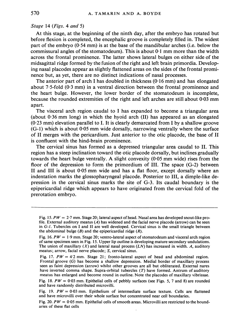
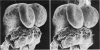
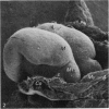
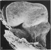
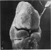
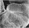
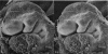
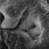
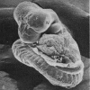
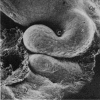
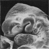
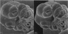
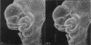
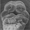
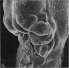
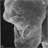
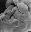
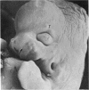
The morphogenesis of the face and visceral arch region were studied by scanning electron microscopy in 63 mouse embryos between 8 and 13 days post coitum. The developmental stages were determined by reference to Theiler's (1971) scheme for the categorization of the normal stages of development. After the cervical fold is formed (stage 12) the mandibular primordium appears (stage 13) followed by the primordia of arches II and III (stage 14 with arch IV forming last (stage 15). Arches II, III and IV regress in reverse order, with no evidence that a cervical sinus pouch is formed. Placodes of the sensory ganglia from cranial nerves 7, 9 and 10 are formed in the first, second and third visceral grooves respectively. Formation of the external auditory meatus and its surrounding tubercles were also observed. Morphogenesis of the upper face is dominated by the midsagittal grove, which extends into the stomatodeum; and by the medial nasal, lateral nasal and maxillary processes. The temporal changes in shape and the interrelationships of the structures mentioned are described in detail.
Full text
PDF
Images in this article
Selected References
These references are in PubMed. This may not be the complete list of references from this article.
- Frazer J. E. The Disappearance of the Precervical Sinus. J Anat. 1926 Oct;61(Pt 1):132–143. [PMC free article] [PubMed] [Google Scholar]
- OTIS E. M., BRENT R. Equivalent ages in mouse and human embryos. Anat Rec. 1954 Sep;120(1):33–63. doi: 10.1002/ar.1091200104. [DOI] [PubMed] [Google Scholar]
- Tamarin A., Boyde A. Three-dimensional anatomy of the 8-day mouse concepts: a study by scanning electron microscopy. J Embryol Exp Morphol. 1976 Dec;36(3):575–596. [PubMed] [Google Scholar]
- WEDIN B. Proneuromeres and neuromeres in the cerebral tube of Torpedo ocellata. Acta Anat (Basel) 1954;21(1):59–69. doi: 10.1159/000140919. [DOI] [PubMed] [Google Scholar]