

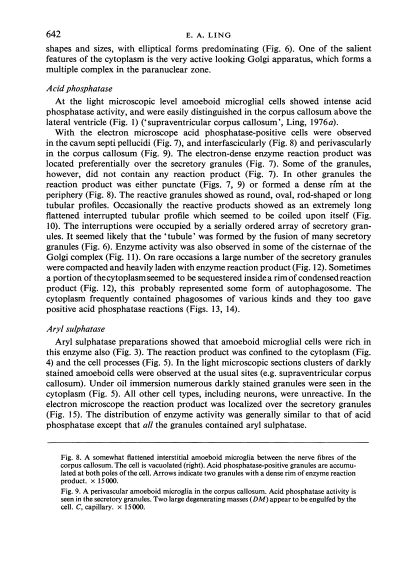
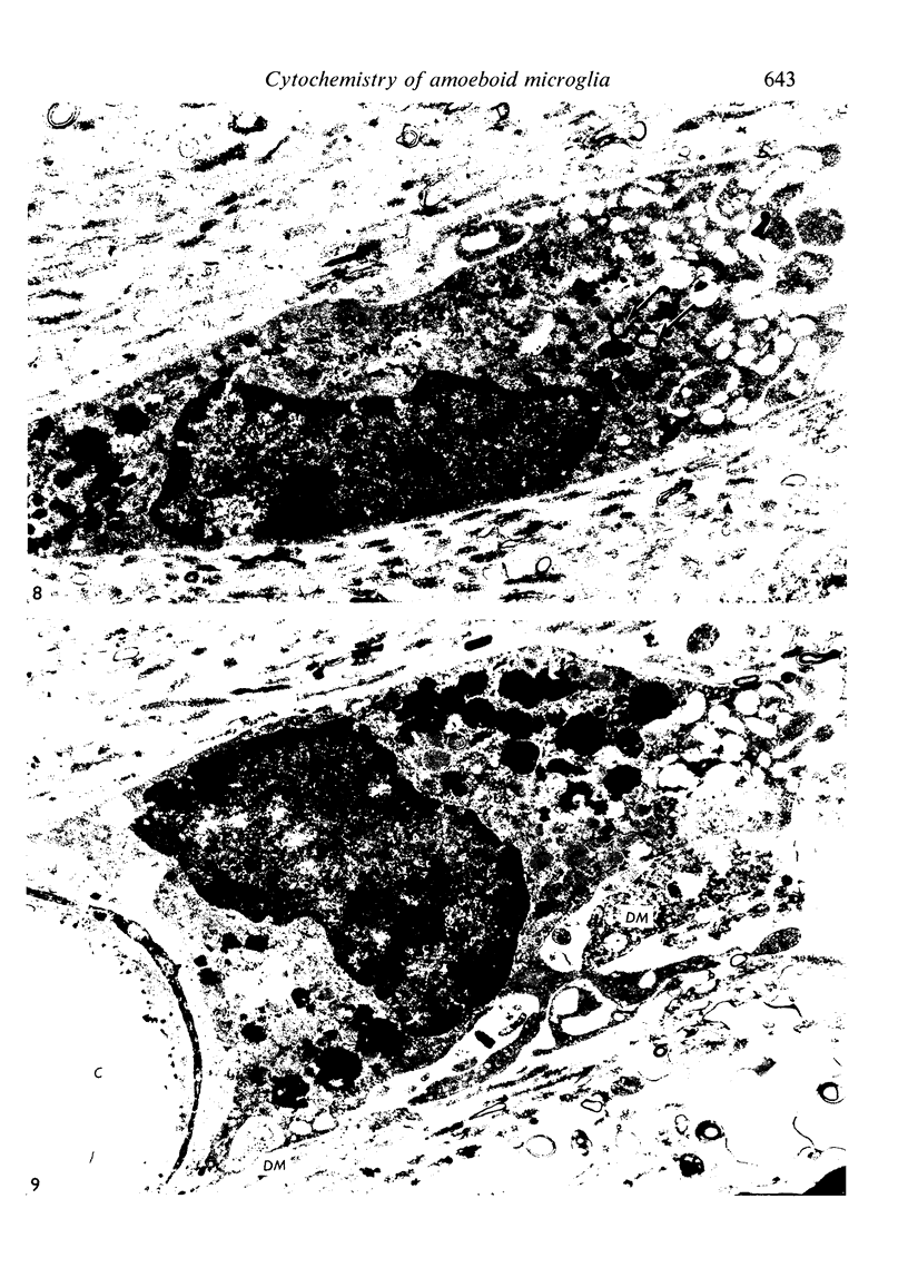
Abstract
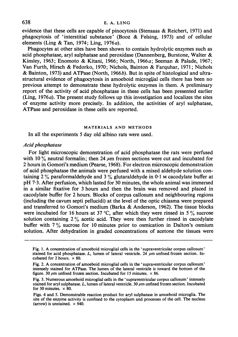
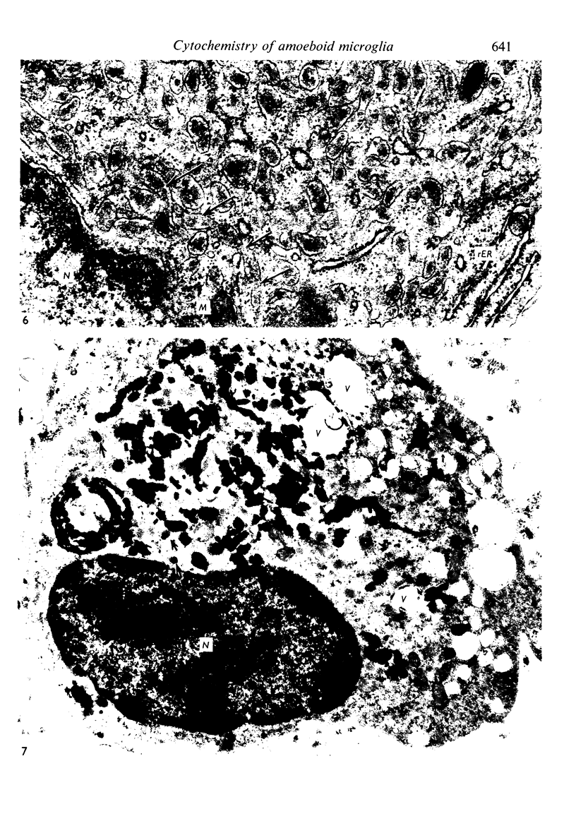
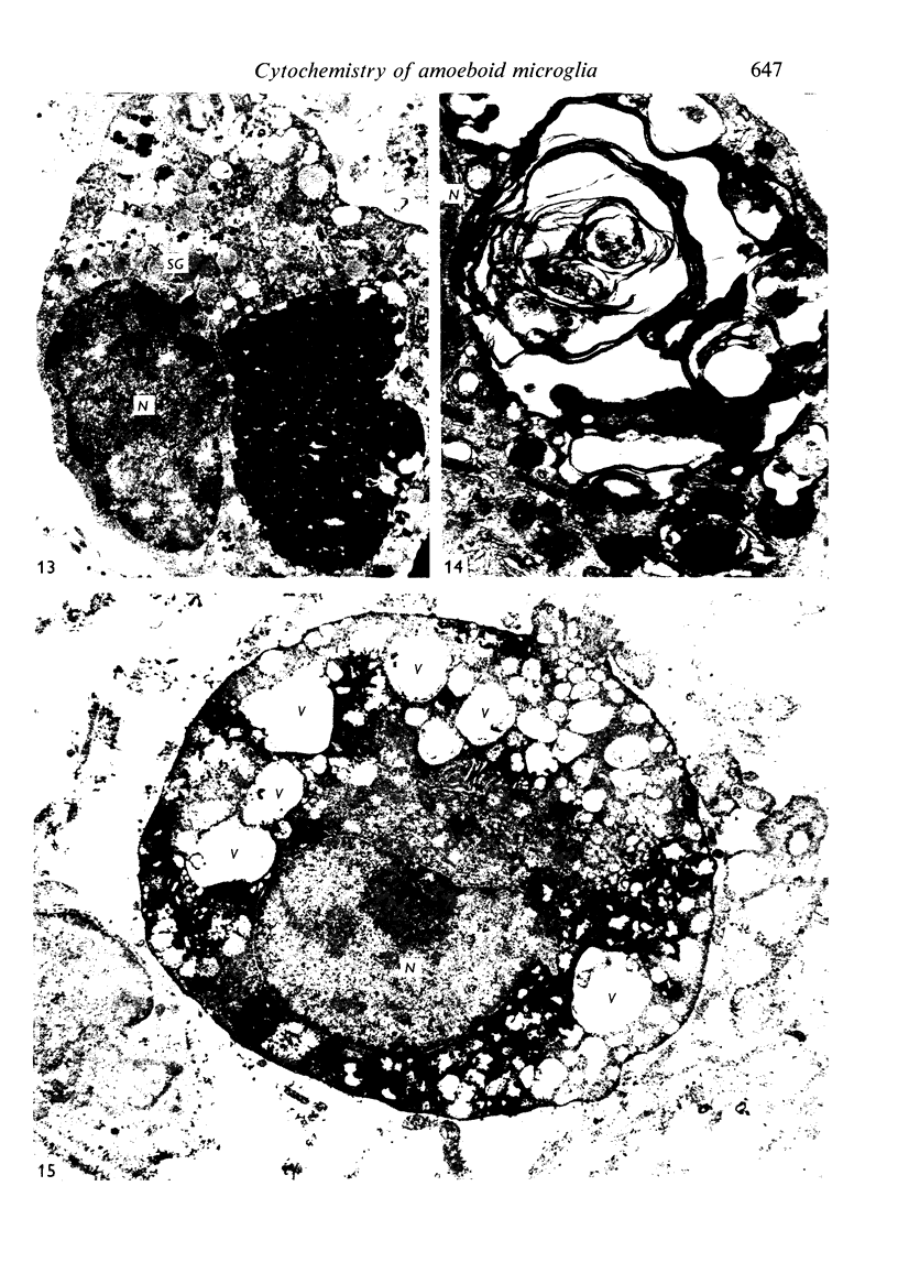
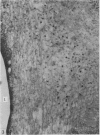
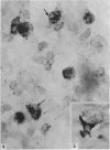
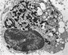
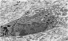
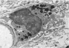
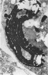
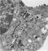
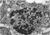
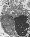
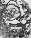
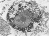
A cytochemical study of the amoeboid microglial cells in the brain of the neonatal rat has shown that these vacuolated cells exhibit strong acid phosphatase, aryl sulphatase and adenosine triphosphatase (ATPase) activities. Endogenous peroxidase, however, was not present. With the electron microscope the reaction product of acid phosphatase was found to be localized in some of the Golgi cisternae, in the majority of the electron-dense secretory granules, and in an occasional long tubular profile. The secretory granules were not uniformly stained for this enzyme, some showing only a focal reaction or none at all. The distribution of the activity of aryl sulphatase corresponded to that of acid phosphatase except that all the granules appeared to contain the former enzyme. With the light microscope the amoeboid microglial cells were intensely stained for ATPase. From these observations it was concluded that amoeboid microglia are active phagocytes and their enzyme-rich secretory granules are lysosomes.
Full text
PDF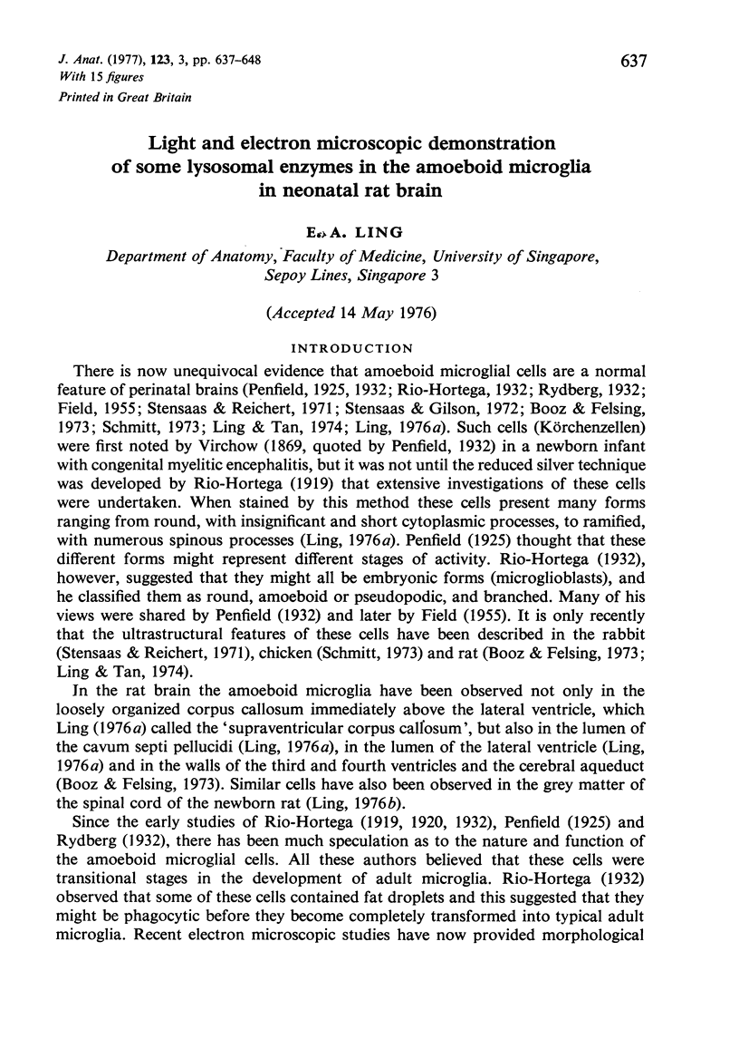


Images in this article
Selected References
These references are in PubMed. This may not be the complete list of references from this article.
- DANNENBERG A. M., Jr, BURSTONE M. S., WALTER P. C., KINSLEY J. W. A histochemical study of phagocytic and enzymatic functions of rabbit mononuclear and polymorphonuclear exudate cells and alveolar macrophages. I. Survey and quantitation of enzymes, and states of cellular activation. J Cell Biol. 1963 Jun;17:465–486. doi: 10.1083/jcb.17.3.465. [DOI] [PMC free article] [PubMed] [Google Scholar]
- Enomoto T., Kitani T. [Electron microscopic studies on peroxidase and acid phosphatase reaction in human leukocytes (in normal and leukemic cells and on phagocytosis)]. Nihon Ketsueki Gakkai Zasshi. 1966 Aug;29(4):554–570. [PubMed] [Google Scholar]
- FIELD E. J. Observations on the development of microglia together with a note on the influence of cortisone. J Anat. 1955 Apr;89(2):201–208. [PMC free article] [PubMed] [Google Scholar]
- Goldfischer S. The cytochemical demonstration of lysosomal aryl sulfatase activity by light and electron microscopy. J Histochem Cytochem. 1965 Jul-Aug;13(6):520–523. doi: 10.1177/13.6.520. [DOI] [PubMed] [Google Scholar]
- Graham R. C., Jr, Karnovsky M. J. The early stages of absorption of injected horseradish peroxidase in the proximal tubules of mouse kidney: ultrastructural cytochemistry by a new technique. J Histochem Cytochem. 1966 Apr;14(4):291–302. doi: 10.1177/14.4.291. [DOI] [PubMed] [Google Scholar]
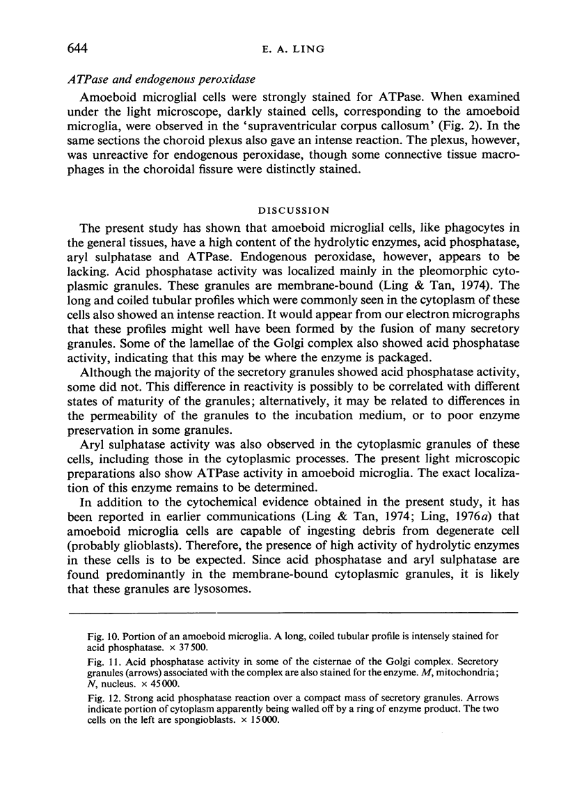
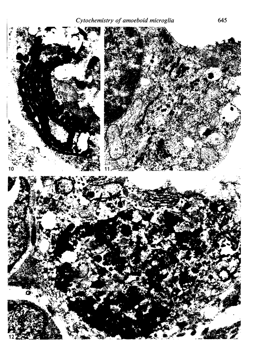
- Ling E. A. Some aspects of amoeboid microglia in the corpus callosum and neighbouring regions of neonatal rats. J Anat. 1976 Feb;121(Pt 1):29–45. [PMC free article] [PubMed] [Google Scholar]
- Ling E. A., Tan C. K. Amoeboid microglial cells in the corpus callosum of neonatal rats. Arch Histol Jpn. 1974 Mar;36(4):265–280. doi: 10.1679/aohc1950.36.265. [DOI] [PubMed] [Google Scholar]
- Nichols B. A., Bainton D. F. Differentiation of human monocytes in bone marrow and blood. Sequential formation of two granule populations. Lab Invest. 1973 Jul;29(1):27–40. [PubMed] [Google Scholar]
- Nichols B. A., Bainton D. F., Farquhar M. G. Differentiation of monocytes. Origin, nature, and fate of their azurophil granules. J Cell Biol. 1971 Aug;50(2):498–515. doi: 10.1083/jcb.50.2.498. [DOI] [PMC free article] [PubMed] [Google Scholar]
- North R. J. The localization by electron microscopy of acid phosphatase activity in guinea pig macrophages. J Ultrastruct Res. 1966 Sep;16(1):96–108. doi: 10.1016/s0022-5320(66)80025-4. [DOI] [PubMed] [Google Scholar]
- North R. J. The localization by electron microscopy of nucleoside phosphatase activity in guinea pig phagocytic cells. J Ultrastruct Res. 1966 Sep;16(1):83–95. doi: 10.1016/s0022-5320(66)80024-2. [DOI] [PubMed] [Google Scholar]
- Penfield W. Microglia and the Process of Phagocytosis in Gliomas. Am J Pathol. 1925 Jan;1(1):77–90.15. [PMC free article] [PubMed] [Google Scholar]
- Schmitt D. Uber glykoproteidhaltige amöboide Zellen im embryonalen Hühnergehirn. Eine licht- und elektronenmikroskopische Untersuchung zur Frage der Volumenreserve bei Wachstumsprozessen im Gehirn. Z Anat Entwicklungsgesch. 1973 Dec 31;142(3):341–358. [PubMed] [Google Scholar]
- Seeman P. M., Palade G. E. Acid phosphatase localization in rabbit eosinophils. J Cell Biol. 1967 Sep;34(3):745–756. doi: 10.1083/jcb.34.3.745. [DOI] [PMC free article] [PubMed] [Google Scholar]
- Stensaas L. J., Gilson B. C. Ependymal and subependymal cells of the caudato-pallial junction in the lateral ventricle of the neonatal rabbit. Z Zellforsch Mikrosk Anat. 1972;132(3):297–322. doi: 10.1007/BF02450711. [DOI] [PubMed] [Google Scholar]
- Stensaas L. J., Reichert W. H. Round and amoeboid microglial cells in the neonatal rabbit brain. Z Zellforsch Mikrosk Anat. 1971;119(2):147–163. doi: 10.1007/BF00324517. [DOI] [PubMed] [Google Scholar]
- WACHSTEIN M., MEISEL E. Histochemistry of hepatic phosphatases of a physiologic pH; with special reference to the demonstration of bile canaliculi. Am J Clin Pathol. 1957 Jan;27(1):13–23. doi: 10.1093/ajcp/27.1.13. [DOI] [PubMed] [Google Scholar]
- van Furth R., Hirsch J. G., Fedorko M. E. Morphology and peroxidase cytochemistry of mouse promonocytes, monocytes, and macrophages. J Exp Med. 1970 Oct 1;132(4):794–812. doi: 10.1084/jem.132.4.794. [DOI] [PMC free article] [PubMed] [Google Scholar]