

Abstract
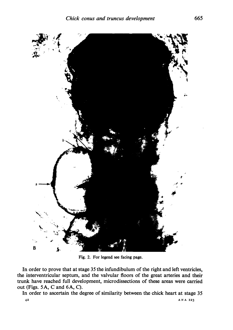
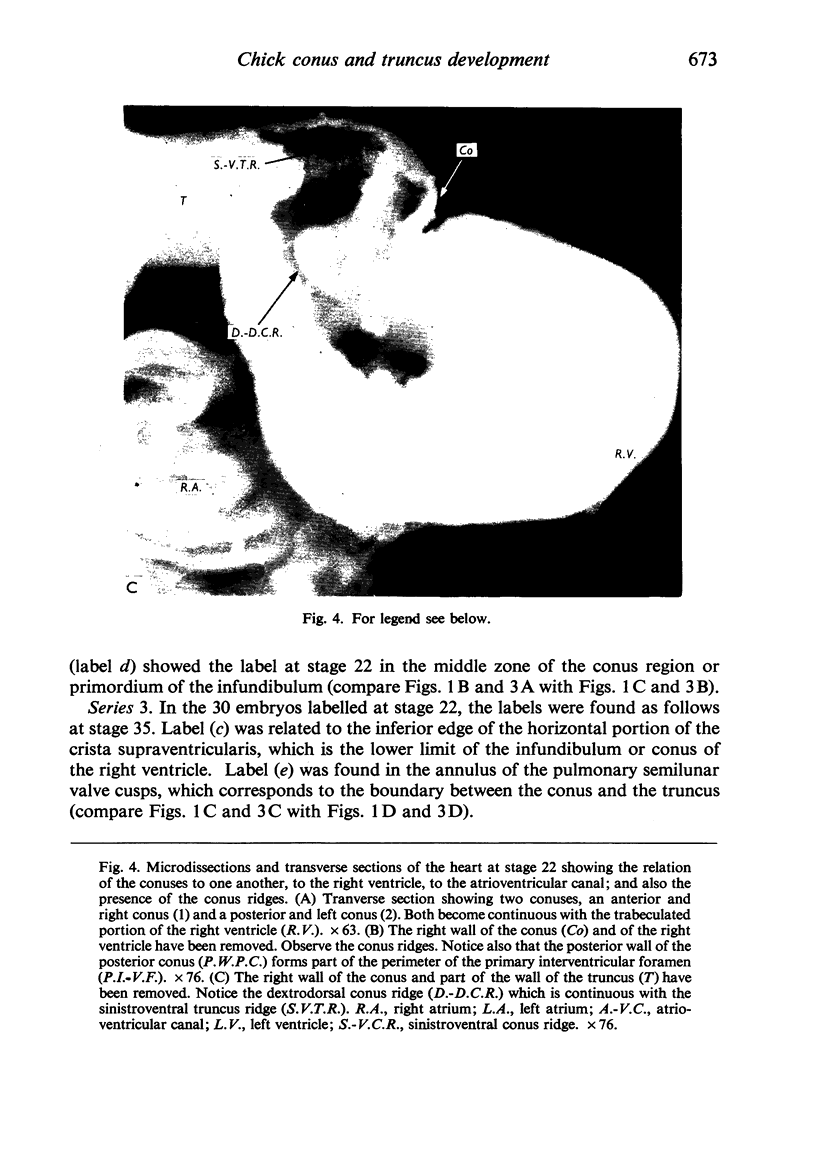
The development of the truncus and the conus was studied in the chick embryo by in vivo labelling techniques. The earliest labels were placed at the stage of fusion of the myocardial troughs (stage 9-) and they were traced until the mature heart stage (stage 35). Microdissections and light microscopic studies were also carried out. The results are discussed in relation to the human heart. Our experiments permit the following conclusions: (1) At stage 9- fusion of the myocardial troughs takes place at the level of the primordium of the trabeculated portion of the right ventricle, when neither the conus nor the truncus are present. (2) At stage 12 (loop stage) there appears the caudal portion of the conus, which constitutes the cephalic end of the cardiac tube. (3) The truncus appears between stages 13 and 22. (4) At stage 22 angular junction between the conus and the truncus is the area where the semilunar valve cusps of the great arteries will develop and that, at this same stage, the junction between the conus and the trabeculated portion of the right ventricle seen from the right surface corresponds to the inferior edge of the crista supraventricularis. (5) It was confirmed that the pulmonary semilunar valve cusps originate from the walls of the truncus. (6) The development of the conus and truncus are similar in chick and man. (7) Histologically, in the chick, the wall of the truncus and the conus contain cardiac muscle as late as stage 28, but from then on the walls of the truncus are transformed into connective tissue and plain muscle.
Full text
PDF


Images in this article


Selected References
These references are in PubMed. This may not be the complete list of references from this article.
- Anderson R. H., Wilkinson J. L., Arnold R., Lubkiewicz K. Morphogenesis of bulboventricular malformations. I. Consideration of embryogenesis in the normal heart. Br Heart J. 1974 Mar;36(3):242–255. doi: 10.1136/hrt.36.3.242. [DOI] [PMC free article] [PubMed] [Google Scholar]
- Argüello C., de la Cruz M. V., Gómez C. S. Experimental study of the formation of the heart tube in the chick embryo. J Embryol Exp Morphol. 1975 Feb;33(1):1–11. [PubMed] [Google Scholar]
- Asami I. Beitrag zur Entwicklung des Kammerseptums im menschlichen Herzen mit besonderer Berücksichtigung der sogenannten Bulbusdrehung. Z Anat Entwicklungsgesch. 1969;128(1):1–17. [PubMed] [Google Scholar]
- Castro-Quezada A., Nadal-Ginard B., De La Cruz M. V. Experimental study of the formation of the bulboventricular loop in the chick. J Embryol Exp Morphol. 1972 Jun;27(3):623–637. [PubMed] [Google Scholar]
- GRANT R. P. The embryology of ventricular flow pathways in man. Circulation. 1962 May;25:756–779. doi: 10.1161/01.cir.25.5.756. [DOI] [PubMed] [Google Scholar]
- Goor D. A., Dische R., Lillehei C. W. The conotruncus. I. Its normal inversion and conus absorption. Circulation. 1972 Aug;46(2):375–384. doi: 10.1161/01.cir.46.2.375. [DOI] [PubMed] [Google Scholar]
- SEICHERT V. STUDY OF THE TISSUE AND ORGAN ANLAGE SHIFTS BY THE METHOD OF PLASTIC LINEAR MARKING. Folia Morphol (Praha) 1965;13:228–238. [PubMed] [Google Scholar]
- Stalsberg H., DeHaan R. L. The precardiac areas and formation of the tubular heart in the chick embryo. Dev Biol. 1969 Feb;19(2):128–159. doi: 10.1016/0012-1606(69)90052-9. [DOI] [PubMed] [Google Scholar]
- VAN MIEROP L. H., ALLEY R. D., KAUSEL H. W., STRANAHAN A. The anatomy and embryology of endocardial cushion defects. J Thorac Cardiovasc Surg. 1962 Jan;43:71–83. [PubMed] [Google Scholar]
- VANMIEROP L. H., ALLEY R. D., KAUSEL H. W., STRANAHAN A. PATHOGENESIS OF TRANSPOSITION COMPLEXES. I. EMBRYOLOGY OF THE VENTRICLES AND GREAT ARTERIES. Am J Cardiol. 1963 Aug;12:216–225. doi: 10.1016/0002-9149(63)90311-4. [DOI] [PubMed] [Google Scholar]