Abstract
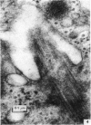
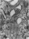
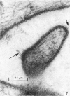
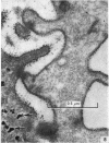
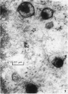
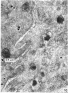
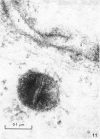
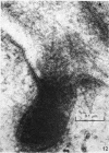
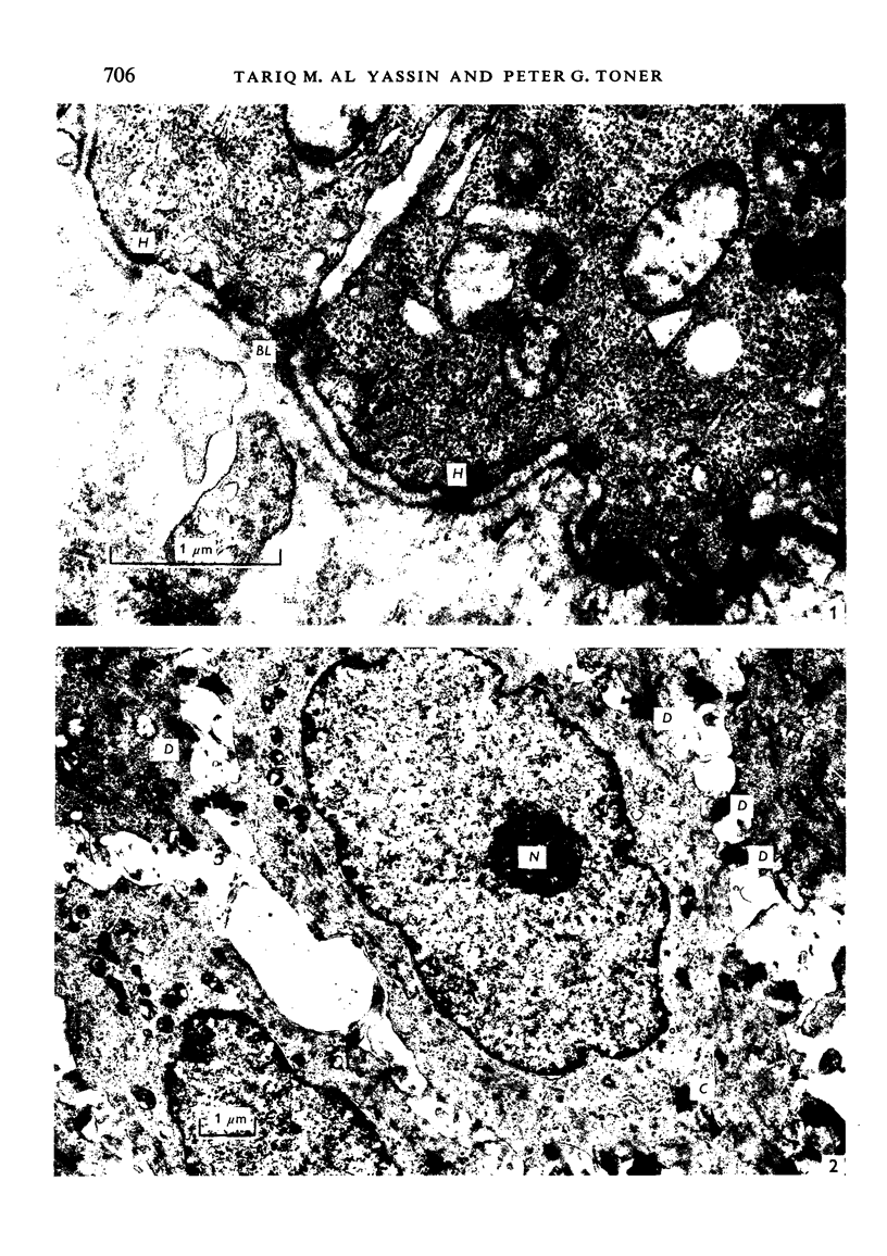
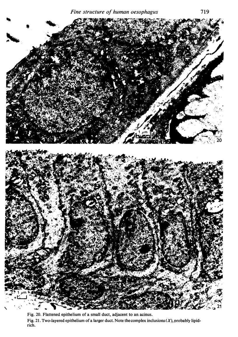
Normal squamous epithelium and submucosal glands of the human oesophagus have been studied in biopsy and resection specimens. The squamous epithelium has a structure similar to that seen in other histologically comparable sites, and contains Langerhans cells. Features of note within the squamous cells include microfibrillar intranuclear bodies and occasional intracytoplasmic desmosomes. The submucosal glands are comparable in many respects to the mucous-secreting minor salivary glands. Myoepithelial cells are observed within the acini and smaller ducts; occasional oncocytic cells are identified at the junction between duct and acinus. The innvervation of these glands appears to be indirect.
Full text
PDF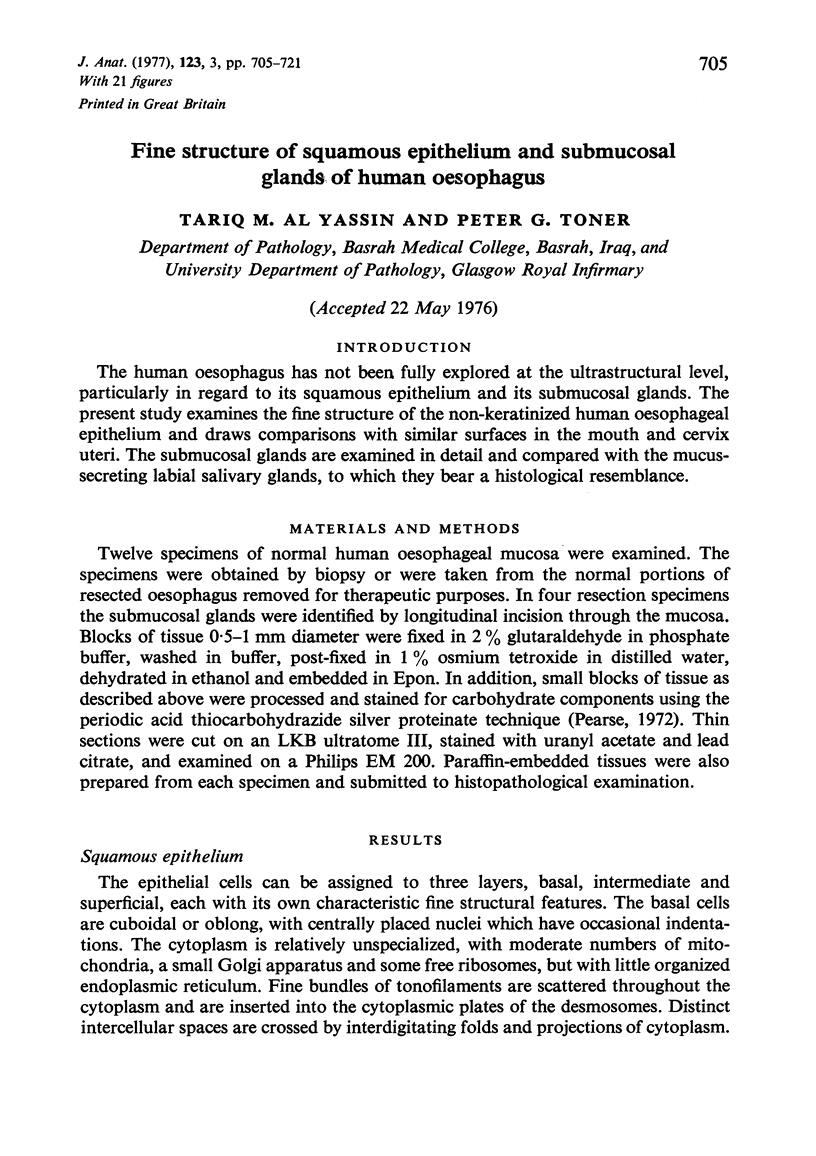
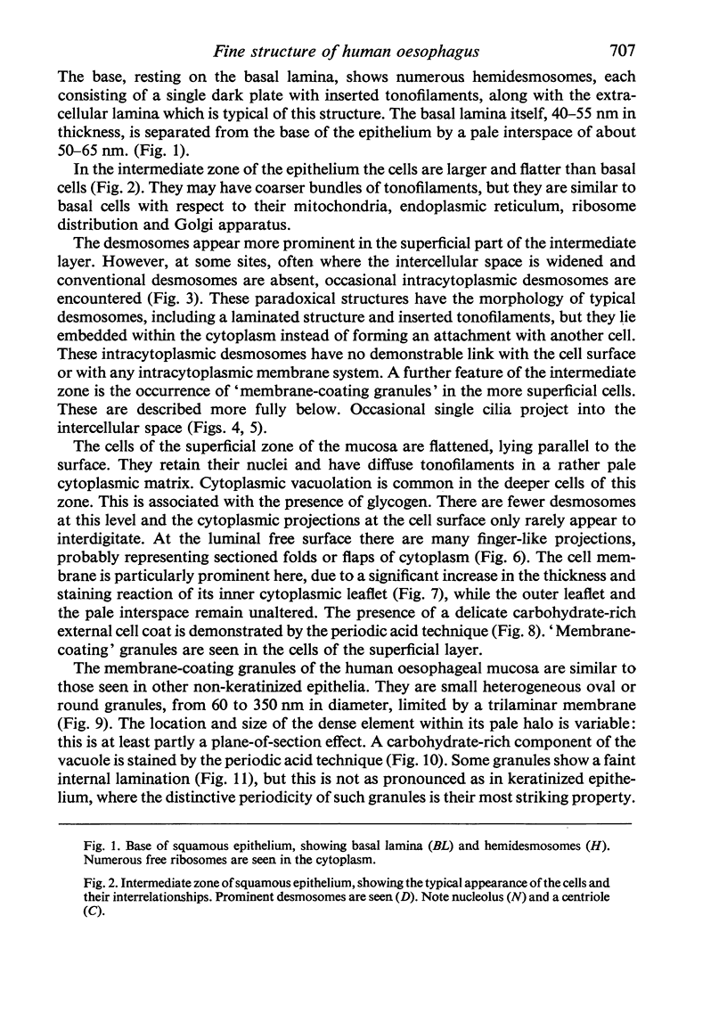
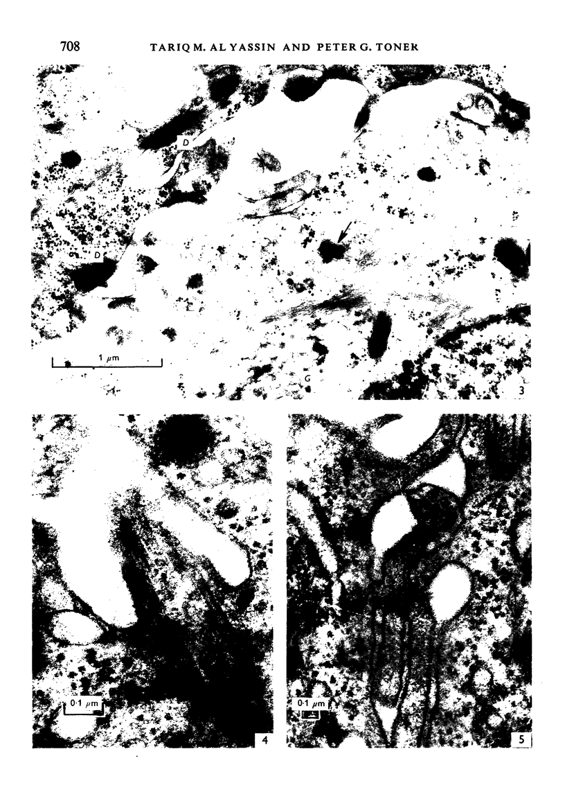
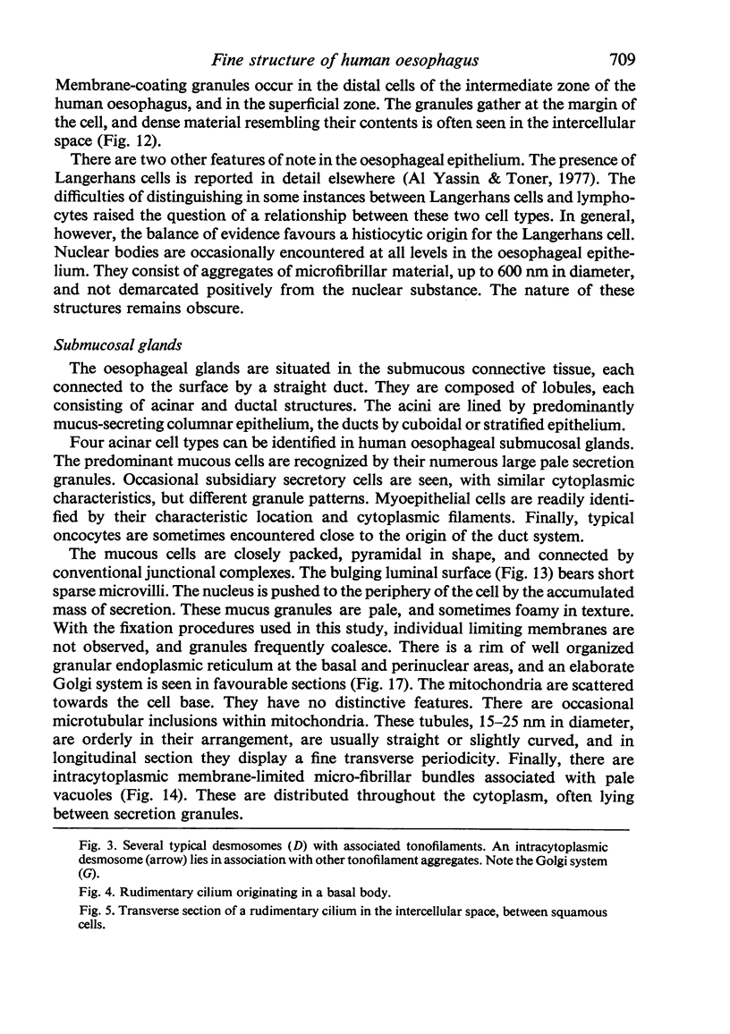
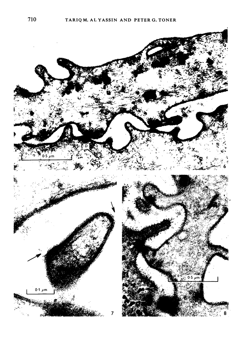
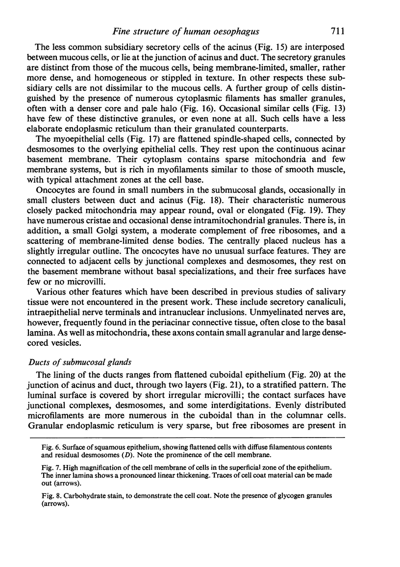
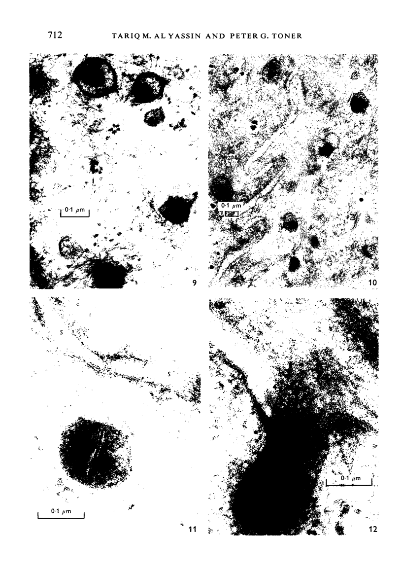
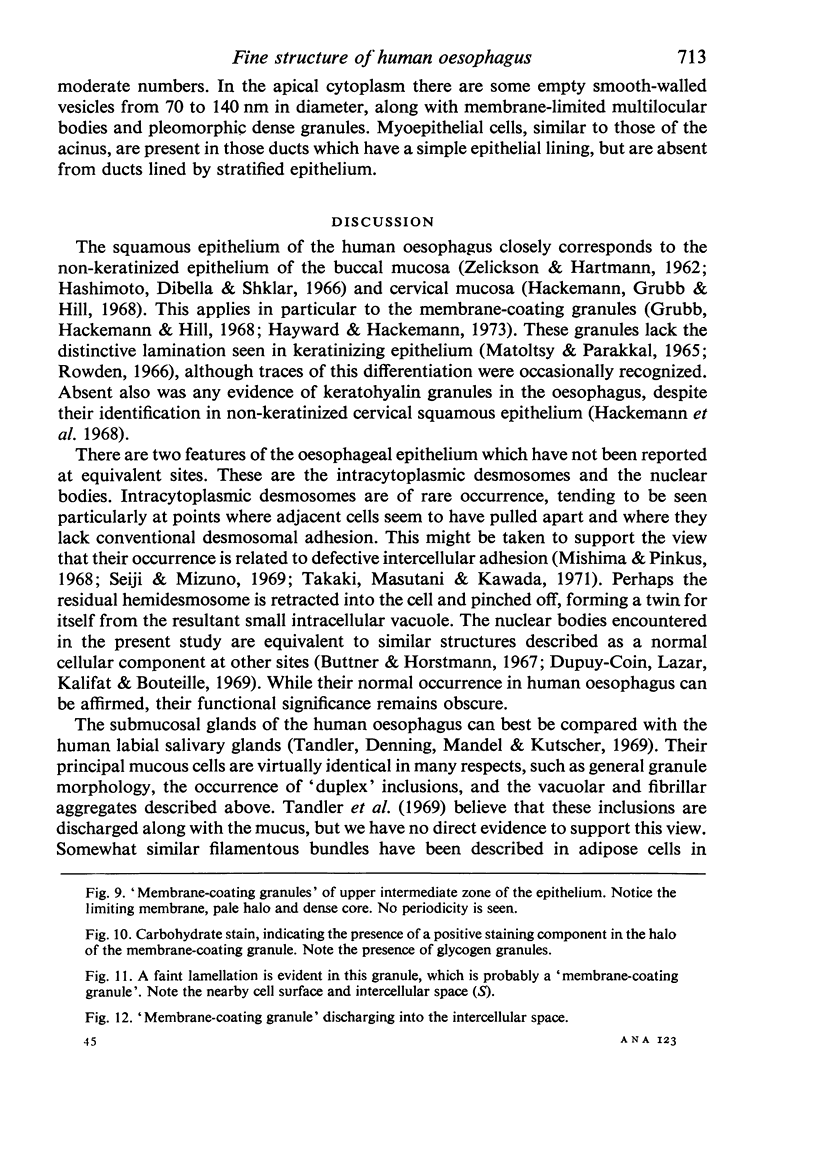
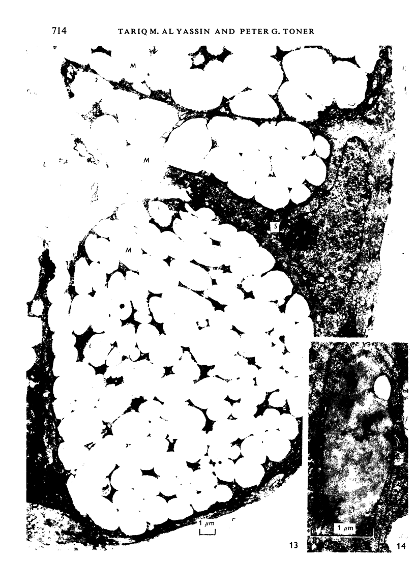
Images in this article
Selected References
These references are in PubMed. This may not be the complete list of references from this article.
- BALOGH K., Jr, ROTH S. I. HISTOCHEMICAL AND ELECTRON MICROSCOPIC STUDIES OF EOSINOPHILIC GRANULAR CELLS (ONCOCYTES) IN TUMORS OF THE PAROTID GLAND. Lab Invest. 1965 Mar;14:310–320. [PubMed] [Google Scholar]
- Bogart B. I. The effect of aging on the rat submandibular gland: an ultrastructural, cytochemical and biochemical study. J Morphol. 1970 Mar;130(3):337–351. doi: 10.1002/jmor.1051300306. [DOI] [PubMed] [Google Scholar]
- Büttner D. W., Horstmann E. Haben die Sphaeridien in den Zellkernen kranker Gewebe eine pathognomonische Bedeutung? Virchows Arch Pathol Anat Physiol Klin Med. 1967;343(2):142–163. [PubMed] [Google Scholar]
- CHAUNCEY H. H., SHKLAR G., BROOKS R. A. Histochemistry of human salivary gland tumors. Oral Surg Oral Med Oral Pathol. 1962 Aug;15:950–964. doi: 10.1016/0030-4220(62)90090-7. [DOI] [PubMed] [Google Scholar]
- Dupuy-Coin A. M., Lazar P., Kalifat S. R., Bouteille M. A method of quantitation of nuclear bodies in electron microscopy. J Ultrastruct Res. 1969 May;27(3):244–249. doi: 10.1016/s0022-5320(69)80015-8. [DOI] [PubMed] [Google Scholar]
- Grubb C., Hackemann M., Hill K. R. Small granules and plasma membrane thickening in human cervical squamous epithelium. J Ultrastruct Res. 1968 Mar;22(5):458–468. doi: 10.1016/s0022-5320(68)90034-8. [DOI] [PubMed] [Google Scholar]
- Hackemann M., Grubb C., Hill K. R. The ultrastructure of normal squamous epithelium of the human cervix uteri. J Ultrastruct Res. 1968 Mar;22(5):443–457. doi: 10.1016/s0022-5320(68)90033-6. [DOI] [PubMed] [Google Scholar]
- Hashimoto K., DiBella R. J., Shklar G. Electron microscopic studies of the normal human buccal mucosa. J Invest Dermatol. 1966 Dec;47(6):512–525. [PubMed] [Google Scholar]
- Hayward A. F., Hackemann M. Electron microscopy of membrane-coating granules and a cell surface coat in keratinized and nonkeratinized human oral epithelium. J Ultrastruct Res. 1973 May;43(3):205–219. doi: 10.1016/s0022-5320(73)80033-4. [DOI] [PubMed] [Google Scholar]
- Hübner G., Paulussen F., Kleinsasser O. Zur Feinstruktur und Genese der Onkocyten. Virchows Arch Pathol Anat Physiol Klin Med. 1967;343(1):34–50. [PubMed] [Google Scholar]
- LILLIBRIDGE C. B. THE FINE STRUCTURE OF NORMAL HUMAN GASTRIC MUCOSA. Gastroenterology. 1964 Sep;47:269–290. [PubMed] [Google Scholar]
- MATOLTSY A. G., PARAKKAL P. F. MEMBRANE-COATING GRANULES OF KERATINIZING EPITHELIA. J Cell Biol. 1965 Feb;24:297–307. doi: 10.1083/jcb.24.2.297. [DOI] [PMC free article] [PubMed] [Google Scholar]
- MCGAVRAN M. H. THE ULTRASTRUCTURE OF PAPILLARY CYSTADENOMA LYMPHOMATOSUM OF THE PAROTID GLAND. Virchows Arch Pathol Anat Physiol Klin Med. 1965 Jan 15;338:195–202. doi: 10.1007/BF00963377. [DOI] [PubMed] [Google Scholar]
- Mishima Y., Pinkus H. Electron microscopy of keratin layer stripped human epidermis. J Invest Dermatol. 1968 Feb;50(2):89–102. doi: 10.1038/jid.1968.12. [DOI] [PubMed] [Google Scholar]
- Mullison E. G. Silicones in head and neck surgery. Arch Otolaryngol. 1966 Jul;84(1):91–95. doi: 10.1001/archotol.1966.00760030093009. [DOI] [PubMed] [Google Scholar]
- ROTH S. I., OLEN E., HANSEN L. S. The eosinophilic cells of the parathyroid (oxyphil cells), salivary (oncocytes), and thyroid (Huerthle cells) glands. Light and electron microscopic observations. Lab Invest. 1962 Nov;11:933–941. [PubMed] [Google Scholar]
- Rowden G. "Membrane-coating" granules of mouse oesophageal and gastric epithelium. J Invest Dermatol. 1966 Oct;47(4):359–362. [PubMed] [Google Scholar]
- Rubin W., Ross L. L., Sleisenger M. H., Jefries G. H. The normal human gastric epithelia. A fine structural study. Lab Invest. 1968 Dec;19(6):598–626. [PubMed] [Google Scholar]
- SHKLAR G., CHAUNCEY H. H. PAPILLARY CYSTADENOMA LYMPHOMATOSUM, A DEVELOPMENTAL MALFORMATION: HISTOCHEMICAL EVIDENCE. J Oral Surg. 1965 May;23:222–230. [PubMed] [Google Scholar]
- Seiji M., Mizuno F. Electron microscopic study of Bowen's disease. Arch Dermatol. 1969 Jan;99(1):3–16. [PubMed] [Google Scholar]
- TANDLER B., SHIPKEY F. H. ULTRASTRUCTURE OF WARTHIN'S TUMOR. I. MITOCHONDRIA. J Ultrastruct Res. 1964 Oct;11:292–305. doi: 10.1016/s0022-5320(64)90034-6. [DOI] [PubMed] [Google Scholar]
- Takaki Y., Masutani M., Kawada A. Electron microscopic study of keratoacanthoma. Acta Derm Venereol. 1971;51(1):21–26. [PubMed] [Google Scholar]
- Tandler B., Denning C. R., Mandel I. D., Kutscher A. H. Ultrastructure of human labial salivary glands. 3. Myoepithelium and ducts. J Morphol. 1970 Feb;130(2):227–245. doi: 10.1002/jmor.1051300208. [DOI] [PubMed] [Google Scholar]
- Tandler B., Denning C. R., Mandel I. D., Kutscher A. H. Ultrastructure of human labial salivary glands. I. Acinar secretory cells. J Morphol. 1969 Apr;127(4):383–407. doi: 10.1002/jmor.1051270402. [DOI] [PubMed] [Google Scholar]
- Tandler B. Fine structure of oncocytes in human salivary glands. Virchows Arch Pathol Anat Physiol Klin Med. 1966 Dec 14;341(4):317–326. doi: 10.1007/BF00956872. [DOI] [PubMed] [Google Scholar]
- Tandler B., Ross L. L. Observations of nerve terminals in human labial salivary glands. J Cell Biol. 1969 Jul;42(1):339–343. doi: 10.1083/jcb.42.1.339. [DOI] [PMC free article] [PubMed] [Google Scholar]
- VOTH D. [Phases of cell structure in papillary cystadenolymphoma and their effect on the tumor form]. Frankf Z Pathol. 1962;71:523–530. [PubMed] [Google Scholar]
- Yassin T. M., Toner P. G. Langerhans cells in the human oesophagus. J Anat. 1976 Nov;122(Pt 2):435–445. [PMC free article] [PubMed] [Google Scholar]
- ZELICKSON A. S., HARTMANN J. F. An electron microscope study of normal human non-keratinizing oral mucosa. J Invest Dermatol. 1962 Feb;38:99–107. doi: 10.1038/jid.1962.20. [DOI] [PubMed] [Google Scholar]


